Copyright © 2017 Korean Neurological Association 105
Electron Microscopy Pathology of ADSSL1 Myopathy
Dear Editor,
Inherited muscular disorder is a heterogeneous group of genetic disorders that primarily affect skeletal muscle fibers. We have recently reported compound heterozygous mutations in ADSSL1 in two Korean families with adolescent-onset distal myopathy.1 However, ultra- structural changes associated with adenylosuccinate synthetase-like 1 (ADSSL1) myopathy have not been demonstrated previously. Here we report a third Korean family with ADSSL1 mutations and describe the ultrastructural features of ADSSL1 myopathy.
A 15-year-old girl (Supplementary Fig. 1A and B in the online-only Data Supplement, II- 2) presented with gait disturbance. She was the second child of healthy, nonconsanguineous parents. She had first noticed difficulty running when aged 8 years, and the muscle weak- ness had progressed very slowly. When we examined her at an age of 15 years, she could still ambulate independently. A neurological examination revealed diffuse muscle weakness, predominantly in the leg muscles. She did not exhibit facial weakness, sensory deficits, joint contractures, or a high-arch palate. Her serum creatine kinase (CK) level was 281 IU/l (ref- erence value <185 IU/l). The findings of needle electromyography were compatible with generalized myopathy. Her electrocardiography findings were normal. Lower-limb MRI showed subtle fatty replacement of the tibialis anterior and gastrocnemius muscles (Supple- mentary Fig. 1C-G in the online-only Data Supplement).
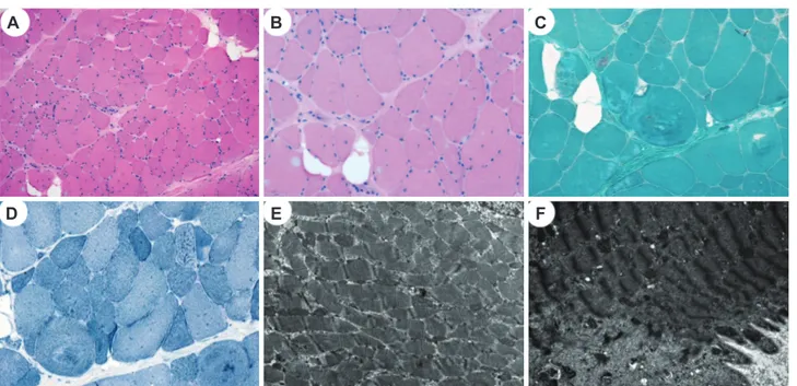
The vastus lateralis muscle was biopsied when she was 15 years old. The myofibers showed marked size and shape variations along with frequent internalization of the sarco- lemmal nuclei and splitting; vacuolization of the sarcoplasm, degenerating myofibers, and abnormal (whorled) arrangement of the myofibrils were also noted (Fig. 1A and B). In ad- dition, focal endomysial fibrosis and fatty infiltration were present. Periodic acid-Schiff and Oil Red O staining did not produce abnormal positive reactions. Modified Gomori tri- chrome and nicotinamide adenine dinucleotide-tetrazolium reductase staining revealed whorled fibers and occasional rimmed vacuoles without ragged red fibers, cytoplasmic bodies, or nemaline rods (Fig. 1C and D). The ATPase reaction with preincubations at dif- ferent pH values and immunostaining with myosin heavy chain (fast), myosin heavy chain (slow), and myosin IIa revealed a predominance of type I fibers. An electron microscopy examination showed myofibers with varying degrees of disorganization, and disorientation of the myofibrils with loss of their normal striation pattern (Fig. 1E). The myofibers show- ing diffuse or localized myofibrillar destruction contained remnants of the myofibrils, ir- regular electron-dense material of Z-line origin, and rounded and swollen mitochondria (Fig. 1F). In addition, various abnormalities of the Z-disc (including thickening) were not- ed, as well as displaced and replicated triads.
To identify the genetic cause, exome sequencing was performed on the proband (Supple- mentary Table 1 in the online-only Data Supplement). Screening of myopathy-related genes revealed 27 functionally significant variants (Supplementary Table 2 in the online-only Data Supplement). We subsequently identified compound heterozygous c.910G>A (p.D340N) and Hyung Jun Parka
Jee Eun Leea Gyeong Seon Choia Heasoo Koob Soo Jeong Hanc Jeong Hyun Yood Young-Chul Choie Kee Duk Parka
a Departments of Neurology,
b Pathology, cRehabilitation Medicine, and dRadiology, Mokdong Hospital, Ewha Womans University School of Medicine, Seoul, Korea
e Department of Neurology,
Yonsei University College of Medicine, Seoul, Korea
pISSN 1738-6586 / eISSN 2005-5013 / J Clin Neurol 2017;13(1):105-106 / https://doi.org/10.3988/jcn.2017.13.1.105
Received April 29, 2016 Revised June 10, 2016 Accepted June 13, 2016 Correspondence Kee Duk Park, MD, PhD Department of Neurology, Ewha Womans University College of Medicine, 1071 Anyangcheon-ro, Yangcheon-gu, Seoul 07985, Korea
Tel +82-2-2650-6010 Fax +82-2-2650-2652 E-mail pkd1165@ewha.ac.kr
cc This is an Open Access article distributed under the terms of the Creative Commons Attribution Non-Com- mercial License (http://creativecommons.org/licenses/by-nc/3.0) which permits unrestricted non-commercial use, distribution, and reproduction in any medium, provided the original work is properly cited.
JCN
Open Access LETTER TO THE EDITOR106 J Clin Neurol 2017;13(1):105-106
Pathology of ADSSL1 Myopathy
JCN
c.1048delA (p.I350fs) mutations in ADSSL1, which we recent- ly reported to be the underlying cause of distal myopathy.1 Subsequent capillary analysis showed that the ADSSL1 muta- tions cosegregated completely with the affected status (Supple- mentary Fig. 1A and B in the online-only Data Supplement).
This study identified compound heterozygous ADSSL1 mutations in a Korean patient with congenital myopathy.
The ADSSL1 protein encoded by ADSSL1 is a muscle-specif- ic enzyme that has a role in purine nucleotide interconver- sion by catalyzing the initial reaction in the conversion of inosine monophosphate to adenosine monophosphate.2 Al- though a previous study using cell and zebra-fish models demonstrated the damaging effect on ADSSL1, the pathogen- esis of ADSSL1 myopathy has remained unclear.1 The muta- tional, clinical, and pathological manifestations of the present patient showed both similarities with and differences from those in previously reported cases of ADSSL1 myopathy (Supplementary Table 3 in the online-only Data Supplement).
Identical ADSSL1 mutations have been identified in three Ko- rean families, which suggests that the c.910G>A and c.1048delA mutations are founder ADSSL1 mutations in the Korean population. Clinically the patients show normal early motor milestones, very slow disease progression, and mild CK elevation. However, the patient in the present study showed diffuse muscle weakness without facial involvement. The
muscle biopsy similarly demonstrated myopathic changes with rare rimmed vacuoles. The present study is the first to reveal pathology findings at the electron microscopy level, including irregular electron-dense material of Z-line origin, and rounded and swollen mitochondria. However, these findings are nonspecific for inherited muscular disorders.
In conclusion, this is the first report on the ultrastructural pathology of ADSSL1 myopathy and it extends the known clinical and pathological features.
Supplementary Materials
The online-only Data Supplement is available with this arti- cle at https://doi.org/10.3988/jcn.2017.13.1.105.
Conflicts of Interest
The authors have no financial conflicts of interest.
Acknowledgements
The authors would like to thank the patients and their families for their es- sential help with this work.
REFERENCES
1. Park HJ, Hong YB, Choi YC, Lee J, Kim EJ, Lee J, et al. ADSSL1 mu- tation relevant to autosomal recessive adolescent onset distal myopa- thy. Ann Neurol 2016;79:231-243.
2. Lipps G, Krauss G. Adenylosuccinate synthase from Saccharomyces cerevisiae: homologous overexpression, purification and character- ization of the recombinant protein. Biochem J 1999;341(Pt 3):537-543.
Fig. 1. Histopathological findings of vastus lateralis muscle biopsy. A and B: Hematoxylin and eosin staining revealed marked variations of the fi- ber size and shape with frequent internalization of sarcolemmal nuclei and splitting; vacuolization of the sarcoplasm, focal endomysial fibrosis, and fatty infiltration were also occasionally noted. C: Modified Gomori trichrome staining showed whorled myofibers and occasional rimmed vac- uoles. D: Nicotinamide adenine dinucleotide-tetrazolium reductase staining revealed a disorganized myofibrillar arrangement. E and F: An electron microscopy examination showed varying degrees of disorganization of the myofibrils with loss of their normal striation pattern (E). Myofibers showing localized myofibrillar destruction contained remnants of the myofibrils, irregular electron-dense material of Z-line origin, and swollen mitochondria (F) (A, ×100; B, C, and D: ×200; E: ×10,000; F: ×15,000).
A B C
D E F