Identification of Genes Connected with the Sensitivity to 5-FU and Cisplatin in Squamous Cell Carcinoma Cell Lines
Na-Young Choi
1, Ok-Joon Kim
2, Geum-Sug Lee
1, Byung-Gook Kim
1, Jae-Hyeong Kim
1, Youn-young Jang
2, Won-bong Lim
2, Min-a Chong
2,
Hong-Ran Choi
2, D.D.S., M.S.D., Ph.D.
Dept. of Oral Medicine
1, Dept. of Oral Pathology
2, School of Dentistry, Dental Science Research Institute, Chonnam National University
Squamous cell carcinoma (SCC) in head and neck show a variability in the response to chemotherapy, even when it present with similar histological tumor type, grade, and clinical stage. The purpose of present study it to identify predictive bio-marker for the sensitivity or resistance to conventional chemotherapeutic agents, 5-fluorouracil (5-FU) and Cisplatin.
Oral cancer cell lines were used in present study. MTT assay was performed to evaluate the sensitivity and/or resistance to 5-FU and Cisplatin. And RT-PCR was carried out for evaluation of the mRNA expressions of various genes associated with mutation, inflammation (COX pathway), cell cycle, senescence and extracellular matrix (ECM).
The molecules which are correlated with the sensitivity to 5-FU are XPA, XPC, OGG, APEX, COX-2, PPAR, Cyclin E, Cyclin B1, CDC2, hTERT, hTR, TIMP-3, TIMP-4 and HSP47. And the molecules are correlated with the sensitivity to Cisplatin are COX-1, iNOS, eNOS, PCNA, collagen 1 and MMP-9.
Taken together, when choosing the appropriate chemotherpeutic agents for patients, considering the molecules which are correlated or reversely correlated is helpful to choose the resonable agents for cancer patients.
Key words : Squamous cell carcinoma, Bio-marker, 5-fluorouracil, Cisplatin
Ⅰ. INTRODUCTION
1)
Squamous cell carcinoma (SCC) in head and neck is the sixth most common solid tumor in the world.
The overall survival rate of patients with this carcinoma has not improved significantly in the last
Corresponding Author : Prof. Hong-Ran Choi
Chair, Department of Oral Pathology, School of Dentistry, Director, Dental Science Research Institute, Chonnam National University 5 Hak-1-Dong Dong-Gu Gwangju 501-746, Korea
E-mail: [email protected] received: 2005-07-15
accepted: 2005-09-01
decade, and the disease continues to be a serious health problem
1,2).
Although patients with oral cancer in the early stages are often treated by radiotherapy or surgery alone, patients in later stages require a multi- modality approach to obtain satisfactory results.
Cisplatin and continuous infusion of 5-fluorouracil
(5-FU) have been established as the standard
induction regimen for advanced cases
3,4). For head
and neck cancers, this regimen has produced
response rates of 60-90% and complete response
rates of 20-50%
3,5,6). And repeated chemotherapeutic
treatment frequently induces chemoresistance of
remaining cancer cells by altering gene expression
and inducing genomic instability because of
mutation, recombination, and gene amplification events.
SCC in head and neck show a variability in the response to chemotherapy, even when it present with similar histological tumor type, grade, and clinical stage. Because conventional clinical and pathologic parameters cannot be used to accurately predict the response to chemotherapy, there exists a great need to identify new markers with which to define the subset of patients who will respond to therapy.
Conventional chemotherpay, usually by 5-FU and cisplatin, exerts its function via the cellular pathways that regulate the cell cycle and apoptosis
7,8). Genetic alterations can have a profound effect on individual patient response to cytotoxic drugs in previous studies
9-11). Therefore, the identification of predictive biomarkers for 5-FU and cisplatin sensitivity or resistance would be highly desirable.
In the progress of present study, mRNA expression level of various genes were evaluated in SCC cell lines in order to identify genes associated with response to 5-FU and cisplatin. These results should contribute, not only to identify genes related to resistance and sensitivity of both agents, but also to a better understanding for clinician to select anti-tumoral agents for the treatment of head and neck cancer.
Ⅱ. MATERIALS AND METHODS 1. Cell lines and culture
The YD cell lines (obtained from Yonsei University) (Table 1) were cultured in F-media (high-Glucose DMEM : F-12 = 3 : 1) (JBI, Korea), supplemented with 10% fetal bovine serum (JBI, Korea) and gentamycin (10 mg/㎖, JBI, Korea) at 37 ℃ in 5% CO
2humidified chamber. The cells were passaged every 4 days. The KB cell were
2. MTT assay
Cell viability was determined by a [3-(4,5-dime- thyltriazol-2-yl)-2,5-diphenyl tetrazolium bromide]
(MTT, Sigma, USA) calorimetric assay. Each cells were seeded into 96-well plates at a density of 1×10
4cells/well in 100 ㎕ of medium and were cultured at 37℃ for 24 hour. The medium was changed new media with 5-Flurouracil (5-FU, 400
㎍/㎖, Sigma, USA) or cis-Diammineplatinum(Ⅱ) dichloride (cisplatin, 333 ㎍/㎖, Sigma, USA) and cultured for 24 hour. The supernatant was removed and 20 ㎕ of the MTT (200 ㎍/㎖) was added to each well of 96-well plates. Four hours later, 50 ㎕ of dimethylsulfoxide(DMSO) was added to dissolve the formazan products. The absorbance was determined using spectrophotometer at 570 nm using an ELISA reader (ELX800uv, BIO TEK INSTRUMENTS, INC).
3. RNA Isolation and RT-PCR
Total RNA was isolated from cultured YDs cell using the guanidinium-phenol-chloroform method.
Cell groups were added with 1 ㎖ of Trizol
Ⓡ(GibcoBRL, UK) and after 10min rest on ice plate, 200 ㎕ of chloroform was mixed. The mixtures were centrifuged with 15000×g at 4℃ for 15 min.
After separation of protein layer, the same amount of isopropanol (Sigma, USA) was added. After 15 min rest on the ice plate, centrifugation was carried
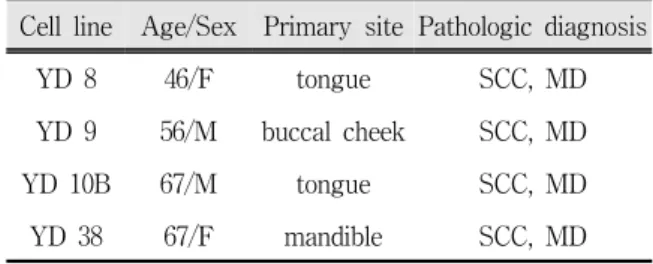
Cell line Age/Sex Primary site Pathologic diagnosis
YD 8 46/F tongue SCC, MD
YD 9 56/M buccal cheek SCC, MD
YD 10B 67/M tongue SCC, MD
YD 38 67/F mandible SCC, MD
Table 1. Information of YD cell lines used in present study.
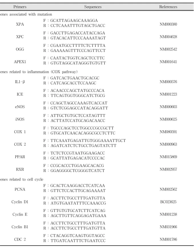
Primers Sequences References Genes associated with mutation
XPA F : GCATTAGAAGCAAAGGA
NM000380 R : CCTCAAATTTGTAGCTGACC
XPC F : GACCTTGAGACCATACCAGA
NM004628 R : GTACACATTCCCAAAATAGT
OGG F : CGAATGCCTTTTCTCTTTTA
NM002542 R : GAAAAAGTTTCCCAGTTCCT
APEX1 F : CAATACTGGTCAGCTCCTTC
NM001641 R : GTGTAGGCATAGGGTGTGTT
Genes related to inflammation (COX pathway)
IL1-β F : GATCACTGAACTGCACGC
NM000576 R : CATCAGCACCTCCAAGC
ICE F : ACAACCCAGCTATGCCCACA
NM001223 R : TTCAGTGGTGGGCATCTGCG
eNOS F : CCAGCTAGCCAAAGTCACCAT
NM000603 R : GTCTCGGAGCCATACAGGATT
iNOS F : ATTGCTGTGCTCCATAGTTT
NM000625 R : ACTTATCCATGCAGACAACC
COX 1 F : TGCCCAGCTCCTGGCCCGCCGCTT
NM080591 R : GTGCATCAACACAGGCGCCTCTTC
COX 2 F : TTCAAATGAGATTGTGGGAAAATTGCT
NM000963 R : AGATCATCTCTGCCTGAGTATCTT
PPAR F : TCTCTCCGTAATGGAAGACC
NM015869 R : GCATTATGAGACATCCCCAC
RXR F : CCGCACCCTGGAAGCACACG
NM002957 R : GGAGGGGCTCGGGGTCATCT
Genes related to cell cycle
PCNA F : GCACTCAAGGACCTCATCAA
NM002562 R : GTTCTCCACTTGCAGAAAAT
Cyclin D1 F : ACCTTCTGCCTTTGATGTTA
BC023625 R : ATGTGAATATTTCCAAACCG
Cyclin E F : GTTGTGTGCATCTTCATCAG
NM001238 R : AGCTTGTTCAGGAGATGAAA
Cyclin B1 F : ACCTTCTGCCTTTGATGTTA
NM031966 R : ACCTTCTGCCTTTGATGTTA
CDC 2 F : CTACAGGTCAAGTGGTAGCC
NM001786 R : TTGATCAATTTCTGAATCCC
Table 2. Primers used in present study.
Primers Sequences References Genes related to senescence
hTERT F : GCCTGAGCTGTACTTTGTCAA
NM003219 R : CGCAAACAGCTTGTTCTCCATGTC
hTR F : GAAGGGCGTAGGCGCCGTGCTTTTGC
U86046 R : GTTTGCTCTAGAATGAACGGTGGAAGG
hTEP F : TCAAGCCAAACCTGAATCTGAG
NM007110 R : CCCCGAGTGAATCTTTCTACGC
TRF1 F : TGTGCGGATGGTAGGGTTGC
NM017489 R : GGGCTGATTCCAAGGGTGTA
TRF2 F : AGTCAATCGCTGGGTGCTCA
NM005652 R : CTTGGTGCTGGCTGTTTATC
MAD F : GAGATGCCTTAAAACGGAGG
NM002357 R : AACCAACAGGGAGAACCTTC
MAX F : TCAATCTGCGGCTGACAAAC
NM002382 R : TGGTGTAGAGGCTGTTGTCT
c-myc F : TAATTCCAGCGAGAGGCAGA
AC103819 R : GTCCCCAAATGGGCAGAATA
Genes related to extracellular matrix
collagen 1 F : AGTGGTTACTACTGGATTGACC
NM000089 R : TTGCCAGTCTCCTCATCC
TGF β F : GGGGAAATTGAGGGCTTTCG
NM000660 R : CCAGGACCTTGCTGTACTGC
MMP 1 F : GTATGCACAGCTTTCCTCCACTGC
NM002421 R : GATGTCTGCTTGACCCTCAGAGACC
MMP 3 F : TGCTTTGTCCTTTGATGCTG
NM002422 R : TTCCTTATCCGAAATGGCTG
MMP 9 F : AGTTCCCGGAGTGAGTTGAA
NM004994 R : CTCCACTCCTCCCTTTCCTC
MMP 13 F : CATTTGATGGGCCCTCTGGCCTGC
NM002427 R : GTTTAGGGTTGGGGTCTTCATCTC
TIMP 3 F : CTGACAGGTCGCGTCTATGA
NM000362 R : GGTCTGTGGCATTGATGATG
TIMP 4 F : ACAGCCAGAAGCAGTATC
NM003256 R : AGAGGTCAGGTGGTAA
HSP 47 F : GCACGGACGGCGCCCTGCTAGTCAACGCCATG
NM001235 R : CAGCCTTCTTCTGCATCTTCCCCATCCAGATC
GAPDH F : TGCATCCTGCACCACCAACT
NM002046 R : CGCCTGCTTCACCACCTTC
out with 15000×g at 4℃ for 15 min. The supernatant was removed, then added 1 ㎖ of 75%
alcohol and dried. The purified RNA was dissolved in DEPC-DW. Optical density(OD) was measured at 260 nm with photospectrometer. Three micrograms of purified RNA was reverse transcribed using RT Premix (Bioneer, Korea) according to the protocol. The amplification of cDNA was carried out using specific primer pairs (Table 2). The conical tubes were placed in a Thermal cycler (Bioneer, Korea). The number of cycles for cDNA amplification was optimized for each primers. The thermal cycle profile was optimized for denaturation (40 s at 95℃), annealing (40s at each anneal temperature) and extension (1 min. 30 s at 72℃). The resultant PCR products were electrophoresed in 1% agarose gels (Bioneer, Korea) containing ethidium bromide together with 100bp ladder (Bioneer, Korea). The agarose gels visualized under UV and scanned for analysis using NIH Scion Image software. The relative expresseion levels of each molecules compared with GAPDH gene expression were determined.
Fig. 1. Viability of 5-FU (Left) and Cisplatin (Right) treatment on human oral cancer cell lines.
Comparison of 5-FU and Cisplatin viability of five human oral cancer cell lines. Viability was determined by means of a MTT assay. Each data point represents the mean±SD(error bars) from five independent experiments.
Ⅲ. RESULTS
1. Evaluation of 5-FU and Cisplatin sensitive or resistant cells in human oral cancer cell lines.
YD10B showed most sensitivity to 5-FU, whereas KB cell lines showed most resistance. For the treatment of Cisplatin, KB cell line showed most sensitivity while YD38 showed most resistance to Cisplatin (Fig. 1). Thereafter, experiment was performed in the focus of most sensitive and resistant cell lines by 5-FU and Cisplain.
2. mRNA expressions of various genes associated with oral cancer
To identify predictive biomarker for sensitivity or
resistance to 5-FU and Cisplatin, the mRNA
expressions of various genes associated with
mutation, inflammation (COX pathway), cell cycle,
senescence and extracellular matrix (ECM) were
evaluated by RT-PCR.
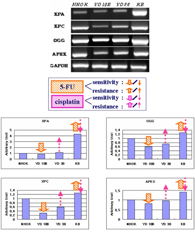
Fig. 2. mRNA expression of genes associated with mutation in human oral cancer cell liens. mRNA expressions of XPA, XPC, OGG, and APEX were evaluated by RT-PCR. All resultants were analyzed by Scion Image Software.
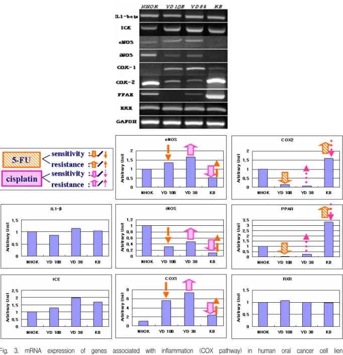
Fig. 3. mRNA expression of genes associated with inflammation (COX pathway) in human oral cancer cell liens.
mRNA expressions of eNOS, COX2, IL-1β, iNOS, PPAR, ICE, COX1 and RXR were evaluated by RT-PCR.
All resultants were analyzed by Scion Image Software.
1) Genes associated with mutation
mRNA expressions of XPA, OGG, XPC and APEX1 were correlated with the sensitivity to 5-FU. In contrast to the results of 5-FU, mRNA expressions of XPA, OGG, XPG and APEX were reversely correlated with the sensitivity to Cisplatin (Fig. 2).
2) Genes related to inflammation (COX pathway)
In an association with inflammation, COX-2 and
PPAR mRNA expressions were correlated with the
sensitivity to 5-FU and mRNA expression of
COX-1, iNOS and eNOS were reversely correlated
with the sensitivity to 5-FU. For the sensitivity to
Cisplatin, COX-1, iNOS and eNOS mRNA
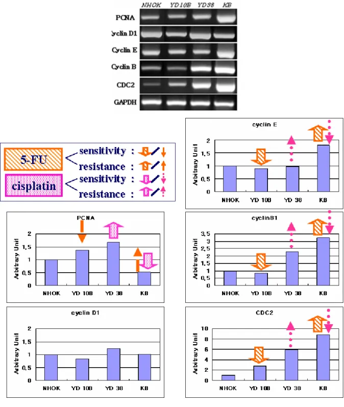
Fig. 4. mRNA expression of genes associated with Cell cycle in human oral cancer cell liens. mRNA expressions of cyclin E, clyclin B1, cyclin D1, PCNA and CDC2 were evaluated by RT-PCR. All resultants were analyzed by Scion Image Software.
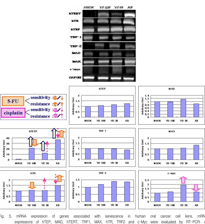
Fig. 5. mRNA expression of genes associated with senescence in human oral cancer cell liens. mRNA expressions of hTEP, MAD, hTERT, TRF1, MAX, hTR, TRF2 and c-Myc were evaluated by RT-PCR. All resultants were analyzed by Scion Image Software.
expression were correlated, but COX-2 and PPAR mRNA expressions were reversely correlated (Fig.
3).
3) Genes related to cell cycle
mRNA expression of Cyclin E and Cyclin B1 and CDC2 were correlated with the sensitivity to 5-FU,
while they were reversely correlated with the
sensitivity to Cisplatin. mRNA expression of PCNA
was reversely correlated with the sensitivity to
5-FU, however, its correlation was in the
sensitivity to Cisplatin (Fig. 4).
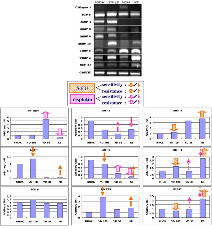
Fig. 6. mRNA expression of genes associated with extracellular matrix in human oral cancer cell liens. mRNA expressions of collagen 1, TFG-β, hsp47, MMP-1, MMP-3, MMP-9, MMP-13, TIMP-3 and TIMP-4 were evaluated by RT-PCR. All resultants were analyzed by Scion Image Software.
4) Genes related to senescence
hTERT and hTR mRNA expression were
5) Genes related to extracellular matrix
mRNA expressions of TIMP3, TIMP4, and
MMP-1, MMP-9 and MMP-13 were reversely correlated with the sensitivity to 5-FU and collagen 1 and MMP-9 were correlated with the sensitivity to Cisplatin (Fig. 6).
Ⅳ. DISCUSSION
In present study, mRNA expression level of various genes were evaluated in oral cancer cell lines in order to identify genes associated with response to 5-FU and Cisplatin. As the result of viability to both agents, YD 10B showed most sensitivity to 5-FU, whereas KB cell lines showed most resistance. For the treatment of Cisplatin, KB cell line showed most sensitivity while YD38 showed most resistance. It meant that every cell line had a variability against chemotherapeutic agents. Therefore, we evaluated the mRNA expressions of various genes in YD10B and KB cell for 5-FU, and YD38 and KB cell for Cisplatin.
Interestingly, both agents, 5-FU and Cisplatin, showed an opposite results in various molecules. In present study, mRNA expressions of XPA, XPC, OGG and APEX are correlated with the sensitivity of 5-FU, while they are reversely correlated with the sensitivity of Cisplatin. The recognition of DNA damage plays an important role in eliminating the damage and maintaining genetic integrity
11-15)and XPA, XPC, OGG and APEX are an important DNA damage recognition protein. Wang et al. reported that XPC protein plays an important role in the Cisplatin treatment-mediated cellular response
16), which was supported the present study. Taken together, genes associated with DNA damage recognition plays an important role in the 5-FU and Cisplatin treatment-mediated sensitivity or resistance.
In an association with inflammation, in present study, COX-2 and PPAR mRNA expressions are correlated with the sensitivity to 5-FU and mRNA expression of COX-1, iNOS and eNOS are reversely correlated with the sensitivity to 5-FU.
For the sensitivity to Cisplatin, COX-1, iNOS and eNOS mRNA expression are correlated, but COX-2
and PPAR mRNA expressions are reversely correlated. COX-2 expression was found to inhibit apoptosis and to stimulate tumor angiogenesis
17,18). In addition, COX-2 upregulation may be related to chemotherapy resistance and poor radiologic responsiveness
19), which is partly supported present study that COX-2 mRNA expression is resistant to Cisplatin, but sensitivity to 5-FU.
iNOS can enhance its ability to promote tumor growth in cooperation with COX-2
20). In present study, response to chemotherapeutic agent of iNOS expression is the opposite to COX-2. These results suggested that when chemotherapeutic agents is chosen, it is better to evaluate the mRNA expressions of iNOS and COX-2.
In terms of cell cycle, mRNA expression of Cyclin E and Cyclin B1 and CDC2 are correlated with the sensitivity to 5-FU, which are reversely correlated with the sensitivity to Cisplatin. Many researchers have suggested that cyclin E is related to tumor aggressiveness
21-24), which meant that tumor aggressiveness is related to the sensitivity to 5-FU. mRNA expression of PCNA is reversely correlated with the sensitivity to 5-FU, however, its correlation is in the sensitivity to Cisplatin.
PCNA is a marker for the proliferation of tumor cells
25,26). These results suggested that proliferation of tumor cells is related to sensitivity to Cisplatin.
According to molecules associated with senescence, hTERT and hTR mRNA expression is correlated with the sensitivity to 5-FU, while they are reversely correlated with the sensitivity to Cisplatin. Strong telomerase activity has been reported in most of cancer cells and immortalized cells
27-29). In present study, mRNA expressions of hTERT in oral cancer cell lines were highly increased comparing its expression in NHOK. Of the molecules related to telomerase, only two molecules, hTERT and hTR, were related to the sensitivity to 5-FU. Because of the highly expressions of hTERT in all cancer cell lines, other molecules should be considered to choose the chemotherapeutic agents.
For the molecules related to extracellular matrix,
mRNA expressions of TIMP-3and TIMP-4, and HSP47 are correlated with the sensitivity to 5-FU, while they are reversely correlated with the sensitivity to Cisplatin. mRNA expressions of MMP-1, MMP-9 and MMP-13 are reversely correlated with the sensitivity to 5-FU, and collagen 1 and MMP-9 are correlated with the sensitivity to Cisplatin. Elevated expression of MMP-13 in head and neck SCCs may reflect increased tumor invasion
30). In present study, MMP-13 mRNA expression is reversely correlated with the sensitivity to 5-FU, which might mean that cancer cells with a highly potential of invasion is resistance to 5-FU. In previous study, oral SCCs with high levels of MMP-2 and -9 show greater invasion capacities compared with those with low levels of expression
31). Overexpression of MMP-1 and MMP-9 mRNA is associated with progression of oral dysplasia to cancer
32). In present study, MMP-1 and MMP-9 mRNA expression was correlated with the resistance to 5-FU, which was also supported our results that cancer with highly invasiveness showed the resistant response to 5-FU. So, Cisplatin may be recommended in cancer having a potential of invasion. TIMP-3 transcripts observed in head and neck SCCs
33), which is an important marker in sensitivity to 5-FU.
Ⅴ. CONCLUSIONS
To identify predictive bio-marker for the sensitivity or resistance to 5-FU and Cisplatin, the mRNA expressions of various genes associated with mutation, inflammation(COX pathway), cell cycle, senescence and extracellular matrix (ECM) were evaluated by RT-PCR.
Human primary fibroblasts were used in the present study. MTT assay was performed to evaluated the sensitivity and/or resistance to 5-FU and Cisplatin. And RT-PCR was carried out for evaluation.
TIMP-3, TIMP-4 and HSP47. And the molecules had correlated with the sensitivity to Cisplatin were COX-1, iNOS, eNOS, PCNA, collagen 1 and MMP-9.
Taken together, when choosing the appropriate chemotherpeutic agents for patients, considering the molecules which are correlated or reversely correlated is helpful to choose the resonable agents for cancer patients.
REFERENCES
1. Franceschi S, Barra S, Vecchia C, Bidoli E, Negri E, and Talamini R. Risk factors for cancer of the tongue and the mouth. A case control study from northern Italy. Cancer 1992;70:2227-2233.
2. Field JK, Kiaris H, Risk JM, et al. Allelotype of squamous cell carcinoma of the head and neck fractional allele loss correlates with survival. Br J Cancer 1995;72:1180-1188.
3. Paccagnella A, Orlando A, Marchiori C, et al. Phase III trial of initial chemotherapy in stage III or IV head and neck cancers: a study by the Gruppo di Studio sui Tumori della Testa e del Collo. J Natl Cancer Inst. 1994;16;86:265-272.
4. Pignon JP, Bourhis J, Domenge C, and Designe L.
Chemotherapy added to locoregional treatment for head and neck squamous-cell carcinoma: three meta-analyses of updated individual data.
MACH-NC Collaborative Group. Meta-Analysis of Chemotherapy on Head and Neck Cancer. Lancet.
2000;18;355:949-955.
5. Lefebvre JL, Chevalier D, Luboinski B, Kirkpatrick A, Collette L, Sahmoud T. Larynx preservation in pyriform sinus cancer: preliminary results of a European Organization for Research and Treatment of Cancer phase III trial. EORTC Head and Neck Cancer Cooperative Group. J Natl Cancer Inst.
1996;3;88:890-899.
6. Veterans Affairs Laryngeal Cancer Study Group.
Induction chemotherapy plus radiation compared with surgery plus radiation in patients with advanced laryngeal cancer. N Engl J Med. 1991; 13;324(24):
1685-1690.
7. Zaffaroni N, Silvestrini R, Orlandi L, Bearzatto A,
ovarian cells. Br J Cancer. 1998;77:1378-1385.
8. Park JS, Yoon SY, Kim JM, Yeom YI, Kim YS, and Kim NS. Identificatin of novel genes associated with the response to 5-FU treatment in gastric cancer cell lines using a cDNA microarray. Cancer Lett.
2004;214:19-23.
9. Buttitta F, Marchetti A, Gadducci A, et al. p53 alterations are predictive of chemoresistance and aggressiveness in ovarian carcinomas: a molecular and immunohistochemical study. Br J Cancer.
1997;75:230-235.
10. Righetti SC, Della Torre G, Pilotti S, et al. A comparative study of p53 gene mutations, protein accumulation, and response to cisplatin-based chemotherapy in advanced ovarian carcinoma. Cancer Res. 1996; 15;56:689-693.
11. Bergh J, Norberg T, Sjogren S, Lindgren A, and Holmberg L. Complete sequencing of the p53 gene provides prognostic information in breast cancer patients, particularly in relation to adjuvant systemic therapy and radiotherapy. Nat Med. 1995;1:1029-1034.
12. Sanchez Y, Bachant J, Wang H, et al. Control of the DNA damage checkpoint by chk1 and rad53 protein kinases through distinct mechanisms. Science. 1999;
5;286:1166-1171.
13. Wakasugi M, and Sancar A. Order of assembly of human DNA repair excision nuclease. J Biol Chem.
1999;274:18759-18768.
14. Batty DP, and Wood RD. Damage recognition in nucleotide excision repair of DNA. Gene.
2000;241:193-204.
15. Zou L, and Elledge SJ. Sensing DNA damage through ATRIP recognition of RPA-ssDNA complexes.
Science. 2003;300:1542-1548.
16. Wang G, Dombkowski A, Chuang L, Xin S, and XU X. The involvement of XPC protein in the cisplatin DNA damaging treatment mediated cellular response.
Cell Res. 2004;14:303-314.
17. Smith WL, DeWitt DL, and Garavito RM, Cyclooxygenases: Structural, cellular, and molecular biology. Annu Rev Biochem. 2000; 69:145–182.
18. Williams CS, Mann M, and DuBois RN, The role of cyclooxygenases in inflammation, cancer, and development. Oncogene. 1999;18:7908–7916.
19. Kim YB, Kim GE, Pyo HR, et al. Differential cyclooxygenase-2 expression in squamous cell carcinoma and adenocarcinoma of the uterine cervix.
Clin Invest 2004;60:822-829.
20. Kim SA, Ahn SG, Kim DK, et al. Sequential
expression of inducible nitric oxide synthase and cyclooxygenase-2 during DMBA-induced hamster buccal pouch carcinogenesis. In Vivo. 2004;18:
609-614.
21. Keyomarsi K, O'Leary N, Molnar G, Lees E, Fingert HJ, and Pardee AB. Cyclin E, a potential prognostic marker for breast cancer. Cancer Res. 1994;54:
380-385.
22. Tahara E. Genetic alterations in human gastroi- ntestinal cancers. Cancer(suppl.) 1995;75:1410-1417.
23. Akama Y, Yasui W, Kuniyasu H, et al. Genetic status and expression of the cyclin-dependent kinase inhibitors in human gastric carcinoma cell lines. Jpn J Cancer Res. 1996;87:824-830.
24. Porter PL, Malone KE, Heagerty PJ, et al. Expression of cell-cycle regulators p27Kip1 and cyclin E, alone and in combination, correlate with survival in young breast cancer patients. Nat Med. 1997;3:222-225.
25. Robbins BA, de la Vega D, Ogata K, Tan EM, and Nakamura RM. Immunohistochemical detection of proliferating cell nuclear antigen in solid human malignancies. Arch Pathol Lab Med.
1987;111:841-845.
26. Hall PA, Levison DA, Woods AL, et al. Proliferating cell nuclear antigen (PCNA) immunolocalization in paraffin sections: an index of cell proliferation with evidence of deregulated expression in some neoplasms. J Pathol. 1990 Dec;162:285-294.
27. Counter CM, Hirte HW. Bacchetti S, and Harley CB.
Telomerase activity in human ovarian carcinoma.
Proc Natl acad Sci USA. 1994;91:2900-2904.
28. Counter CM, Gupta J, Harley CB, Leber B, and Bacchetti S. Telomerase activity in normal leukocytes and in hematologic malignancies. Blood.
1995;85:2315-2320.
29. Fujimoro r, Kamata N, Taki M, et al. Gene expression of telomerase related proteins in human normal oral and ectocervical epithelial cells. Oral Oncolo.
2003;39:445-452.
30. Culhaci N, Metin K, Copcu E, and Dikicioglu E.
Elevated expression of MMP-13 and TIMP-1 in head and neck squamous cell carcinoma may reflect increased tumor invasiveness. BMC Caner. 2004;4:42.
31. Robinson CM, Stone AM, Shields JD, et al Functional significance of MMP-2 and MMP-9 expression by human malignant oral keratinocyte cell lines. Arch Oral Biol 2003;48:779-786.
32. Richard CKJ, Maricris M, Caroline HS, et al.
Overexpression of matrix metalloproteinase-1 and -9
mRNA is associated with progression of oral dysplasia to cancer. Clin Cancer Res. 2004;10:
6460-6465.
국문요약
편평세포암 세포주에서 5-FU와 Cisplatin에의 감수성과 관련된 유전자의 동정
전남대학교 치의학전문대학원 구강내과학교실
1, 구강병리학교실
2전남대학교 치의학연구소
최나영
1․김옥준
2․이금숙
1․김병국
1․김재형
1장윤영
2․임원봉
2․정민아
2․최홍란
2두경부에서 발생하는 편평세포암종은 같은 조직학적 분류, 조직학적 단계 및 임상 단계에 있다하더라도 항암 제 투여에 따른 감수성은 환자마다 다양하게 나타난다. 따라서, 본 연구의 목적은 기존에 가장 많이 사용하는 항암제인 5-FU 와 Cisplatin의 감수성을 예측할 수 있는 생물학적 표지자를 찾아 환자에게 맞는 항암제를 선택 하기 위해서이다.
5종의 구강 편평암세포암주를 이용하였으며, 5-FU 와 Cisplatin의 항암제 감수성을 측정하기 위해 MTT assay를 시행하였다. 각 세포 주의 전 RNA를 추출하였으며 이어서 cDNA를 합성하였다. 다양한 유전자를 1) 돌연변이 2) 염증(COX pathway) 3) 세포주기 4) 노화 5) 세포외기질 과 관련되게 분류하였고 RT-PCR을 시행하 여 유전자의 발현량을 살펴보았다. 결과물은 Scion image 프로그램을 통해 분석하였고, Sigma plot을 이용해 표 시하였다.
5-FU의 감수성과 비례하는 유전자들은 XPA, XPC, OGG, APEX, COX-2, PPAR, Cyclin E, Cyclin B1, CDC2, hTERT, hTR, TIMP-3, TIMP-4 및 HSP47이었으며, Cisplatin의 감수성과 비례하는 유전자들은 COX-1, iNOS, eNOS, PCNA, Col-1 및 MMP-9 으로 알 수 있었다.
위 결과는 암환자에게 알맞은 항암제를 선택 시 항암제의 감수성과 관련한 유전자들의 발현 정도를 파악한다 면 올바른 항암제를 선택하는데 도움이 되리라 사료된다.
주제어 : 편평세포암, 생물학적 표지자, 5-fluorouracil, cisplatin
33. Birkedal-Hansen B, Pavelic ZP, Gluckman JL, Stambrook P, Li YQ, and Stetler-Stevenson WG.
MMP and TIMP gene expression in head and neck squamous cell carcinomas and adjacent tissues. Oral Dis. 2000 ;6:376-382.