Expression of Bcl-2 Family in 4-Nitroquinoline 1-Oxide-Induced Tongue Carcinogenesis of the Rat
Jae-Wook Choi
1, Sung-Su Chung
2, Geum-Sug Lee
1, Byung-Gook Kim
1,
Jae-Hyeong Kim
1, Eun-Byul Kook
3, Mi-Sun Jang
3, Mi-Kyeong Ko
3, Kwon Jung
3, Hong-Ran Choi
3, Ok-Joon Kim
3, D.D.S., M.S.D., Ph.D.
Dept. of Oral Medicine
1, Dept. of Anesthesiology
2, Dept. of Oral Pathology
3, School of Dentistry, Dental Science Research Institute, Chonnam National University
The number of patients with tongue carcinoma is increasing rapidly among young individuals in many parts of the world. Oral carcinoma progresses from hyperplastic lesion through dysplasia to invasive carcinoma and the concept of "field cancerization" with molecular alteration has been suggested for oral cavity carcinogenesis.
Significant improvement in treatment and prognosis will depend on more detailed understanding of the multi-step process leading to cancer development.
To induce tongue carcinoma in rat by 4-NQO, each drinking water was made to 10 ppm, 25 ppm, 50 ppm and control (only D.W. without 4-NQO). Specimens were classified into 4 groups such as control, I (mild & moderate dysplasia), II (severe dysplasia and carcinoma in situ), III (carcinoma). The mRNA expressions of Bcl-2 family were evaluated by RT-PCR technique.
For anti-apoptotic Bcl-2 family, mRNA expression of Bcl-w was down-regulated in all stages of tongue carcinogenesis model. However, mRNA expression of Bcl-2 was up-regulated. For pro-apoptotic Bcl-2 family, all members were down-regulated in all stages of tongue carcinogenesis model except for Bad mRNA in group III. In terms of BH3 only protein, mRNA expressions of Bok and Mcl-1 were down regulated in all stages of specimen, but Bmf in group II and BBC3 in group III were up-regulated.
Our current findings demonstrated the involvements of mRNA expression of Bcl-2 family in multi-step tongue carcinogensis. This highlights the necessity for continued efforts to discover suitable biomakers (Bcl-2 family) for early diagnosis of the disease, and to understand its pathogenesis as a first step in improving methods of treatment. The discovery of these potential biomarkers and molecular targets for cancer diagnostics and therapeutics has the potential to significantly change the clinical approach and outcome of the disease.
Key words : Oral cavity carcinogenesis, 4-nitroquinoline 1-oxide, mRNA expressions of Bcl-2 family, RT-PCR technique
표 1
Corresponding Author : Assist. Prof. Ok-Joon Kim Department of Oral Pathology, School of Dentistry, Chonnam National University 5 Hak-1-Dong, Dong-Gu Gwangju 501-746, Korea
E-mail: [email protected] received: 2005-07-15
accepted: 2005-09-02
Ⅰ. INTRODUCTION
Oral cancer progresses from hyperplastic
epithelial lesions and epithelial dysplasia to invasive
carcinoma. The concept of "field cancerization" with
molecular alterations can be applied to oral cavity
carcinogenesis
1-3). Also, significant improvement in
treatment and prognosis will depend on more detailed understanding of the multi-step process leading to cancer development
4).
The most frequently used animal models in oral cancer research studies have been the hamster buccal pouch model, rat and less frequently, mouse
4-6).
A fat-soluble 7,12-dimethylbenz[α]anthracene (DMBA) and a water-soluble 4-nitroqinoline 1- oxide (4-NQO) are the most frequently used carcinogens in the studies. Since 4-NQO is water-soluble, it is well suited in examining the role of xenobiotics in experimental oral carcinogenesis. Topical application of 4-NQO and administration of 4-NQO in drinking water both induce premalignant and malignant transformation in the rat oral cavity. This results in papilloma and invasive squamous cell carcinoma, resembling the clinical and histologic changes observed in these neoplasms in humans
6-10).
Cell proliferation, apoptosis, and tumor cell implants were used to monitor malignant properties.
Cell proliferation plays an important role in multi-step carcinogenesis with multiple genetic changes
11). Therefore, control of cell proliferations is important for cancer prevention
12). Furthermore, apoptosis induction is one of the important characteristics of candidate cancer chemopreventive agents
13).
Bcl-2 family serve as critical regulators of pathway involved in apoptosis, acting to either inhibit or promote cell death. Bcl-2 family members can be grouped into three categories, the anti-apoptotic member including Bcl-2, Bcl-xL, and Mcl-1, the multidomain pro-apoptotic members such as Bax and Bak, and the BH3 domain only proteins such as Bim, Bid, Bad, and Bik
14). This family of Bcl-2 converges on the mitochondrial membrane depolarization that ultimately determine cell fate.
The Bax subfamily has been implicated as the
"gateway" to apoptosis
15). These Bcl-2 family are required to initiate most forms of apoptosis
15,16). During apoptosis , Bax translocates to the
mitochondria and oligomerizes, causing cytochrome c release from the mitochondria
17,18). Translocation and oligomerization of Bax are preceded by a conformational change
19).
Bcl-2 plays an important role in cancer and resistance of cancer to conventional therapies.
Bcl-2 can contribute to neoplastic cell expansion by preventing normal cell turnover caused by physiological cell death mechanisms
20,21). High expression of Bcl-2 is found in a wide variety of human cancers and mediates the resistance of cancers to a wide spectrum of chemotherapeutic drugs and irradiation which act by inducing apoptosis in tumor cells
22).
Anti-apoptostic Bcl-2 family members function at least in part by inhibiting cytochrome c release from the mitochondria. They perform this task by preventing translocation and/or activation of Bax-like proteins on the mitochondria
23,24), however, the mechanism of this inhibition is not entirely clear. In addition, it is not certain that the role of anti-apoptotic Bcl-2 family members is limited to the mitochondria. Much emphasis has been placed on Bcl-2 function on the mitochondria, although it has been reported that wild type Bcl-2 localizes to the mitochondria, endoplamic reticulum (ER), and nuclear membranes
25). And there is growing evidence that the ER is important in apoptosis
26-28). Our current findings demonstrated the involvements of mRNA expression of Bcl-2 family in multi-step tongue carcinogensis.
Ⅱ. MATERIALS AND METHODS 1. Chemicals and Animals
Animal experiment care and use of all animal
laboratory accompanied system and morals
experiment that Chonnam Dental college animal
laboratory provision of “Guidelines and Regulations
for Use and Care of Animals” based on “Guide for
the care and use of laboratory animals (National
Research Council, USA)” and “Policy on humane
care and use of laboratory animals (United States
Histologic grade Experimental group
Normal Control
Dysplasia
mild I
moderate severe Carcinoma in situ II
Cancer III
Table 1. Experimental Criteria
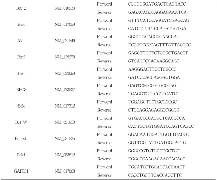
Bcl-2 NM_016933 Forword CCTGTGGATGACTGAGTACC
Reverse GAGACAGCCAGGAGAAATCA
Bax NM_017059 Forword GTTTCATCCAGGATCGAGCAG
Reverse CATCTTCTTCCAGATGGTGA
Mcl NM_021846 Forword GGCGTGCAGCGCAACCAC
Reverse TCCTGCCCCAGTTTGTTACGCC
Bmf NM_139258 Forword GAGCTTGCTCTCTGCTGACCT
Reverse GTCACCCCACAAGGCAGC
Bad NM_022698 Forword AAGGGACTTCCTCGCCC
Reverse GATCCCACCAGGACTGGA
BBC3 NM_173837 Forword GAGTCGCCCGTGCCCAG
Reverse TGAGGTCGTCCGCCATCC
Bok NM_017312 Forword TGGAGGTGCTGCGGCGC
Reverse CTCCAGGAGAGGCCGGCG
Bcl-W NM_021850 Forword GTGACCCCAGGCTCAGCCCA
Reverse CACTGCTGTGGATCCAGTCAGCC
Bcl-xL NM_031535 Forword GGACAATGGACTGGTTGAGCC
Reverse GGTTGCCATTGATGGCACTG
Bak1 NM_053812 Forword GGGCCGTGTGGTGGCTCT
Reverse TGGCCCAACAGAACCACACC
GAPDH NM_017008 Forword TGCATCCTGCACCACCAACT
Reverse CGCCTGCTTCACCACCTTC
Table 2. Bcl-2 family mRNA primer.
Public Health service, USA)”.
Male Sprague-Dawley rats weighing about 30 g
and 4 weeks old were used and 4-NQO was
obtained from TCI Co., Ltd. (Tokyo, Japan). 4-NQO
solution was given to rats to induce multi-step
tongue carcinogenesis. 4-NQO was given in
drinking water for 4 to 8 months and each drinking
water made 10 ppm, 25 ppm, 50 ppm and control
(only D.W. without 4-NQO). The rats were
sacrificed every week from 4 to 8 months after
4-NQO drinking. All animals were housed in wire
cages with free access to drinking and under controlled conditions of humidity (50 ± 5%), lighting (12hr light/dark cycle) and temperature (22
± 2℃). A half of tongue was kept frozen in liquid nitrogen and another half prepared for H&E staining. Experimental group was classified according to histomorphological findings (Table 1).
2. Histologic study
Rats were sacrificed and the tongue were longitudinally cut, followed by fixation in 10%
phosphate-buffered formalin for 48 h, routinely embedded in paraffin, and serially sectioned at 3-4
㎛. The sections were used for H&E staining. And the remaining samples were frozen at deep freezer for RNA study.
3. RNA isolation and RT-PCR
Total RNA was isolated from control and 4-NQO-induced tongue by means of Tri
ⓇReagent (Invitrogen, USA.), according to the manufacturer's instructions. 3 ㎍ of total RNA was reverse- transcribed in a total volume of 50 ㎕, using Accupower
®RT premix (Bioneer, seoul, korea).
Oligonucleotide primers (5→3) were generated consulting NCBI (Genebank). The primers used in present study are listed in table 2. 2 ㎕ of cDNA was mixed PCR premix (Bioneer, korea) a final total volume of 20 ㎕, performed using Mygenic 96 Thermal Block (Bioneer, korea). Amplofocation profiles included denaturation for 40 s at 94℃,
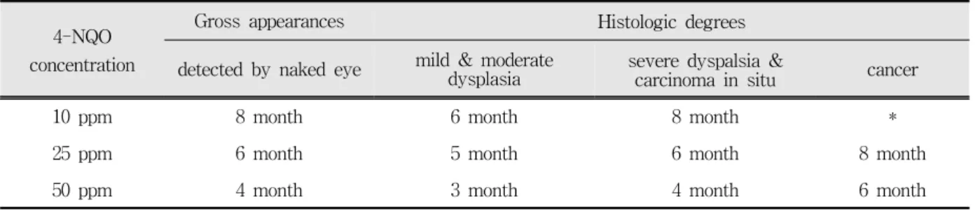
4-NQO concentration
Gross appearances Histologic degrees
detected by naked eye mild & moderate
dysplasia severe dyspalsia &
carcinoma in situ cancer
10 ppm 8 month 6 month 8 month *
25 ppm 6 month 5 month 6 month 8 month
50 ppm 4 month 3 month 4 month 6 month
Table 3. Time intervals of lesion induced by 4-NQO.
annealing for 40 s at 56℃: Bcl-2, Bax and Bad ; annealing for 40 s at 66℃:Mcl, Bmf, Bak1, BBC3, Bok, Bcl-w, Bcl-xL and primer extension for 90 s at 72℃ and the final step extended to 10 min at 72
℃. The PCR precedure was performed at least three times for each sample.
Ⅲ. RESULTS 1. 4-NQO induced oral carcinogenesis
4-NQO was treated to induce multi-step carcinogenesis by time and dose dependent manner.
Observing the histologic findings, histologic degrees (dysplasia, carcinoma in situ and SCC) were different to time and dose dependent manner (Table 3). With 25ppm, 8month needed to induce SCC, while 6month at 50ppm. And all specimen was followed the multi-step carcinogenesis. Fig. 1 to 5 showed the gross and histologic findings.
2. RT-PCR
RT-PCR were performed to investigate a variety of Bcl-2 mRNA expression in 4-NQO induced tongue carcinogenesis of rat. Bcl-2 family.
covariance anaylsis has been used to normalize data using values obtained for mRNA expression of the house keeping gene GAPDH.
1) Bcl-2
mRNA expression of Bcl-2 was increased in
multistep carcinogenesis than in control (Fig. 6).
using values of mRNA expression shows 1 in control and other using values of mild or/ and moderate dysplasia shows 1.25 (SD=±0.04) compare with control. using values of severe dysplasia shows 1.53 (SD=±0.17) and squamous cell carcionoma shows 1.21 (SD=±0.05) compare with control (Fig. 6). Image data show a little different band.
2) Bax
mRNA expression of Bax decreased in multistep
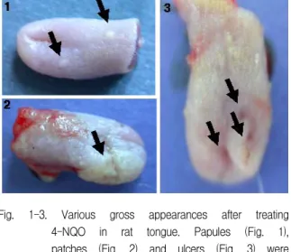
Fig. 1-3. Various gross appearances after treating 4-NQO in rat tongue. Papules (Fig. 1), patches (Fig. 2) and ulcers (Fig. 3) were examined.
Fig. 4-5. Histologic appearances after treating 4-NQO in rat tongue. Moderate dysplasia (Fig. 4) and well- differentiated squamous cell carcinoma (Fig. 5) were observed.
carcinogenesis than in control (Fig. 7). using values of mRNA expression shows 1 in control and using value of the resr mRNA expression of Bax show each 0.5 (SD=±0.12), 0.71 (SD=±0.08), 0.39 (SD=±0.02) compare with control. mRNA expression of squamous cell carcinoma were significant difference and decrease 2.5 folds compare with control (Fig. 7). Image data show that normal be seen band but other band be seen a glimmer.
3) Bcl - W
Bcl-W result shows oppsition result of Bcl-2.
Bcl-W gradually decreases mRNA expression than in control (Fig. 8). using values of mRNA expression shows 1 in control and mild or/and moderate dysplasia shows 0.79 (SD=±0.18) as using value of mRNA expression compare with control.
severe dysplasia shows that mRNA expression
decread than in other dysplasia and using values of
mRNA expression shows 0.49 (SD=±0.11) compare
with control. mRNA expression of squamous cell
carcinoma decrease than in control and uing values
of mRNA expression shows decrease 0.25 folds
compare with control (Fig. 8). Image data show
that normal be seen band but other band be seen a
glimmer.
A . B.
Bcl - 2
Control
% ratio of GAPDH
0 20 40 60 80 100 120 140 160 180
M -Dysplasia S - Dysplasia SCC
C.
Bcl - 2 Control M-Dysplasia S-Dysplasia SCC
Fold Increase
(Bcl-2/GAPDH) 1 1.25
(SD=±0.04)
1.53 (SD=±0.17)
1.21 (SD=±0.05) Fig. 6. mRNA expression of Bcl-2 in multistep carcinoma were decrease and the latter phase inceased mRNA
expression (a). RT-PCR result shows image (b) and anaylysis has been used to data that using values obtained for mRNA expressin compare with control (c).
A. B.
0 20 40 60 80 100 120
Bax
Control
% ratio of GAPDH
M -Dysplasia S - Dysplasia SCC
C.
Bax Control M-Dysplasia S-Dysplasia SCC
Fold Increase
(Bcl-2/GAPDH) 1 0.5
(SD=±0.12)
0.71 (SD=±0.08)
0.39 (SD=±0.02) Fig. 7. mRNA expression of Bax in multistep carcinoma were decrease and the latter phase inceased mRNA
expression (a). RT-PCR result shows image (b) and anaylysis has been used to data that using values obtained for mRNA expressin compare with control (c).
A. B.
0 20 40 60 80 100 120
Bcl - W
Control
% ratio of GAPDH
M -Dysplasia S - Dysplasia SCC
C.
Bcl - W Control M-Dysplasia S-Dysplasia SCC
Fold Increase
(Bcl-2/GAPDH) 1 0.79
(SD=±0.18)
0.49 (SD=±0.11)
0.39 (SD=±0.17) Fig. 8. mRNA expression of Bcl-w in multistep carcinoma were decrease and the latter phase inceased mRNA
expression (a). RT-PCR result shows image (b) and anaylysis has been used to data that using values obtained for mRNA expressin compare with control (c).
A. B.
0 20 40 60 80 100 120
Bcl - XL
Control
% ratio of GAPDH
M -Dysplasia S - Dysplasia SCC
C.
Bcl - xL Control M-Dysplasia S-Dysplasia SCC
Fold Increase
(Bcl-2/GAPDH) 1 0.91
(SD=±0.11)
0.89 (SD=±0.06)
0.93 (SD=±0.08) Fig. 9. mRNA expression of Bcl-xL in multistep carcinoma were decrease and the latter phase inceased mRNA
expression (a). RT-PCR result shows image (b) and anaylysis has been used to data that using values obtained for mRNA expressin compare with control (c).
4) Bcl - XL
mRNA expression of Bcl-XL shows reveals almost same mRNA expression in multistep carcinogenesis including the control (Fig. 9). using calues of mRNA expression shows 2 in control and using value of the rest mRNA expression of Bcl-XL shows each 0.91 (SD=±0.11), 0.89 (SD=±0.06) and 0.93 (SD=±0.08) compare with control. in other words, each dysplasia and squamous cell carcinoma shows a little difference mRNA expression and almost it is smilar. Image shows almost similar (Fig. 9).
5) Bak
mRNA expression of Bak decrease in dyplasia and mRNA expressin in sqmous cell carcinoma is more increased than in dysplasia (Fig. 10). Image data show upward band in normal, squamous cell carcinoma. Using values of mRNA expression shows 1 in control and mild or/and moderate using values shows 0.29 (SD=±0.04) compare with control. using values of mRNA expression in severe dysplasia show 0.22 (SD=±0.12) and squamous cell carcinoma shows 0.62 (SD=±0.02) (Fig. 10).
A. B.
0 20 40 60 80 100 120
Bak
Control
% ratio of GAPDH
M -Dysplasia S - Dysplasia SCC
C.
Bak Control M-Dysplasia S-Dysplasia SCC
Fold Increase
(Bcl-2/GAPDH) 1 0.29
(SD=±0.04)
0.22 (SD=±0.12)
0.62 (SD=±0.02) Fig. 10. mRNA expression of Bak in multistep carcinoma were decrease and the latter phase inceased mRNA
expression (a). RT-PCR result shows image (b) and anaylysis has been used to data that using values obtained for mRNA expressin compare with control (c).
6) Bad
mRNA expression of Bad shows reveals almost same mRNA expression in multistep carcinomage- nesis including the control and alone mRNA expression of squsmous cell carcinoma slight increase (Fig. 11). using values of mRNA expression shows 1 in control and using value of the reat mRNA expression of Bad shows each 1.02 (SD=±0.13), 0.94 (SD=±0.03), 1.24 (SD=±0.03) compare with control. also Image data show similar (Fig. 11).
7) Bok
mRNA expression of Bok decrese in mild or/and
moderate dysplasia and grdually increase in severe
dysplaisa, in squamous cell carcinoma than in mild
or/and moderate dysplasia (Fig. 12). Using values
of mRNA expressoin of mRNA expression shows 1
in control and mild or/and moderate dysplasia
shows 0.37 (SD=±0.03) as using values of mRNA
expression compare with control. severe dysplasia
shows that mRNA expression decrease than in
control also increase than in mild or/and moderate
dysplasia. using values of mRNA expression shows
0.57 (SD=±0.07) compare with control. mRNA expression of squamous cell carconoma increase than in control and using values of mRNA expression shows increased 1.23 folds compare with control. Image data show decreased in mild dysplasia (Fig. 12).
8) Bmf
mRNA expression of Bmf shows a smilar tendency in Bcl-2 (Fig. 13). mRNA expression of Bmf was increased in multistep carcinogenesis than in control. using values of mRNA expression shows 1 in control and other using values of mild or/ and moderate dysplasia shows 1.12 (SD=±0.03) compare with control. using values of severe dysplasia shows 1.49 (SD=±0.01) and squamous cell carcionoma shows 1.08 (SD=±0.04) compare with control (Fig. 13). Image data show a little different band.
9) Mcl
Mcl result shows oppsition result of Bmf or BCl-2. Mcl gradually decreases mRNA expression
A. B.
0 20 40 60 80 100 120 140
Bad
Control
% ratio of GAPDH
M -Dysplasia S - Dysplasia SCC
C.
Bad Control M-Dysplasia S-Dysplasia SCC
Fold Increase
(Bcl-2/GAPDH) 1 1.02
(SD=±0.13)
0.94 (SD=±0.03)
1.24 (SD=±0.03) Fig. 11. mRNA expression of Bad in multistep carcinoma were decrease and the latter phase inceased mRNA
expression (a). RT-PCR result shows image (b) and anaylysis has been used to data that using values obtained for mRNA expressin compare with control (c).
than in control (Fig. 14). using values of mRNAexpression shows 1 in control and mild or/and moderate dysplasia shows 0.69 (SD=±0.17) as using value of mRNA expression compare with control. severe dysplasia shows that mRNA expression decread than in other dysplasia and using values of mRNA expression shows 0.65 (SD=±0.24) compare with control. mRNA expression of squamous cell carcinoma decrease than in control and using values of mRNA expression shows decrease 0.2 folds compare with control (Fig. 14). Image data show that normal be seen band but other band be seen a glimmer.
10) BBC3
mRNA expression of BBC3 shows some increased mRNA expression in multistep carcinomagenesis including the control (Fig. 15).
Using values of mRNA expression shows 1 in control and using value of the rest mRNA expression of Bad shows each 1.08 (SD=±0.06), 1.11 (SD=±0.04), 1.43 (SD=±0.03) compare with control.
Image data shows similar (Fig. 15).
A. B.
0 20 40 60 80 100 120 140 160
B ok
C ontrol
% ratio of GAPDH
M -Dysplasia S - Dysplasia S CC
C.
Bok Control M-Dysplasia S-Dysplasia SCC
Fold Increase
(Bcl-2/GAPDH) 1 0.37
(SD=±0.03)
0.57 (SD=±0.07)
1.23 (SD=±0.28) Fig. 12. mRNA expression of Bok in multistep carcinoma were decrease and the latter phase inceased mRNA
expression (a). RT-PCR result shows image (b) and anaylysis has been used to data that using values obtained for mRNA expressin compare with control (c).
A. B.
0 20 40 60 80 100 120 140 160
Bmf
Control
% ratio of GAPDH
M -Dysplasia S - Dysplasia SCC
C.
Bmf Control M-Dysplasia S-Dysplasia SCC
Fold Increase
(Bcl-2/GAPDH) 1 1.12
(SD=±0.03)
1.49 (SD=±0.01)
1.08 (SD=±0.04) Fig. 13. mRNA expression of Bmf in multistep carcinoma were decrease and the latter phase inceased mRNA
expression (a). RT-PCR result shows image (b) and anaylysis has been used to data that using values obtained for mRNA expressin compare with control (c).
A. B.
0 20 40 60 80 100 120
Mcl
Control
% ratio of GAPDH
M -Dysplasia S - Dysplasia SCC
C.
Mcl Control M-Dysplasia S-Dysplasia SCC
Fold Increase
(Bcl-2/GAPDH) 1 0.69
(SD=±0.17)
0.65 (SD=±0.24)
0.43 (SD=±0.17) Fig. 14. mRNA expression of Mcl in multistep carcinoma were decrease and the latter phase inceased mRNA
expression (a). RT-PCR result shows image (b) and anaylysis has been used to data that using values obtained for mRNA expressin compare with control (c).
A. B.
0 20 40 60 80 100 120 140 160
BBC3
Control
% ratio of GAPDH
M -Dysplasia S - Dysplasia SCC
C.
BBC3 Control M-Dysplasia S-Dysplasia SCC
Fold Increase
(Bcl-2/GAPDH) 1 1.08
(SD=±0.06)
1.11 (SD=±0.04)
1.43 (SD=±0.03) Fig. 15. mRNA expression of BBC3 in multistep carcinoma were decrease and the latter phase inceased mRNA
expression (a). RT-PCR result shows image (b) and anaylysis has been used to data that using values obtained for mRNA expressin compare with control (c).
Ⅳ. DISCUSSION
In present study, bcl-2 mRNA expression level in multistep tongue carcinogenesis of rat was evaluated by RT-PCR. This is the first trial, as far as we've known, to examine the all bcl-2 family in multistep tongue carcinogenesis. The results for bcl-2 family mRNA expression could be largely categorized into 3 patterns such as ‘increase’,
‘decrease’ and ‘fluctuant’. The Bcl-2 family showing
‘increase’ pattern are bcl-2, bmf and bbc3. bax, bcl-W, bcl-XL, bak and mcl showed ‘decrease’
pattern in present study. The ‘fluctuant’ pattern was showed in bad and bok. We could also observed the pattern of steadily ‘increase’ or
‘decrease’ as the histologic grade was advanced.
bbc3 was the only one whose expression was increased and bcl-W and mcl showed decreased pattern as the histologic grade was advanced.
In present study, we performed delivery of 4-NQO in the drinking water at the doses, resulted in easily incidence of oral cavity carcinogenesis and the duration of cancer incidence came to be short at dose dependent manner.
In the literature, there are partly divergent results regarding the expression of bcl-2 in oral squamous cell carcinoma
41,42). and in other epithelial tumors
43,44)
. In present study, mRNA expression of bcl-2 was increased comparing with control. Considering the highest expression in severe dysplasia group, bcl-2 mRNA expression can be used as a bio-marker to differentiate severe dysplasia from other dysplastic grade and oral squamous cell carcinoma.
In protein level, Bax is also detected in many normal tissues
45), including normal oral epithelium
46)
, and in neoplastic tissues, including cancers
42). The result of present study, mRNA expression for bax protein was down-regulated in all groups comparing with the expression of control. A milestone in understanding how Bcl-2 functions came when it was discovered that Bcl-2 was capable of heterodimerizing with its pro-apoptotic relative Bax
31,33). Apoptosis of cancer emerged that
anti-apoptotic proteins such as Bcl-2 and pro-apoptotic proteins such as Bax do battle with each other by hand-combat, with the Bcl-2:Bax ratio dictating the relative sensitivity or resistance of cells to a wide variety of apoptotic stimuli
31,32,33). The result of their ratio (control : M-dysplasia : S-dysplasia : SCC = 1 : 2.5 : 2.2 : 3.1) in present study was supported by other's study
31,32,33), but was not consistent with Loro's et al
47). The highest score was observed in SCC group, which indicated the anti-apoptotic tendency.
Bcl-w was expressed at relatively high levels in certain tumor cell lines of an epithelial origin, such as colonic, cervical, and breast cancer cells in protein level
48). To our knowledge, there is no report regarding bcl-w mRNA expression in oral squamous cell carcinoma. In present study, its mRNA expression was decreased as the histologic grade was advanced, which was not consistent with the fact that expression of Bcl-w was increased in various cancer cell line and colorectal
49)and gastric adenocarcinoma
50). This might be due to the different site involved in carcinoma. Also, bcl-w mRNA could be used as a bio-marker to trace the multistep carcinogenesis in tongue.
Bcl-XL has known to be an anti-apoptotic molecules in Bcl-2 family. Investigators have sensitized the cancer cells line and carcinoma by down-regualting Bcl-XL protein
51-54). In present study, its expression was not significantly changed in all group. However, considering the Bcl-xL/Bax ratio in transcriptional level, mild, severe dysplasia and SCC were scored at 1.82, 1.25 and 2.38 in one another comparing with control. This meant that dysplasia and SCC were tended to be anti- apoptosis.
Bak immunoreactivity has been demonstrated in
a wide variety of human tissues, including
nasopharyngeal and esophageal epithelium, and
epidermal keratinocytes
55,56). Xie et al. suggest it
shows a different pattern of expression in oral
dysplasias and carcinomas than other sites and its
expression , particularly in combination with Bax
expression has prognostic value in tongue SCC
59).
Bak immunoreactivity in diseased oral mucosal lesions appeared uniform from basal to surface layer, as opposed to upper layer staining, as reported for normal nasopharynx, esophagus, and epidermis
56). In contrast, Bak levels were found to be reduced in primary colorectal adenocarcinomas and gastric adenocarcinomas, as compared to normal gut mucosa
57,58), suggesting that Bak expression may be site dependent. In transcriptional level, in present study, its expression was decreased in all group comparing with the expression of control. Especially, Bak mRNA expression in severe dysplasia was the lowest in all Bcl-2 family, which was the opposite to bcl-2, the highest one in Bcl-2 family. Considering the fold increase of bak mRNA expression in severe dysplasia, it can be used as a bio-marker to discriminate severe dysplasia and SCC. Lindsten et al. reported Bax/Bak have been shown to be essential for programmed cell death. Considering the ratio of Bax/Bak, their ratio was contrast to Bcl-xL/Bax in SCC group. On our basic knowledge, the ratio of Bax/Bak was not favorable to predict the apoptosis.
Bad shares homology with Bcl-2 family only in the BH3 region, also plays an essential role in the regulation of cell death
60). Scarcity of literature regarding Bad, it is hard to discuss. The result for Bad mRNA in present study showed its expression was not changed in dysplasia group, but slightly increased in SCC.
There was no report regarding Bok, Bmf, Mcl and BBC3 in oral squamous cell carcinoma, as far as we've known. Bok is a pro-apoptotic Bcl-2 family protein identified in the ovary based on its dimerization with the anti-apoptotic Bcl-2 proteins and cell killing
34). In present study, Bok mRNA expression was decreased in dysplasia group comparing with the expression of control. But, in SCC group, its expression is not significant. Bok mRNA can be useful to discriminate dysplasia group from normal and SCC.
The Bcl-2 modifying factor, Bmf, is a pro- apoptotic member of the Bcl-2 family of
apoptosis-related protein that has been shown to initiate apoptosis in response to the loss of attachment of cells from their basal lamina
35). In present study, mRNA expression of bmf, like bcl-2, increased comparing with its expression in control.
And a trend of expression level was similar to bcl-2.
Mantle cell lymphoma (MCL) is a mature B-cell proliferation characterized by the presence of translocation t(11;14)(q13;q32), an aggressive clinical course, and poor response to chemotherapy
61)
. In present study, Mcl mRNA expression, like bcl-W, was decreased as histologic grade advanced. It can be used as a bio-marker together with bcl-w to discriminate all group of mulistep oral carcinogenesis.
BBC3 encodes a BH3-only protein that is induced by the p53 tumor suppressor and other apoptotic stimuli
62,63). In contrast to decreasing pattern of mcl mRNA, BBC3 mRNA expression was increased as the histologic grade was advanced in present study, which suggest that it could be used as a bio- marker to discriminate the all group of multistep carcinogenesis together with mcl and bcl-2.
Taken together, Bcl-2 family mRNA expressions were complicated in mulitstep tongue carcino- genesis. And several Bcl-2 family in transcriptional level can be used as a biomarker to proper diagnosis for normal to malignant process through dysplasia stages.
Ⅴ. CONCLUSIONS
The number of patients with tongue carcinoma is increasing rapidly among young individuals in many parts of the world. Oral carcinoma progresses from hyperplastic lesion through dysplasia to invasive carcinoma and the concept of "field cancerization" with molecular alteration has been suggested for oral cavity carcinogenesis.
Significant improvement in treatment and prognosis will depend on more detailed understanding of the multi-step process leading to cancer development.
To induce tongue carcinoma in rat by 4-NQO,
each drinking water made 10 ppm, 25 ppm, 50 ppm and control (only D.W. without 4-NQO). Specimens were classified into 4 groups such as control, I (mild & moderate dysplasia), II (severe dysplasia and carcinoma in situ), III (carcinoma). mRNA expressions of Bcl-2 family were evaluated by RT-PCR.
For anti-apoptotic Bcl-2 family, mRNA expression of Bcl-w was down-regulated in all stages of tongue carcinogenesis model, however, up-regulation of Bcl-2 mRNA was evaluated. For pro-apoptotic Bcl-2 family, all members were down-regualted in all stages of tongue carcinogenesis model except Bad mRNA in group III. In terms of BH3 only protein, mRNA expressions of Bok and Mcl 1 were down regulated in all stages of specimen, but Bmf in group II and BBC3 in group III were up-regualted.
Our current findings demonstrated the involvements of mRNA expression of Bcl-2 family in multi-step tongue carcinogensis. This highlights the necessity for continued efforts to discover suitable biomakers (Bcl-2 family) for early diagnosis of the disease, and to understand its pathogenesis as a first step in improving methods of treatment. The discovery of these potential biomarkers and molecular targets for cancer diagnostics and therapeutics has the potential to significantly change the clinical approach and outcome of the disease.
REFERENCES
1 .Calfano, J., van der Riet, P., Westra, W., Nawroz, H., Clayman,G., Piantadosi, S., Corio, R., Lee, D., Greenberg, B., Koch, W. and Sidransky, D. Genetic progression model for head and neck cancer:
implications for field cancerization. Cancer Res.
1996;56: 2488-2492.
2. Slaughter, D.L., Southwick, H.W., Smejkal, W. Filed cancerization in oral stratified squamous epithelium;
clinical implications of multicentric origin. Cancer.
1953;6: 963-968.
3. Patridge, M., Emilion, G., Pateromichelakis, S., Phillips, E., and Langdon, J. Field cancerisation of the oral cavity: comparison of the spectrum of molecular
alrerations in cases presention with both dysplastic and malignant lesions. Oral. Oncel. 1997;33: 332-337.
4. Shklar, G. Development of experimental oral carcinogenesis and its impact on current oral cancer research. J. Dent. Res. 1999;78: 1768-1772.
5. Eveson, J.W. Animal models of intra-oral chemical carcinogenesis: a review. J. Oral. Pathol. 1981;10:
129-146.
6. Fisker, A.V. Experimental oral carcinogenesis. A basic rat model for the study of oral carcinogenesis using the carcinogen 4-nitroquinoline 1-oxide. Dan.
Med. Bull. 1990;15:43-47.
7. Steidler, N.E., and Reade, P.C. Initiation and promotion of experimental oral mucosal carcino- genesis in mice. J. Oral. Pathol. 1986;15: 43-47.
8. Tanaka, T., Kawamori, T., Ohnishi, M., Okamoto, K., Mori, H. and Hara, A. Chemoprevention of 4-nitroquinoline 1-oxide-induced oral carcinogenesis by dietary protocatechuic acid during initiation and postinitiation phases. Cancer Res. 1994;54: 2359-2365.
9. Ohne, M., Satoh, T., Yamada, S. and Takai, H.
Experimental tongue carcinoma of rats induced by oral administration of 4-nitroquinoline 1-oxide (4-NQO) in drinking water. Oral Surg. Oral Med.
Oral Pathol. 1985;59: 600-607.
10. Wallenius, K. and Lekholm, U. Oral cancer in rats induced by the water soluble carcinogen 4-nitroqui- noline 1-oxide. Odont. Rev. 1973;24: 39-48.
11. Moore, M.A. and Tsuda, H. Chronically elevated proliferation as a risk factor for neoplasia. Eur. J.
Cancer Prev. 1998;7: 353-385.
12. Mori, H., Sugie, S., Yoshimi, N., Hara, A. and Tanaka, T. control of cell proliferation in cancer prevention.
Mutat Res. 1999;428: 291-298.
13. Sharma, R.A., Manson, M.M., Gescher, A. and Steward, W.P. Colorectal cancer chemoprevention:
biochemical targets and clinical development of promising agents. Eur. J. Cancer. 2001;37: 12-22.
14. Adams, J.M., Cory, S. The Bcl-2 protein family:
arbiters of cell survival. Science. 1998;281: 1322-1326.
15. Wei, M.C., Zong, W.X., Cheng, E.H., Lindsten, T., Panoutsakopoulou, V., Ross, A.J., Roth, K.A., Macgregor, G.R., Thompson, C.B., and Korsmeyer, S.J. Proapoptotic BAX and BAK: a requisite gateway to mitochondrial dysfunction and death. Science.
1998;292: 727-730.
16. Zong, W.X.,, Lindsten, T., Ross, A.J., MacGregor.
G.R., Thompson, C.B. BH3-only proteins that bind pro-survival Bcl-2 family members fail to induce
apoptosis in the absence of Bax and Bak. Genes Dev.
2001;15: 1481-1486.
17. Antonsson, B., Montessuit, S., Lauper, S., Eskes, R., Martinou, J.C. Bax oligomerization is required for channel-forming activity in liposomes and to trigger cytochrome c release from mitochondria. Biochem J.
2000 ;345 Pt 2: 271-278.
18. Jurgensmeier, J.M., Xie, Z., Deveraux, Q., Ellerby, L., Bredesen, D., Reed, J.C. Bax directly induces release of cytochrome c from isolated mitochondria.Proc Natl Acad Sci U S A. 1998;95: 4997-5002.
19. Wei, M.C., Lindsten, T., Mootha, V.K., et al. tBID, a membrane-targeted death ligand, oligomerizes BAK to release cytochrome c. Genes Dev. 2000;14: 2060-71.
20. Reed, J.C. Bcl-2 and the regulation of programmed cell death. J Cell Biol. 1994;124: 1-6.
21. Reed, J.C., Miyashita, T., Takayama, S., et al. BCL-2 family proteins: regulators of cell death involved in the pathogenesis of cancer and resistance to therapy.J Cell Biol. 1996;60: 23-32.
22. Ziwei, H. Bcl-2 proteins as target for anticancer durg design. Oncogene. 2000;19: 6627-6631
23. Mikhailov, V., Mikhailova, M., Pulkrabek, D.J., Dong, Z., Venkatachalam, M.A., Saikumar, P. Bcl-2 prevents Bax oligomerization in the mitochondrial outer membrane. J Biol Chem. 2001;276:18361-18374.
24. Murphy, K.M., Streips, U.N., Lock, R.B. Bcl-2 inhibits a Fas-induced conformational change in the Bax N terminus and Bax mitochondrial translocation. J Biol Chem. 2000;275:17225-17228.
25. Akao, Y., Otsuki, Y., Kataoka, S., Ito, Y., Tsujimoto, Y. Multiple subcellular localization of bcl-2: detection in nuclear outer membrane, endoplasmic reticulum membrane, and mitochondrial membranes. Cancer Res. 1994;54: 2468-2471.
26. Foyouzi-Youssefi, R., Arnaudeau, S., Borner, C., et al.
Bcl-2 decreases the free Ca2+ concentration within the endoplasmic reticulum. Proc Natl Acad Sci U S A. 2000;97: 5723-5728.
27. He, H., Lam, M., McCormick, T.S., Distelhorst, C.W.
Maintenance of calcium homeostasis in the endoplasmic reticulum by Bcl-2. J Cell Biol. 1997;138:
1219-28.
28. Lam, M., Dubyak, G., Chen, L., Nunez, G., Miesfeld, R.L., Distelhorst, C.W.. Evidence that BCL-2 represses apoptosis by regulating endoplasmic reticulum-associated Ca2+ fluxes. Proc Natl Acad Sci U S A. 1994;91: 6569-6573.
29. Nutt, L.K., Pataer, A., Pahler, J., et al. Bax and Bak
promote apoptosis by modulating endoplasmic reticular and mitochondrial Ca2+ stores. J Biol Chem.
2002;277: 9219-9225.
30. Pinton, P., Ferrari, D., Magalhaes, P., et al. Reduced loading of intracellular Ca(2+) stores and downregulation of capacitative Ca(2+) influx in Bcl-2-overexpressing cells. J Cell Biol. 2000;148:
857-862.
31. Oltvai, Z.N., Milliman, C.L., Korsmeyer, S.J. Bcl-2 heterodimerizes in vivo with a conserved homolog, Bax, that accelerates programmed cell death. Cell.
1993 ;74: 609-19.
32. Oltvai, Z.N., Korsmeyer, S.J. Checkpoints of dueling dimers foil death wishes. Cell. 1994 ;79: 189-92.
33. John, CR. Bcl-2 family proteins. Oncogene. 1998;17:
3225-3236.
34. Hsu, S.Y., Hsueh, A.J. A splicing variant of the Bcl-2 member Bok with a truncated BH3 domain induces apoptosis but does not dimerize with antiapoptotic Bcl-2 proteins in vitro. J Biol Chem. 1998;273:
30139-30146.
35. Show, M.D., Folmer, J.S., Anway, M.D., Zirkin, B.R.
Testicular expression and distribution of the rat bcl2 modifying factor in response to reduced intrates- ticular testosterone. Biol Reprod. 2004;70: 1153-1161.
36. Yu, J., Zhang, L., Hwang, P.M., Kinzler, K.W., Vogelstein, B. PUMA induces the rapid apoptosis of colorectal cancer cells. Mol. Cell. 2001;7: 673-682.
37. Nakano, K., Vousden,K.H. PUMA, a novel proapo- ptotic gene, is induced by p53. Mol. Cell. 2001; 7:
683-694.
38. Reed JC. Mechanisms of apoptosis avoidance in cacner. Curr Opin Oncol 1999; 11:68-75.
39. Okazaki Y, Tanaka Y, Tonogi M, Yamane G.
Ivestigation of environmental factors for diagnosting malignant potential in oral epithelial dysplasia.
40. Wyllie AH, Kerr JF, Currie AR. Cell death: the significance of apoptosis. Int Rev Cytol 1980;68:
251-306.
41. Ravi D, Nalinakumari KR, Rajaram RS, Nair MK, Pillai MR. Expression of programmed cell death regulatory p53 and bcl-2 proteins in oral lesions.
Cancer Lett. 1996;105(2):139-46.
42. Jordan RC, Catzavelos GC, Barrett AW, Speight PM.
Differential expression of bcl-2 and bax in squamous cell carcinomas of the oral cavity. Eur J Cancer B Oral Oncol. 1996;32(6):394-400.
43. Koide N, Koike S, Adachi W, Amano J, Usuda N, Nagata T. Immunohistochemical expression of bcl-2
protein in squamous cell carcinoma and basaloid carcinoma of the esophagus. Surg Today.
1997;27(8):685-91.
44. Nakopoulou L, Vourlakou C, Zervas A, Tzonou A, Gakiopoulou H, Dimopoulos MA. The prevalence of bcl-2, p53, and Ki-67 immunoreactivity in transitional cell bladder carcinomas and their clinicopathologic correlates. Hum Pathol. 1998;29(2):146-54.
45. Krajewski S, Krajewska M, Shabaik A, Miyashita T, Wang HG, Reed JC. Immunohistochemical determination of in vivo distribution of Bax, a dominant inhibitor of Bcl-2. Am J Pathol. 1994
;145(6):1323-36.
46. Chrysomali E, Greenspan JS, Dekker N, Greenspan D, Regezi JA. Apoptosis-associated proteins in oral hairy leukoplakia. Oral Dis. 1996 ;2(4):279-84.
47. Loro LL, Vintermyr OK, Liavaag PG, Jonsson R, Johnnessen AC. Oral squamous cell carcinoma is associated with decreased bcl-2/bax expression ratio and incresed apoptosis. Hum Pathol ;30:1097-1105.
48. O’Reilly L. A., Print C., Hausmann G., et al. Tissue expression and subcellular localization of the pro-survival molecule Bcl-w. Cell Death Differ., 2001;8:486-494.
49. Wilson JW, Nostro MC, Balzi M, et al. Bcl-w expression in colorectal adenocarcinoma. Br J Cancer.
2000;82(1):178-85.
50. Lee HW, Lee SS, Lee SJ, Um HD. Bcl-w is expressed in a majority of infiltrative gastric adenocarcinomas and suppresses the cancer cell death by blocking stress-activated protein kinase/c-Jun NH2-terminal kinase activation. Cancer Res. 2003;63(5):1093-100.
51. Li D, Ueta E, Kimura T, Yamamoto T, Osaki T.
Reactive oxygen species (ROS) control the expression of Bcl-2 family proteins by regulating their phosphorylation and ubiquitination. Cancer Sci.
2004;95(8):644-50.
52. Masuda M, Suzui M, Lim JT, Weinstein IB Epigallocatechin-3-gallate inhibits activation of HER-2/neu and downstream signaling pathways in human head and neck and breast carcinoma cells.
Clin Cancer Res. 2003;9(9):3486-91.
53. Itoh M, Noutomi T, Chiba H, Mizuguchi J. BcI-xL antisense treatment sensitizes Bcl-xL-overexpre- ssing squamous cell carcinoma cells to carboplatin.
Oral Oncol. 2002 ;38(8):752-6.
54. Mihara M, Shintani S, Kiyota A, Matsumura T, Wong DT. Cyclin-dependent kinase inhibitor (roscovitine) suppresses growth and induces apoptosis by regulating Bcl-x in head and neck squamous cell carcinoma cells. Int J Oncol. 2002 ;21(1):95-101.
55. M.C. Kiefer, M.J. Brauer, V.C. Powers et al., Modulation of apoptosis by the widely distributed Bcl-2 homologue Bak. Nature 2002 ;374: 736 - 739.
56. S. Krajewski, M. Krajewska and J.C. Reed, Immunohistochemical analysis of in vivo patterns of Bak expression, a proapoptotic member of the Bcl-2 protein family. Cancer Research 1996; 56(12): 2849–
2855.
57. M. Krajewska, S.F. Moss, S. Krajewski, K Song, P.R.
Holt and J.C. Reed, Elevated expression of Bcl-X and reduced Bak in primary colorectal adenocarcinomas.
Cancer Research 1996; 56(10) : 2422–2427.
58. M. Krajewska, C.M. Fenoglio-Preiser, S. Krajewski et al., Immunohistochemical analysis of Bcl-2 family proteins in adenocarcinomas of the stomach.
American Journal of Pathology 1996; 149(5): 1449–
1457.
59. Xie X, Clausen OP, Boysen M. Prognostic value of Bak expression in oral tongue squamous cell carcinomas. Oncol Rep. 2003 ;10(2):369-74.
60. Huang DC, Strasser A. BH3-Only proteins-essential initiators of apoptotic cell death. Cell.
2000;103(6):839-42.
61. Ferrer A, Marce S, Bellosillo B, et al. Activation of mitochondrial apoptotic pathway in mantle cell lymphoma: high sensitivity to mitoxantrone in cases with functional DNA-damage response genes.
Oncogene. 2004; 23(55) : 8941-9.
62. Nakano K, Vousden KH. PUMA, a novel porapoptotic gene, is induced by p53. Molecular Cell 2001;7:
683-694.
63. Han J, Flemington C, Houghton AB, et al. Expression of bbc3, a pro-apoptotoic Bch3-only gene, is regulated by diverse cell death and survival signals.
Proceedings of the National Academy of Sciences of the USA. 2001;98:11318-11323.