Apoptotic Response of Human Oral Squamous Carcinoma Cells to Etoposide
Gyoo-Cheon Kim
1, Ph.D., Kyoung-Duk Lee
1, D.D.S.,M.S.D., Jae-Hyun Park
1, D.D.S.,M.S.D., Duk-Han Kim
1, D.D.S.,M.S.D., Jeong-Kil ParK
2, D.D.S.,M.S.D.,
June-Sang Park
3, D.D.S.,M.S.D.,Ph.D., Bong-Soo Park
1, D.D.S.,M.S.D.,Ph.D.
Dept. of Oral Anatomy1, Dept. of Oral Conserve dentistry2, Dept. of Oral Medicine3, College of Dentistry and Research Institute for Oral Biotechnology, Pusan National University
Anti-cancer drugs have been shown to target diverse cellular functions in mediation cell death in chemosensitive tumors.
Most antineoplastic drugs used in chemotherapy of leukemias and solid tumors induce apoptosis in drug-sensitive target cells. However, the precise molecular requirements that are central for drug-induced cell death are largely unknown.
Etoposide is used for the treatment of lung and testicular cancer. This study was performed to examine whether etoposide promote apoptosis in human oral squamous carcinoma cells (OSC9) as well as in lung and testicular cancer.
Etoposide had a significant dose- and time-dependent inhibitory effect on the viability of OSC9 cells. TUNEL assay showed the positive reaction on condensed nuclei. Hoechst stain demonstrated that etoposide induced a change in nuclear morphology. The expression of p53 was increased at 48 hour, suggesting that the nuclear of OSC9 cell was damaged, thereby inducing apoptosis. Etoposide treatment induced caspase-3 cleavage and activation. Intact PARP protein 116-kDa and 85-kDa cleaved product were observed. The activated caspase-3 led cleavage of the PARP. These results demonstrate that etoposide-induced apoptosis in OSC9 cells is associated with caspase-3 activation.
Key words : Apoptosis, etoposide, human oral squamous carcinoma cells
1)
Ⅰ. INTRODUCTION
Apoptosis or programmed cell death is an active process in cell auto-destruction which is essential for tissue homeostasis
1). Diverse death signals activate different pathways that converge toward a
Corresponding author : Bong-Soo Park
Ami-Dong, Seo-Ku, Pusan 602-739 Dept. of Oral Anatomy, College of Dentistry and Research Institute for Oral Biotechnology, Pusan National university
E-mail: [email protected] Received: 2005-05-11 Accepted: 2005-06-10
* This work was supported by Pusan National University research grant.
conserved execution machinery composed of specific apoptotic proteases
2). The hierarchical activation of these proteases provokes the cleavage of key nuclear and cytoplamic proteins as well as the activation of DNases, leading the nuclear and cytoplasmic lesions characteristic of apoptotic cell death
3).
Apoptotic nuclear modifications are characterized
by a progressive condensation of chromatin at the
periphery of the nucleus
1). Condensed chromatin
masses further bleb off the nuclear surface and
ultimately disperse into the cytoplasm. In parallel
with these morphological changes, chromatin is
cleaved into nucleosomal and polynucleosomal
fragments due to the activation of a specific
nuclease
4,5).
Multiple lines of evidence indicate that apoptosis can be triggered by the activation of caspases
6). Among them caspase-3 has been studied the most intensively, which is activated proteolytically when cells are signalled to undergo apoptosis
7). Several substrates of caspase-3 have been demonstrated, including poly (adenosine diphosphate ribose) polymerase (PARP) and DNA fragmentation factor (DFF). It is not known yet whether the cleavage of the substrates plays a causal role in apoptosis.
Cellular stresses that can induce p53-mediated apoptosis include DNA damage
8), deregulated oncogene expression
9), hypoxia
10), ribonucleotide depletion
11), and oxidative stress
12). The most well-studied gene in terms of its impact on DNA damage-induced cell death is p53
13). The p53 gene is one of the most frequently altered genes in a wide variety of tumor types, indicating its importance in cell growth control and tumorigenesis
14). The loss of wild type p53 activity removes important controls on cell cycle regulation, apoptosis, and maintenance of genomic integrity in cultured cells
15), and contributes to tumor development
16). Some targets of p53 appear to negatively regulate its function through regulation of the half-life of p53.
Chemotherapy and radiotherapy are ideal for killing malignant cells in cancer patients, because such therapies induce apoptosis of these cells
17), and because the therapy-induced apoptotic cells are believed to be safely scavenged by phagocytoic cells such as marcophages
18). Apoptosis is regulated by a set of proteins including p53, whose mutation appears to explain much of the failure of such therapies
19).
Etoposide is one of the chemotherapeutic agents which are currently used in clinics, and is well known to inhibit topoisomerase II, thereby inducing apoptosis of a variety of cells
20, 21). The drug is used for the treatment of lung and testicular cancer. This study was performed to examine whether etoposide has cytotoxic effects and promote apoptosis in human oral squamous carcinoma cells (OSC9) as well as in lung and testicular cancer.
Ⅱ. MATERIALS AND METHODS 1. Cell culture
OSC9 cell line that established from oral squamous carcinoma patient was used in this experiment. The cells were cultured in Dulbecco‘s modified Eagles medium (DMEM/F12, 3:1) supplement with 10% heat-inactivated fetal bovine serum (FBS), 1% glutamine, 100 ㎍/㎖
penicillin/streptomycin at 37℃ in a humidified atmosphere containing 5% CO
2.
2. Antibodies
The following reagents were obtained comme- rcialy: anti-human p53, anti-human caspase-3 and PARP antibodies were from Santa Crux Biotechnology (Santa Cruz, CA, USA) ; Peroxidase labelled anti-mouse antibody was from Amersham Biosciences (Buckinghamshire, UK)
3. Agents
Etoposide, dimethyl sulfoxide (DMSO), leupeptin, aprotinin, propidium iodide, phenyl-methylsulfonyl fluoride (PMSF), 3-[4,5-dimethylthiazol-2-yl]-2,5- diphenyltetrazolium-bromide (MTT), hoechst 33342, Ponceau S were purchased from Sigma (St Louis, MO). ECL western blotting detection reagents and Hybond nitrocellulose membrane from Amersham Biosciences (Buckinghamshire, UK).
TUNEL reaction mixture kit was purchased from Boehringer Mannheim (Mannheim, Germany).
4. Cell viability assay
The viability of cultured cells was estimated by
MTT assay. In the MTT assay, cells were placed
in a 96-well plate and incubated for 24 h. Cells
were treated with different concentrations of
etoposide for proper times. And then, the media
was aspirated and replaced with 0.2 ml of MTT (1
mg/ml in PBS), followed by incubation at 37℃ for
4 h. MTT was then aspirated and the wells were air dried for 5 min. The crystal were dissolved with 200 ㎕ DMSO, until the solution turned purple and absorbance analyzed in an enzyme-linked immunosorbent assay (ELISA) plate reader (Quant, Bio-Tek, Highland Park, USA) at 540 nm. The assay was performed in triplicate.
5. Hoechst stain
Cells were washed with cold PBS and incubated with 4 ㎍/ml of hoechst 33258, a DNA-binding fluorescent dye, for 30 min at 37℃. And then fixed in 4% paraformaldehyde for 10 min. The morphological characteristics of apoptotic cells were identified with the aid of a fluorescent microscope (ECLIPSE E800, Nikon, Tokyo, Japan) with excitation at 540 nm. The cells with fragmented and/or condensed nuclei were classified as apoptotic cells.
6. TUNEL assay
To detect DNA breaks in situ, the TUNEL assay was employed with an TUNEL reaction mixture kit. After etoposide treatment, cells were washed twice with PBS, fixed in 4% paraformaldehyde for 30 min, and applied permeabilisation solution for 2 min at 4℃, and washed again with PBS. This was followed by in situ end labeling according to the manufacturer's instructions. Apoptotic cells were detected under a fluorescent microscope with excitation at 450 nm.
7. Western blot analysis
For protein analysis, cells were lysed with RIPA buffer (10 mM Tris/HCl, pH 7.2, 1% Triton X-100, 1% sodium deoxycholate, 0.1% SDS, 158 mM NaCl, 1 mM EGTA, 0.1 mM phenylmethylsulfonyl fluoride) on ice for 1 h. Lysate were clarified by centrifugation at 12,000 revolution for 20 min at 4℃, and then the supernatant was obtained. The protein contents of the lysate were determined using the
Bio-Rad Protein Assay (Bio-Rad laboratoris Hercules, CA). The 30 ㎍ protein was mixed with equal volume of electrophoresis buffer (10 nM Tris/HCl, 200 mM DTT, 4% SDS, 0.2%
bromophenol blue, 20% glycerol). After heating, the protein was resolved on polyacrylamide SDS gels and transferred to nitrocellulose membrane. After transfer, the membranes were blocked with blocking reagent (5% non-fat milk in distilled water) for 1 h and then the membranes were incubated with primary antibody. The membranes were incubated for 1 h with the corresponding secondary antibody, diluted in the above blocking reagent. After three final washes, the membranes were treated with chemiluminescence reagent. All the procedures were done at room temperature.
Ⅲ. RESULTS
1. The cytotoxic effect of etoposide on cell viability
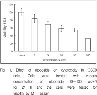
The effect of the exposure to etoposide on the cell viability of OSC9 cells, as assessed by the MTT test, is shown in Fig. 1. After cells were incubated for 24 h in medium (control) with various concentrations of etoposide (0∼100 ㎍/㎖), the
0 20 40 60 80 100 120
control 1 5 10 50 100
concentration (µg/ml)
viability (%)
Fig. 1. Effect of etoposide on cytotoxicity in OSC9 cells. Cells were treated with various concentration of etoposide (0∼100 ㎍/㎖) for 24 h and the cells were tested for viability by MTT assay.
0 20 40 60 80 100 120
0h 24h 48h 72h
Time (hour)
viability (%)
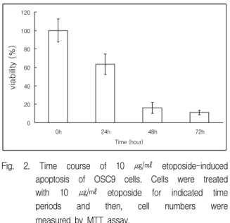
Fig. 2. Time course of 10 ㎍/㎖ etoposide-induced apoptosis of OSC9 cells. Cells were treated with 10 ㎍/㎖ etoposide for indicated time periods and then, cell numbers were measured by MTT assay.
number of cells was decreased according to increasing concentrations. The growth of OSC9 cells was inhibited in time-dependent manner by same concentration of etoposide (Fig. 2). At 10 ㎍/
㎖ of etoposide, the viability dropped to 63.3%, 16%
and 10.9% of the control on treatment times 24 h, 48 h and 72 h, respectively.
2. Morphological changes of cells treated with etoposide
The incubation of OSC9 cells with 10 ㎍/㎖
etoposide resulted in several morphological associated with apoptosis. Hoechst stain demonstrated that etoposide induced a change in nuclear morphology (Fig. 3). Compared to the typical round nuclei of the control cells, apoptotic OSC9 cells displayed condensed and fragmented nuclei. In other to confirm nuclear event on etoposide-induced apoptotic cells, TUNEL stain was conducted (Fig. 4). TUNEL stain showed that the percentage of cell death was increased compared to control cells and the positive reaction on condensed nuclei in the cells treated with etoposide.
(a) (b) Fig. 3. Immunofluorescent micrographs after
Hoechst staining. Control cells showing typical round nuclei (a). Cells treated with 10 ㎍/㎖ etoposide show atypical fragmented nuclei (b).
(a) (b)
5 60
85 95
0 20 40 60 80 100 120
0 24 48 72
Time (h)
% of cell death
(c)
Fig. 4. TUNEL staining. OSC9 cells were stimulated with etoposide (10 ㎍/㎖) for 24 h. Cells were fixed and analyzed by TUNEL staining.
While control cells showing negative reaction (a), cells treated with etoposide show positive reaction (b). The time course quantification of etoposide-induced cell death of OSC9 cells is depicted (c).
0 24 48 72 (h)
Fig. 5. The expression of p53 protein levels. Cells were treated with 10 ㎍/㎖ etoposide for 24, 48 and 72 h.
3. Western blot analysis
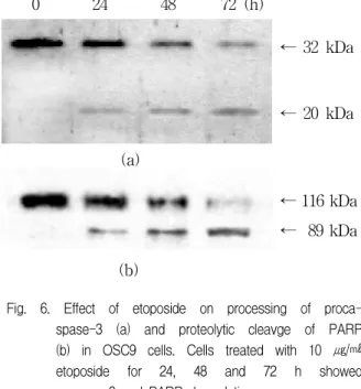
To establish a relationship between the change in the various molecules and apoptotic pathways, we examined tumor suppressor gene p53 and other proteins such as caspase-3 and PARP by western blot analysis. In order to investigate the change in molecules expression according to time, OSC9 cell was treated with 10 ㎍/㎖ etoposide and then incubated for 24 h, 48 h and 72 h. The expression of p53 protein was increased at 48 h in OSC9 cells (Fig. 5). We observed the proteolytic activation of procaspase-3 induced by etopside by Western blot (Fig. 6). 32 kDa precursor of caspase-3 was degraded, and 17 kDa cleavage products were produced. Activation of caspase-3 leads to the cleavage of a number of proteins, one of which is PARP. Treatment of OSC9 cells with 10 ㎍/㎖
etoposide induced proteolytic cleavage of 116 kDa PARP with accumulation of the 89 kDa cleaved products.
Ⅳ. DISCUSSION
Anti-cancer drugs have been shown to target diverse cellular functions in mediation cell death in chemosensitive tumors. Cytotoxic drugs which are currently used for the treatment of malignant diseases such as cytarabine, doxorubicin, metho- trexate, etoposide and cisplatin are thought to exert their effects through inhibition of DNA-polymerase (cytarabine), DNA-intercalation (doxorubicin), antagonization folic acid (methotrexate), inhibition of topoisomerase II (etoposide) and DNA-crosslinking
(b)
0 24 48 72 (h)
← 32 kDa
← 20 kDa
← 116 kDa
← 89 kDa (a)
Fig. 6. Effect of etoposide on processing of proca- spase-3 (a) and proteolytic cleavge of PARP (b) in OSC9 cells. Cells treated with 10 ㎍/㎖
etoposide for 24, 48 and 72 h showed caspase-3 and PARP degradation.
(cisplatin)
22). In addition, it has been recently shown that most antineoplastic drugs used in chemotherapy of leukemias and solid tumors induce apoptosis in drug-sensitive target cells.
Apoptosis is typical accompanied by the activation of a class of death proteases (caspase) and widespread biochemical and morphological changes to the cell
23). These changes almost invariably involve chromatin condensation and its margination at the nuclear periphery, extensive double-stranded DNA fragmentation, and cellular shrinkage and blebbing
23). However, apoptosis can also occur in the absence of DNA fragmentation
24). It can be activated intracellulary through a genetically defined developmental program or extracellulary by endogenous proteins, cytokines and hormones, as well as drugs, xenobiotic compounds, radiation, oxidative stress, and hypoxia
25, 26).
This study focused on the etoposide-induced
apoptosis in oral carcinoma cells as well as lung and
testicular cancer. Etoposide-induced apoptosis
variously confirmed by MTT assay, Hoechst stain,
TUNEL stain and western blot analysis. Etoposide
had a significant dose- and time-dependent
inhibitory effect on the viability of OSC9 cells.
Hoechst stain demonstrated that etoposide induced a change in nuclear morphology and TUNEL assay showed the positive reaction on condensed nuclei in cells treated with etoposide.
Apoptosis has been shown to be a significant mode of cell death following cytotoxic drug treatment in a variety of tumour types. In this process, p53 plays a key role
26). p53 is a regulatory protein that determines the cell regulation in response to DNA damage and other cellular stresses
27). Under DNA damage and other stimulation the wild type p53 level rapidly increases in the cell and this increased level is required for the functioning of p53 as a guardian of the genome. In the present study, the expression of p53 was increased at 48 hour, suggesting that the nuclear of OSC9 cell was damaged, thereby inducing apoptosis p53-dependently.
Caspases are synthesized as inactive proenzymes that are processed in cells undergoing apoptosis of self-proteolytic cascade, capable of cleaving and activating specific substrates, including an enzyme involved in DNA repair and genomic maintenance, PARP and DFF, etc. PARP plays the active role of
"nick senor" during DNA repair and apoptosis, when it synthesizes ADP-ribose from NAD+ in the presence of DNA strand breaks. Moreover, PARP becomes a target of apoptotic caspases, which originate two proteolytic fragments of 89 and 24 kDa
28). Since the specific proteolytic cleavage of PARP is considered to be a biochemical characteristic of apoptosis. Figure 4B shows that PARP is cleaved into 89 kDa fragment suggesting that capase-3 is activated. In this study, etoposide treatment induced caspase-3 cleavage and activation. Intact 116-kDa and 89-kDa cleaved product were indicated. The data of Figure 4 give evidences that etoposide-induced cell death is apoptosis.
In summary, we present evidence that incubation of OSC9 cells with etoposide leads to an activation of the caspase-3 and indcution of apoptosis.
However, based on presented evidence, it is impossible to differentiate direct epoposide action
from indirection as a stage in caspase's cascade.
Although more investigation is needed, etoposide can be considered as a good candidate with specific activity on oral squamous carcinom cells.
V. CONCLUSIONS
Etoposide showed apoptotic effect on OSC9 cells.
The death of cells was further demonstrated to be due to apoptosis characterized by chromatin condensation and nuclear fragment by Hoechst staining and TUNEL method. Apoptosis-related factors were analyzed by Western blot. Etoposide- induced apoptosis in OSC9 cells is associated with caspase-3 activation.
REFERENCES
1. Wyllie, A.H., Kerr, J.F., and Currie, A.R.: Cell death:
the significance of apoptosis. Int Rev Cytol 68 : 251-306, 1980.
2. Golstein P: Controlling cell death. Science 275(5303) : 1081-1082, 1997.
3. Orth, K., O'Rourke, K., Salvesen, G.S., and Dixit, V.M.: Molecular ordering of apoptotic mammalian CED-3/ICE-like proteases. J Biol Chem 271(35) : 20977-20980, 1996.
4. Enari, M., Sakahira, H., Yokoyama, H. et al. : A caspase-activated DNase that degrades DNA during apoptosis, and its inhibitor ICAD. Nature 391(6662) : 43-50, 1998.
5. Sakahira, H., Enari, M. and Nagata, S.: Cleavage of CAD inhibitor in CAD activation and DNA degradation during apoptosis. Nature 391(6662) : 96-99, 1998.
6. Thornberry, N.A., Rosen, A. and Nicholson, D.W.:
Control of apoptosis by proteases, in Apoptosis (Kaufmann SH eds). Adv Pharmacol 44 : 155-177, 1997.
7. Yuan, J.: Evolutionary conservation of a genetic pathway of programmed cell death. J Cell Biochem 60 : 4-11, 1996.
8. Clarke, A.R., Purdie, C.A., Harrison, D.J. et al. : Thymocyte apoptosis induced by p53-dependent and independent pathways. Nature 362(6423) : 849-852, 1993.
9. Wagner, A.J., Kokontis, J.M. and Hay, N.: Myc-
mediated apoptosis requires wild-type p53 in a manner independent of cell cycle arrest and the ability of p53 to induce p21waf1/cip1. Genes Dev 8 : 2817-2830, 1994.
10. Graeber, T.G., Osmanian, C., Jacks, T., Housman, D.E., Koch, C.J., Lowe, S.W. and Giaccia, A.J.:
Hypoxia-mediated selection of cells with diminished apoptotic potential in solid tumours. Nature 379 : 88-91, 1996.
11. Linke, S.P., Clarkin, K.C., Di, Leonardo, A., Tsou, A.
and Wahl, G.M.: A reversible, p53-dependent G0/G1 cell cycle arrest induced by ribonucleotide depletion in the absence of detectable DNA damage. Genes Dev 10 : 934-947, 1996.
12. Yin, Y., Terauchi, Y., Solomon, G.G. et al. : Invol- vement of p85 in p53-dependent apoptotic response to oxidative stress. Nature 391 : 707-710, 1998.
13. Schmitt, C.A. and Lowe, S.W. : Apoptosis and Therapy. J Pathol 187 : 127-137, 1999.
14. Attardi, L.D., Reczek, E.E., Cosmas, C. et al. : PERP, an apoptosis-associated target of p53, is a novel member of the PMP-22/gas3 family. Genes Dev 14 : 704-718, 2000.
15. Ko, L.J. and Prives, C.: p53: puzzle and paradigm.
Genes Dev 10 : 1054-1072, 1996.
16. Levine, A.J.: The tumor suppressor genes. Annu Rev Biochem 62 : 623-651, 1993.
17. Fisher, D.E.: Apoptosis in cancer therapy: crossing the threshold. Cell 78(4) : 539-542, 1994.
18. Steller, H.: Mechanisms and genes of cellular suicide.
Science 267(5203) : 1445-1449, 1995.
19. Lowe, S.W., Ruley, H.E., Jacks, T. and Housman, D.E.: p53-dependent apoptosis modulates the cytotoxicity of anticancer agents. Cell 74(6) : 957-967, 1993.
국문요약
Etoposide에 대한 사람구강편평상피암종세포의 세포자멸사 반응
부산대학교 치과대학 및 구강생물공학연구소 구강해부학교실
1, 구강보존과학교실
2, 구강내과학교실
3김규천
1․이경덕
1․박재현
1․김덕한
1․박정길
2․박준상
3․박봉수
1항암제의 연구는 화학물질에 민감한 암세포를 죽음에 이르게 하는 세포자멸사와 같은 다양한 세포기능에 초점 을 맞추어 왔다. 그러나 약물이 유도한 세포의 죽음에 있어서 핵심적인 분자적 기작은 아직 잘 이해되지 않고
20. Burden, D.A., Kingma, P.S., Froelich-Ammon, S.J. et al. : Topoisomerase II.etoposide interactions direct the formation of drug-induced enzyme-DNA cleavage complexes. J Biol Chem 271(46) : 29238-29244, 1996.
21. Ramirez, C.D., Sleiman, R.J., Catchpoole, D.R. and Stewart, B.W.: Morphological and molecular evidence of differentiation during etoposide-induced apoptosis in human lymphoblastoid cells. Cell Death Differ 7(6) : 548-555, 2000.
22. Ucker, D.S., Obermiller, P.S., Eckhart, W. et al. : Genome digestion is a dispensable consequence of physiological cell death mediated by cytotoxic T lymphocytes. Mol Cell Biol 12 : 3060-3069, 1992.
23. Nicholson, D.W. and Thornberry, N.A.: Caspases:
killer proteases. Trends Biochem Sci 22 : 299-306, 1997.
24. Schulze-Osthoff, K., Walczak, H., Droge, W. and Krammer, P.H.: Cell nucleus and DNA fragmentation are not required for apoptosis. J Cell Biol 127 : 15-20, 1994.
25. Williams, G.T. : Programmed cell death: apoptosis and oncogenesis. Cell 65 : 1097-1098, 1991.
26. Tolis, C., Peters, G.J., Ferreira, C.G., Pinedo, H.M. and Giaccone, G.: Cell cycle disturbances and apoptosis induced by topotecan and gemcitabine on human lung cancer cell lines. Eur J Cancer 35(5) : 796-807, 1999.
27. Harris, C.C.: Structure and function of the p53 tumor suppressor gene: clues for rational cancer therapeutic strategies. J Natl Cancer Inst 88(20) : 1442-1455, 1996.
28. Soldani, C., Lazze, M.C., Bottone, M.G. et al. : Poly(ADP-ribose) polymerase cleavage during apoptosis: when and where? Exp Cell Res 269(2) : 193-201, 2001.