ISSN 2234-3806 • eISSN 2234-3814
Ann Lab Med 2018;38:242-248
https://doi.org/10.3343/alm.2018.38.3.242
Spectrum of MNX1 Pathogenic Variants and
Associated Clinical Features in Korean Patients with Currarino Syndrome
Seungjun Lee, M.D.1,2, Eun Jin Kim, M.T.3, Sung Im Cho, M.T.3, Hyunwoong Park, M.D.4, Soo Hyun Seo, M.D.5,
Moon-Woo Seong, M.D.3, Sung Sup Park, M.D.3, Sung-Eun Jung, M.D.6, Seong-Cheol Lee, M.D.6,*, Kwi-Won Park, M.D.6,*, and Hyun-Young Kim, M.D.6
Department of Laboratory Medicine1, Kangbuk Samsung Hospital, Sungkyunkwan University School of Medicine, Seoul; Department of Laboratory Medicine2, Seoul National University College of Medicine, Seoul; Department of Laboratory Medicine3, Seoul National University Hospital, Seoul National University College of Medicine, Seoul; Department of Laboratory Medicine4, Gyeongsang National University Changwon Hospital, Changwon; Department of Laboratory Medicine5, Seoul National University Bundang Hospital, Seongnam; Department of Pediatric Surgery6, Seoul National University College of Medicine, Seoul, Korea
Background: The major genetic cause of Currarino syndrome (CS), a congenital malfor- mation syndrome typically characterized by sacral agenesis, anorectal malformation, and presence of a pre-sacral mass, is known to be pathogenic variants in motor neuron and pancreas homeobox 1 (MNX1), which exist in almost all familial cases and 30% of spo- radic cases. Less commonly, a large deletion or a complex rearrangement involving the 7q36 region is associated with CS. We investigated the spectrum of MNX1 pathogenic variants and associated clinical features in the Korean patients with CS.
Methods: We enrolled 25 patients with CS, including 24 sporadic cases and one familial case. Direct sequencing of MNX1 and multiplex ligation-dependent probe amplification were performed. We also analyzed clinical phenotypes and evaluated genotype-phenotype correlations.
Results: We identified six novel variants amongst a total of six null variants, one missense variant, and one large deletion. The null variants included four frameshift variants (p.Gly98- Alafs*124, p.Gly145Alafs*77, p.Gly151Leufs*67, and p.Ala216Profs*5) and two nonsense variants (p.Tyr186* and p.Gln212*). The missense variant, p.Lys295Gln, was located in the highly-conserved homeobox domain and was predicted to be deleterious. A large de- letion involving the 7q36 region was detected in one patient. Pathogenic variants in MNX1 were detected in 28% of all CS cases and 25% of sporadic cases. The clinical phenotype was variable in patients with and without pathogenic variants; no significant genotype-phe- notype correlation was observed.
Conclusions: This study revealed the spectrum and phenotypic variability of MNX1 patho- genic variants in the Korean population.
Key Words: Currarino syndrome, MNX1, Pathogenic variant, Korean
Received: April 9, 2017
Revision received: September 20, 2017 Accepted: December 11, 2017 Corresponding author: Hyun-Young Kim Department of Pediatric Surgery, Seoul National University College of Medicine, 103 Daehak-ro, Jongno-gu, Seoul 03080, Korea
Tel: +82-2-2072-2478 Fax: +82-2-747-5130 E-mail: [email protected]
*Professor emeritus
© Korean Society for Laboratory Medicine This is an Open Access article distributed under the terms of the Creative Commons Attribution Non-Commercial License (http://creativecom- mons.org/licenses/by-nc/4.0) which permits unrestricted non-commercial use, distribution, and reproduction in any medium, provided the original work is properly cited.
INTRODUCTION
Currarino syndrome (CS; OMIM #176450), first described in
1981, is a rare congenital disorder characterized by the triad of sacral agenesis, anorectal malformation, and the presence of a presacral mass [1]. The most frequent type of sacral agenesis is
2017-03-16 https://crossmark-cdn.crossref.org/widget/v2.0/logos/CROSSMARK_Color_square.svg
hemisacrum, which is a pathognomonic sign of CS and is also known as “scimitar sacrum” because of peculiar radiological findings such as sickle-shaped or crescent-shaped sacral defor- mities. Besides these major signs, developmental delays, neural tube defects, sensorineural deafness, and other features such as depressed nasal bridge, joint hyperextensibility, cataracts, ptosis, microcephaly, and facial dysmorphism have been re- ported in patients with CS [2-4]. Phenotypic variability has also been observed in these patients, with some of the major signs not being observed in incomplete cases [5-12].
The autosomal dominant inheritance pattern of CS was noted by Yates et al [13]. CS is caused by haploinsufficiency of motor neuron and pancreas homeobox 1 (MNX1), located on chromo- some 7q36.3 [5, 14-16]. This gene encodes a transcription fac- tor that contains a homeobox domain. Pathogenic variants in MNX1 have been revealed in almost all cases of familial CS, and in approximately 30% of sporadic CS cases [5, 17, 18]. At least 82 MNX1 heterozygous pathogenic variants have been reported to date [18]; two studies on MNX1 pathogenic variants in Ko- rean patients with CS have been conducted, which included three familial cases and two sporadic cases [9, 19]. However, these pathogenic variants have been identified only in familial cases; moreover, reduced penetrance and variable expressivity of the MNX1 pathogenic variants in CS cases have been reported, even in familial cases [6-12, 18, 19]. In this study, we investi- gated a large number of patients with CS to determine the spec- trum of MNX1 pathogenic variants, and described, for the first time, the MNX1 pathogenic variants in Korean patients with spo- radic CS.
METHODS
1. Study subjectsIn total, 25 index patients were included in this study between July 2014 and June 2015. These Korean patients were diag- nosed as having CS and treated at the Department of Pediatric Surgery at Seoul National University Children’s Hospital, Seoul, Korea. Medical records were reviewed to collect clinical infor- mation. Among these patients, only one patient had a family history of CS, while the remaining 24 patients were classified as sporadic cases of CS upon thorough investigation of family his- tory. This study was approved by the Institutional Review Board of Seoul National University Hospital (No. 1703-125-841). Writ- ten informed consent was obtained from all patients and/or their legal representatives.
2. Genetic analysis
Genetic analysis was performed on archival samples. Genomic DNA was extracted from peripheral blood samples using the Gentra Puregene blood kit (Qiagen, Valencia, CA, USA). PCR was performed using newly designed primers specific to the three exons and flanking regions of MNX1. Direct sequencing with in-house primers was performed on an Applied Biosystems 3730xl DNA Analyzer (Applied Biosystems, Foster City, CA, USA) using a BigDye Terminator v3.1 Cycle Sequencing Kit (Applied Biosystems). Sequence variants were identified according to the NM_005515.3 and NP_005506.3 reference sequences using Sequencher 5.0 (Gene Codes Corporation, Ann Arbor, MI, USA).
If the result of sequence analysis was negative, gene dosage analysis was performed by using a multiplex ligation-dependent probe amplification (MLPA) kit (SALSA MLPA probemix P277- B2 Human Telomere-10; MRC-Holland, Amsterdam, Nether- lands). The MLPA kit contains 14, 11, 11, and 11 probes in the terminal region of chromosomes 5q, 6q, 7q, and 8q, respec- tively. In silico prediction of missense variants was carried out using Polyphen-2, SIFT, and Mutation Taster [20-22]. The splic- ing effect of variants was predicted using Human Splicing Finder [23]. The variants were classified according to the latest Ameri- can College of Medical Genetics guidelines [24].
RESULTS
1. Clinical features of patients with CS
The study group consisted of 25 index patients (11 males and 14 females). Most patients (21/25) presented the complete CS triad, while the remaining four patients showed only two major CS features without sacral anomalies (Table 1). Patient 1 was the only familial case, and she presented the complete CS phe- notype.
Hemisacrum (17/25) was the predominant sacral anomaly phenotype, followed by partial sacral agenesis (4/25). Anorectal malformations, such as imperforate anus (13/25) and anorectal stenosis (12/25), were observed in all patients. Imperforate anus (9/11) was the most common anorectal malformation in males, while anorectal stenosis (10/14) was predominant in females.
Sex-dependent differences were not observed for the other two major CS phenotypes. Among the 13 imperforate anus cases, high type (11/13) was more common than low type (2/13). All patients presented with presacral mass. Teratoma (9/25) was observed predominantly, followed by cystic formation (5/25), li- poma (4/25), lipomeningomyelocele (4/25), and others.
Twelve patients presented with other signs in addition to the
Currarino triad. Urogenital malformations, including kidney mal- formation, uterus didelphys, micropenis, hypospadia, bifid scro- tum, bladder diverticula, and vesicoureteral reflux were most commonly observed. Musculoskeletal deformities (Sprengel’s deformity, clubfoot, pes planus, and developmental dislocation of the hip), eye complications (coloboma, ptosis, and exotropia), and tethered cord were observed in some cases. Microcephaly and dysmorphic facial features were identified only in one case.
2. Genetic analysis
From MNX1 sequence analysis, we identified seven different pathogenic or likely pathogenic variants, including four frame- shift variants (p.Gly98Alafs*124, p.Gly145Alafs*77, p.Gly151- Leufs*67, and p.Ala216Profs*5), two nonsense variants (p.Tyr186*
and p.Gln212*), and one missense variant (p.Lys295Gln) (Fig.
1 and Table 2). Except for one missense variant located in exon 3, the other six variants were located in exon 1.
Pathogenic variants were more commonly identified in females (6/14) than in males (1/11). Of these, only one variant, c.634C>T Table 1. Clinical data of patients with Currarino syndrome
No. case Sex Age* Sacral anomaly Anorectal
malformation Presacral mass Other signs
1† F 3 yr Hemisacrum IA (High type) Teratoma −
2 F 7 yr Hemisacrum Anorectal stenosis Lipoma Kidney malformation, Tethered cord
3 F 35 m Hemisacrum Anorectal stenosis Teratoma −
4 F 17 yr Hemisacrum Anorectal stenosis Meningocele Uterus didelphys with double vagina
5 M 18 yr Hemisacrum IA (High type) Teratoma −
6 F 28 m Hemisacrum Anorectal stenosis Teratoma −
7 F 10 yr − Anorectal stenosis Cystic formation Anterior angulation of coccyx
8 F 13 m Hemisacrum Anorectal stenosis Teratoma −
9 F 25 m Hemisacrum Anorectal stenosis Lipoma Bilateral optic and iris coloboma
10 M 3 m Hemisacrum IA (High type) Lipoma Pes planus
11 M 4 yr Hemisacrum IA (Low type) Myelomeningocele Horseshoe kidney, Clubfoot deformity, Hip abduction contracture 12 M 14 yr Partial sacral agenesis Anorectal stenosis Lipoma Microcephaly, Dysmorphic face, Hypotelorism, Micropenis,
Tethered cord
13 F 3 yr Hemisacrum Anorectal stenosis Teratoma −
14 F 11 yr − Anorectal stenosis Cystic formation −
15 F 17 yr Hemisacrum IA (High type) Lipomeningomyelocele Kidney malformation, Uterus didelphys
16 M 31 m − IA (High type) Teratoma Congenital ptosis, Bifid uvula, Choanal atresia, Laryngomalacia, Sprengel’s deformity, Cardiac malformation, Umbilical hernia, Anterior angulation of coccyx
17 M 27 m − IA (High type) Cystic formation Exotropia
18 F 6 yr Hemisacrum IA (Low type) Lipomeningomyelocele −
19 M 13 yr Hemisacrum Anorectal stenosis Teratoma Tracheoesophageal fistula, Kidney malformation, Developmental dislocation of hip
20 F 8 yr Partial sacral agenesis IA (High type) Cystic formation Renal pelvic ectasia, Cloacal anomaly
21 M 19 yr Hemisacrum IA (High type) Lipomeningomyelocele −
22 M 18 yr Partial sacral agenesis IA (High type) Lipomeningomyelocele Tethered cord
23 F 17 yr Hemisacrum Anorectal stenosis Teratoma −
24 M 28 m Hemisacrum IA (High type) Cystic formation Tethered cord, Hypospadia, Bifid scrotum, Bladder diverticula, Vesicoureteral reflux
25 M 29 yr Partial sacral agenesis IA (High type) Lipomeningocele −
*Age is expressed in years (yr) or months (m); †Only patient 1 had family history of Currarino syndrome.
Abbreviations: F, female; M, male; IA, imperforate anus; −, absent.
(p.Gln212*), was previously reported in a Swedish patient [10].
The other variants were identified for the first time in this study.
Of the seven variants, p.Lys295Gln was the only missense variant. This variant was predicted to be “probably damaging”
by Polyphen-2, “deleterious” by SIFT, and “disease causing” by Mutation Taster. In addition, this missense variant was located in a highly conserved homeobox domain between codons 244 and 297.
Almost all previously reported missense variants are located in the homeobox domain. Only a few of them (p.Met1Ile, p.Pro- 27Leu, and p.Arg243Trp) have been reported outside of this re- gion [18, 25, 26]. Therefore, we classified p.Lys295Gln as a likely pathogenic variant. A synonymous variant, c.852G>A (p.Gln284=), was identified in patient 8. This variant was at the last nucleotide position of exon 2. The Human Splicing Finder predicted the
wild type motif with a score of 87.86 and the mutant motif with a score of 77.28. The difference of the score between them (–12.04%) was under –10%, suggesting that the variant breaks the splice site. However, these findings were not sufficient to conclusively determine pathogenicity of the variant. Therefore, this synonymous variant was classified as a variant with uncer- tain significance.
We performed gene dosage analysis for 18 patients with neg- ative results in the direct sequencing. MLPA analysis revealed a large deletion in patient 12 (Fig. 2). A reduced ratio was detected in all 11 probes in the 7q terminal region. This finding suggested that patient 12 had at least a 5.1-Mb 7q36 deletion. The exact breakpoints and range of the deletion could not be determined by MLPA results alone.
Fig. 1. Distribution of the seven different MNX1 variants. Length of the exons and location of the variants are displayed in proportion to ac- tual length and location, except introns. Black boxes indicate coding regions and gray boxes non-coding regions. Boxes with diagonal pat- terns indicate a homeobox domain (p.244-p.297). The missense variant, c.883A>C, is located in the homeobox domain.
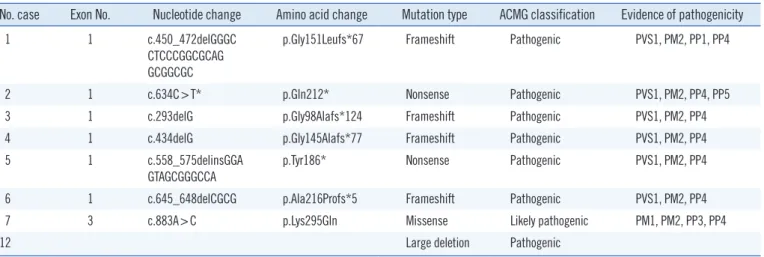
Table 2. Pathogenic variants identified in patients with Currarino syndrome
No. case Exon No. Nucleotide change Amino acid change Mutation type ACMG classification Evidence of pathogenicity
1 1 c.450_472delGGGC
CTCCCGGCGCAG GCGGCGC
p.Gly151Leufs*67 Frameshift Pathogenic PVS1, PM2, PP1, PP4
2 1 c.634C>T* p.Gln212* Nonsense Pathogenic PVS1, PM2, PP4, PP5
3 1 c.293delG p.Gly98Alafs*124 Frameshift Pathogenic PVS1, PM2, PP4
4 1 c.434delG p.Gly145Alafs*77 Frameshift Pathogenic PVS1, PM2, PP4
5 1 c.558_575delinsGGA
GTAGCGGGCCA
p.Tyr186* Nonsense Pathogenic PVS1, PM2, PP4
6 1 c.645_648delCGCG p.Ala216Profs*5 Frameshift Pathogenic PVS1, PM2, PP4
7 3 c.883A>C p.Lys295Gln Missense Likely pathogenic PM1, PM2, PP3, PP4
12 Large deletion Pathogenic
All variants in the above eight patients are heterozygous.
*Only c.634C>T was previously reported [10], and the other variants were first reported in this study.
Abbreviations: ACMG, American College of Medical Genetics and Genomics; PVS, pathogenic very strong; PM, pathogenic moderate; PP, pathogenic sup- porting.
3. Genotype-phenotype correlations
Seven of eight patients with pathogenic variants displayed the complete Currarino triad. Only three of 17 patients without pathogenic variants showed an incomplete Currarino triad with- out sacral anomaly. Patients with pathogenic variants presented predominantly with hemisacrum, anorectal stenosis, and tera- toma for each Currarino triad. However, no significant pheno- typic differences were observed between the patients with and without pathogenic variants. Patients in both groups exhibited variable expression of all the three major phenotypes and asso- ciated anomalies. The pathogenic variants were classified as six null variants, one missense variant, and one large deletion. The missense variant was associated with milder phenotypes (ano- rectal stenosis, cystic formation, and anterior angulation of coc- cyx) compared with phenotypes of the other null variants. The large deletion case presented with multiple associated malfor- mations in addition to the classic CS triad.
A 3-year-old girl with suspected CS (Patient 1) was referred for genetic study. She did not excrete meconium within 24 hours of birth. Imaging studies suggested hemisacrum, high type im- perforate anus, and teratoma. Pena operation and teratoma ex-
cision were performed at the age of five months. Direct sequenc- ing revealed a frameshift variant, c.450_472del (p.Gly151Leufs*67), in the heterozygous state. She had no siblings. The patient’s mother presented with fecal and urinary incontinence since birth. She underwent neurosurgical intervention for ligation of the anterior sacral meningocele and spinal cord untethering at the age of five years. After three months, Pena operation was performed to correct her low type imperforate anus. Imaging studies revealed sacral spina bifida and horseshoe kidney. There was no family history of CS in the patient’s mother’s parents or siblings. The same pathogenic variant as her daughter was iden- tified by sequence analysis.
DISCUSSION
The MNX1 sequence analysis identified six novel and one previ- ously reported pathogenic variants. The overall detection rate of sequence analysis was 28% (7/25) in all patients and 25% (6/24) in sporadic patients. Haploinsufficiency of MNX1 causes CS;
according to previous reports, pathogenic variants of MNX1 were detected in approximately 30% of CS sporadic patients [5, 17, Fig. 2. Gene dosage analysis and mapping of the 7q36 deletion in patient 12. (A) Result of multiplex ligation-dependent probe amplifica- tion analysis. Arrows indicate the reduced ratio of all 11 probes in the 7q terminal region. These probes are located in MNX1, DPP6, RNF32, SHH, NCAPG2, WDR60, HTR5A, VIPR2, PTPRN2, UBE3C, and PTPRN2, in the order of increasing probe length. (B) Schematic representation of the 7q36 deletion. Genomic coordinates are based on GRCh38/hg38 assembly. Box with a diagonal pattern indicates a 7q36 deletion longer than 5.1 Mb. The exact breakpoints and range of the 7q36 deletion were not available. Black boxes indicate genes af- fected by 7q36 deletion.
A
B
18]. According to Cretolle et al [17], frameshift variants (45%) are the most frequent type of variants, followed by missense vari- ants (25%) and nonsense variants (12%). Similarly, four frame- shift variants, two nonsense variants, and one missense variant were identified in this study.
We identified a 7q36 deletion in a sporadic CS patient through gene dosage analysis. Large deletions or chromosome rearrange- ment involving 7q36 are related with holoprosencephaly (HPE) and CS due to haploinsufficiency of sonic hedgehog (SHH) and MNX1. Some previous studies reported patients with a deletion or chromosome rearrangement involving 7q36 [3, 4, 14, 17, 27-34]. In these patients, microcephaly was the predominant craniofacial finding of HPE, and hypotelorism, ptosis, and mid- face hypoplasia were commonly associated. However, patients presenting with a complete CS triad were rare. Sacral anomaly was most frequently observed, followed by presacral mass and anorectal malformation. Pavone et al [3] described the first re- ported case with complete CS triad and microcephaly. A 10.3- Mb duplication of 7q34-q35 and an 8.8-Mb deletion on 7q36 were identified by comparative genomic hybridization (CGH) ar- ray in the patient. Another study reported a patient with a 2.7- Mb deletion 7q36.3 excluding SHH [28]. The patient presented with microcephaly and incomplete CS triad without anorectal malformation. In this study, we reported a patient with 7q36 de- letion including loss of SHH and MNX1. This patient presented with complete CS triad and microcephaly, which was a rare phe- notype spectrum upon 7q36 deletion.
CS shows characteristically diverse phenotypes with variable expressivity [5, 17, 31], and there is no clear relationship be- tween the genotype and phenotype. The 25 index patients re- vealed variable phenotypes in this study. Sacral anomalies var- ied from hemisacrum to partial sacral agenesis; likewise, presa- cral mass and anorectal malformation exhibited various patho- logical and clinical diagnoses. The clinical phenotypes were di- verse, even in the familial case: patient 1 and her mother exhib- ited differences in all the three major phenotypes and their as- sociated anomalies. Furthermore, phenotypic variability in famil- ial cases has been previously reported [7, 8, 10-12, 18, 31].
The fact that clinical variation was observed in individuals with the same variant suggests that factors other than MNX1 affect the CS phenotype. Other modifier genes might influence the MNX1 protein, partners, or transcriptional factors [5, 6, 16].
Some candidates, such as proneuronal basic-helix-loop-helix proteins and LIM (named after Lin-11, Isl-2, and Mec-3 protein) domain proteins, have been found to regulate MNX1 expression in other vertebrates [35-37]. A recent study using whole exome
sequencing suggested three candidate genes, ETV3L, ARID3A, and NCAPD3, were involved in CS pathogenesis [38].
The most common malformations in patients with CS are uro- genital anomalies and terminal spinal cord malformations. In this study, nine patients displayed these malformations. How- ever, other associated malformations were also observed in some patients. Patient 12, in particular, with the complete Currarino triad, displayed severe associated malformations, including mi- crocephaly and facial dysmorphism. Moreover, patient 16 also exhibited multiple associated anomalies including ptosis, bifid uvula, choanal atresia, laryngomalacia, Sprengel’s deformity (congenital elevation of the scapula), cardiac anomalies, and umbilical hernia. According to the diagnostic algorithm proposed by Cuturilo et al [4], patients with CS exhibiting growth delay and/or developmental delay and/or facial dysmorphic features should be evaluated by a chromosomal microarray. Large dele- tions and complex rearrangements involving the 7q36 region have been identified in many previous reports [3, 4, 14, 17, 27- 34]. Almost all cases described in these studies exhibited vari- ous associated anomalies. In this study, we performed genetic analysis using direct sequencing and MLPA. Therefore, further studies using chromosomal microarray or whole exome sequenc- ing will be needed to investigate patients with negative results, especially in cases with multiple associated malformations.
To our knowledge, only four studies have reported genetic analysis of a large series of patients with CS [5, 16-18]. This study described the clinical and genetic aspects of 25 index pa- tients with CS. We identified seven different pathogenic variants, six of which were novel, including a large deletion. The clinical variability of CS, including intrafamilial variability, was also con- firmed. In conclusion, CS should be considered for early diag- nosis even if the cases exhibit an incomplete CS phenotype. For adequate genetic counseling, genetic analysis should be con- sidered for patients with suspected CS.
Authors’ Disclosures of Potential Conflicts of Interest
There are no conflicts of interest to be declared.
REFERENCES
1. Currarino G, Coln D, Votteler T. Triad of anorectal, sacral, and presacral anomalies. AJR Am J Roentgenol 1981;137:395-8.
2. Horn D, Tonnies H, Neitzel H, Wahl D, Hinkel GK, von Moers A, et al.
Minimal clinical expression of the holoprosencephaly spectrum and of Currarino syndrome due to different cytogenetic rearrangements delet-
ing the Sonic Hedgehog gene and the HLXB9 gene at 7q36.3. Am J Med Genet A 2004;128A:85-92.
3. Pavone P, Ruggieri M, Lombardo I, Sudi J, Biancheri R, Castellano-Chi- odo D, et al. Microcephaly, sensorineural deafness and Currarino triad with duplication-deletion of distal 7q. Eur J Pediatr 2010;169:475-81.
4. Cuturilo G, Hodge JC, Runke CK, Thorland EC, Al-Owain MA, Ellison JW, et al. Phenotype analysis impacts testing strategy in patients with Currarino syndrome. Clin Genet 2016;89:109-14.
5. Hagan DM, Ross AJ, Strachan T, Lynch SA, Ruiz-Perez V, Wang YM, et al. Mutation analysis and embryonic expression of the HLXB9 Currarino syndrome gene. Am J Hum Genet 2000;66:1504-15.
6. Kochling J, Karbasiyan M, Reis A. Spectrum of mutations and genotype- phenotype analysis in Currarino syndrome. Eur J Hum Genet 2001;9:599- 605.
7. Wang RY, Jones JR, Chen S, Rogers RC, Friez MJ, Schwartz CE, et al.
A previously unreported mutation in a Currarino syndrome kindred. Am J Med Genet A 2006;140:1923-30.
8. Cretolle C, Sarnacki S, Amiel J, Genevieve D, Encha-Razavi F, Zrelli S, et al. Currarino syndrome shown by prenatal onset ventriculomegaly and spinal dysraphism. Am J Med Genet A 2007;143A:871-4.
9. Kim IS, Oh SY, Choi SJ, Kim JH, Park KH, Park HK, et al. Clinical and genetic analysis of HLXB9 gene in Korean patients with Currarino syn- drome. J Hum Genet 2007;52:698-701.
10. Zu S, Winberg J, Arnberg F, Palmer G, Svensson PJ, Wester T, et al. Mu- tation analysis of the motor neuron and pancreas homeobox 1 (MNX1, former HLXB9) gene in Swedish patients with Currarino syndrome. J Pediatr Surg 2011;46:1390-5.
11. Markljung E, Adamovic T, Cao J, Naji H, Kaiser S, Wester T, et al. Novel mutations in the MNX1 gene in two families with Currarino syndrome and variable phenotype. Gene 2012;507:50-3.
12. Wang Y and Wu Y. A novel HLXB9 mutation in a Chinese family with Currarino syndrome. Eur J Pediatr Surg 2012;22:243-5.
13. Yates VD, Wilroy RS, Whitington GL, Simmons JC. Anterior sacral defects:
an autosomal dominantly inherited condition. J Pediatr 1983;102:239- 42.
14. Lynch SA, Bond PM, Copp AJ, Kirwan WO, Nour S, Balling R, et al. A gene for autosomal dominant sacral agenesis maps to the holoprosen- cephaly region at 7q36. Nat Genet 1995;11:93-5.
15. Ross AJ, Ruiz-Perez V, Wang Y, Hagan DM, Scherer S, Lynch SA, et al.
A homeobox gene, HLXB9, is the major locus for dominantly inherited sacral agenesis. Nat Genet 1998;20:358-61.
16. Belloni E, Martucciello G, Verderio D, Ponti E, Seri M, Jasonni V, et al.
Involvement of the HLXB9 homeobox gene in Currarino syndrome. Am J Hum Genet 2000;66:312-9.
17. Cretolle C, Pelet A, Sanlaville D, Zerah M, Amiel J, Jaubert F, et al. Spec- trum of HLXB9 gene mutations in Currarino syndrome and genotype- phenotype correlation. Hum Mutat 2008;29:903-10.
18. Merello E, De Marco P, Ravegnani M, Riccipetitoni G, Cama A, Capra V.
Novel MNX1 mutations and clinical analysis of familial and sporadic Cur- rarino cases. Eur J Med Genet 2013;56:648-54.
19. Kim AY, Yoo SY, Kim JH, Eo H, Jeon TY. Currarino syndrome: variable imaging features in three siblings with HLXB9 gene mutation. Clin Im- aging 2013;37:398-402.
20. Adzhubei IA, Schmidt S, Peshkin L, Ramensky VE, Gerasimova A, Bork P, et al. A method and server for predicting damaging missense muta- tions. Nat Methods 2010;7:248-9.
21. Kumar P, Henikoff S, Ng PC. Predicting the effects of coding non-syn- onymous variants on protein function using the SIFT algorithm. Nat Pro-
toc 2009;4:1073-81.
22. Schwarz JM, Cooper DN, Schuelke M, Seelow D. MutationTaster2: mu- tation prediction for the deep-sequencing age. Nat Methods 2014;11:
361-2.
23. Desmet FO, Hamroun D, Lalande M, Collod-Beroud G, Claustres M, Beroud C. Human Splicing Finder: an online bioinformatics tool to pre- dict splicing signals. Nucleic Acids Res 2009;37:e67.
24. Richards S, Aziz N, Bale S, Bick D, Das S, Gastier-Foster J, et al. Stan- dards and guidelines for the interpretation of sequence variants: a joint consensus recommendation of the American College of Medical Genet- ics and Genomics and the Association for Molecular Pathology. Genet Med 2015;17:405-24.
25. Garcia-Barcelo M, So MT, Lau DK, Leon TY, Yuan ZW, Cai WS, et al.
Population differences in the polyalanine domain and 6 new mutations in HLXB9 in patients with Currarino syndrome. Clin Chem 2006;52:46- 52.
26. Garcia-Barcelo MM, Lui VC, So MT, Miao X, Leon TY, Yuan ZW, et al.
MNX1 (HLXB9) mutations in Currarino patients. J Pediatr Surg 2009;
44:1892-8.
27. Benzacken B, Siffroi JP, Le Bourhis C, Krabchi K, Joye N, Maschino F, et al. Different proximal and distal rearrangements of chromosome 7q associated with holoprosencephaly. J Med Genet 1997;34:899-903.
28. Coutton C, Poreau B, Devillard F, Durand C, Odent S, Rozel C, et al.
Currarino syndrome and HPE microform associated with a 2.7-Mb de- letion in 7q36.3 excluding SHH gene. Mol Syndromol 2014;5:25-31.
29. Frints SG, Schoenmakers EF, Smeets E, Petit P, Fryns JP. De novo 7q36 deletion: breakpoint analysis and types of holoprosencephaly. Am J Med Genet 1998;75:153-8.
30. Frints SG, Schrander-Stumpel CT, Schoenmakers EF, Engelen JJ, Reek- ers AB, van den Neucker AM, et al. Strong variable clinical presentation in 3 patients with 7q terminal deletion. Genet Couns 1998;9:5-14.
31. Holm I, Monclair T, Lundar T, Stadheim B, Prescott TE, Eiklid KL. A 5.8 kb deletion removing the entire MNX1 gene in a Norwegian family with Currarino syndrome. Gene 2013;518:457-60.
32. Masuno M, Fukushima Y, Sugio Y, Ikeda M, Kuroki Y. Two unrelated cases of single maxillary central incisor with 7q terminal deletion. Jinrui Idengaku Zasshi 1990;35:311-7.
33. Rodriguez L, Cuadrado Perez I, Herrera Montes J, Lorente Jareno ML, Lopez Grondona F, Martinez-Frias ML. Terminal deletion of the chromo- some 7(q36-qter) in an infant with sacral agenesis and anterior myelo- meningocele. Am J Med Genet 2002;110:73-7.
34. Roessler E, Ward DE, Gaudenz K, Belloni E, Scherer SW, Donnai D, et al. Cytogenetic rearrangements involving the loss of the Sonic Hedge- hog gene at 7q36 cause holoprosencephaly. Hum Genet 1997;100:172- 81.
35. Lee SK and Pfaff SL. Synchronization of neurogenesis and motor neu- ron specification by direct coupling of bHLH and homeodomain tran- scription factors. Neuron 2003;38:731-45.
36. Lee SK, Jurata LW, Funahashi J, Ruiz EC, Pfaff SL. Analysis of embryon- ic motoneuron gene regulation: derepression of general activators func- tion in concert with enhancer factors. Development 2004;131:3295- 306.
37. Nakano T, Windrem M, Zappavigna V, Goldman SA. Identification of a conserved 125 base-pair Hb9 enhancer that specifies gene expression to spinal motor neurons. Dev Biol 2005;283:474-85.
38. Holm I, Spildrejorde M, Stadheim B, Eiklid KL, Samarakoon PS. Whole exome sequencing of sporadic patients with Currarino Syndrome: a re- port of three trios. Gene 2017;624:50-5.