서 론
초음파는 근골격계 질환을 평가하는 효과적인 방법으로 고해상 도 변환기(transducer, probe)의 발전에 힘입어 족부와 족관절의 표면에 위치하는 인대와 건 등에 대해서는 자기공명영상 검사에 못지 않은 검사법이다. 초음파의 장점으로 자기공명영상 검사에 비해서 가격이 더 저렴하고 접근성이 좋다는 점이 강조되고 있 으며, 또 다른 장점은 족부와 족관절에 동적 조작(dynamic ma- neuver)을 가한 상태에서 검사할 수 있다는 것으로서 정적인 상태 에서는 진단이 어려운 충돌 증후군과 불안정성, 아탈구 같은 현 상을 확인할 수 있어 스포츠 손상을 진단하는 데 많은 도움이 된 다.1-4) 또한 도플러(Doppler) 검사를 이용하면 염증반응과 혈류량 증가 등을 확인할 수도 있다.1,4)
본 종설에서는 족부와 족관절에서 발생할 수 있는 스포츠 손상 중 대표적인 족관절 염좌, 족저근막염, 족저근막 파열, 족장판 파 열, 아킬레스건 손상, 비복근 내측두 파열, 비골건 손상과 비골건
불안정성 등에 대해서 초음파상의 특징을 알아보았다.
족부 및 족관절 스포츠 손상의 초음파 소견
1. 족관절 염좌(ankle sprain)
족관절의 염좌는 족관절의 스포츠 손상 중 가장 흔한 형태로서5) 대부분 내반 손상에 의해 외측 인대의 파열이 발생한다.6,7) 족관절 염좌는 정확한 진단이 중요하며 손상 정도가 실제 상태에 비해 낮게 평가되어 충분한 치료가 이루어지지 않는다면 만성 통증, 근육 위축, 불안정성 등이 발생하여 추후 수술이 필요하게 될 수 도 있다.6)
초음파는 특히 족관절 인대를 평가하는 데 효율적인 검사법으 로 초음파의 정확도는 전거비인대 파열의 경우 90%-100%, 종비 인대 파열은 87%-92%, 전경비인대 파열의 경우 85% 정도로 알려 져 있다.8,9) 초음파 장축(long axis) 영상에서 인대는 경계가 명확한 섬유 형태로 관찰되는데 정상 인대의 굵기는 2 mm를 넘지 않는 다.8) 초음파 촬영 시 인대는 이방성(anisotropy)이 발생하기 쉬우 므로 변환기를 인대 면에 수직으로 접촉하고 족관절을 약간 내번 시켜 인대에 장력을 가한 채로 검사하는 것이 좋다.
Morvan 등4)은 족관절 염좌를 초음파 소견에 따라 분류하였다.
Copyright © 2019 by The Korean Orthopaedic Association
“This is an Open Access article distributed under the terms of the Creative Commons Attribution Non-Commercial License (http://creativecommons.org/licenses/by-nc/4.0/) which permits unrestricted non-commercial use, distribution, and reproduction in any medium, provided the original work is properly cited.”
The Journal of the Korean Orthopaedic Association Volume 54 Number 5 2019 Received May 16, 2018 Revised August 24, 2018 Accepted October 14, 2018 Correspondence to: Jae Hoon Ahn, M.D.
Department of Orthopaedic Surgery, Seoul St. Mary’s Hospital, College of Medicine, The Catholic University of Korea, 222 Banpo-daero, Seocho-gu, Seoul 06591, Korea TEL: +82-2-2258-2837 FAX: +82-2-535-9834 E-mail: jahn@catholic.ac.kr ORCID: https://orcid.org/0000-0002-4253-3943
Use of Ultrasonography for Musculoskeletal Problems
족부 및 족관절 스포츠 손상에서 초음파의 활용
문영석 • 김종빈 • 안재훈
가톨릭대학교 의과대학 서울성모병원 정형외과학교실
Use of Ultrasonography for Foot and Ankle Sports Injuries
Youngseok Moon, M.D., Chong-bin Kim, M.D., and Jae Hoon Ahn, M.D.
Department of Orthopaedic Surgery, Seoul St. Mary’s Hospital, College of Medicine, The Catholic University of Korea, Seoul, Korea
Sports injuries of the foot and ankle are commonly encountered in clinical practice. Ultrasound is very useful for the diagnosis of such injuries, because it is more economical, readily accessible, and can perform a dynamic study compared to magnetic resonance imaging.
This review focused on the sonographic features of common foot and ankle sports injuries.
Key words: foot, ankle, sports injury, ultrasound
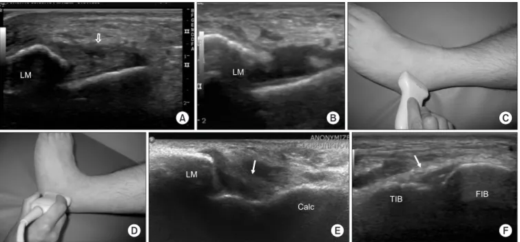
가벼운 급성 손상의 경우 정상 두께 혹은 약간 두꺼워져 보이게 되는데, 고반사의 섬유구조(hyperechoic fibrillation)로 변할 수도 있다. 중등도의 부분 파열 경우 인대 실질 중간 혹은 부착부에 무 반사(anechoic) 영역을 부분적으로 확인할 수 있으나 인대의 장력 은 유지된다(Fig. 1A). 중증 완전 파열의 경우 인대의 연속성이 저 반사 불연속성(hypoechoic gap)에 의해 없어지고 견열된 섬유가 관찰되는데 인대의 장력은 소실되어 물결 모양으로 보이고 인대 에 장력을 가해도 팽팽해지지 않으며 결손된 부위를 통해서 혈액 성 삼출물이 관절 밖 피하조직과 인접 인대로도 퍼져나간다(Fig.
1B). 한편 오래된 파열의 경우 초음파 상에서 인대가 두꺼워지고 골화된 소견이 관찰될 수도 있다.8,10)
전거비인대(anterior talofibular ligament)는 가장 흔히 손상되 는 인대로서 단독으로 손상되거나 혹은 종비인대 손상이 동반된 다.11) 초음파 검사 시에는 인대에 장력을 주기 위해서 족관절을 족저굴곡, 내번시키거나 전방전위시켜야 한다(Fig. 1C).10,12,13) 종 비인대(calcaneofibular ligament)의 단독 손상은 매우 드물며 대 부분의 경우는 전거비인대 손상과 같이 발생하는데 족관절을 족 배굴곡시키면서 발뒤꿈치를 내번시켜 인대에 장력을 가하면 촬 영 시 이방성을 피할 수 있다(Fig. 1D, 1E). 특히 인대가 손상되 지 않으면 족배굴곡 시 그 장력에 의해 비골건이 표면쪽으로 이 동하게 되며 이 이동이 관찰되지 않으면 완전 손상으로 볼 수 있
다.8) 완전 손상이 발생하면 비골 건초에 삼출액이 관찰될 수도 있 다. 전경비인대(anterior tibiofibular ligament)는 원위 경비골 안정 성에 중요한 역할을 하는데 종종 이 인대의 손상을 과소 평가하 는 경우가 있다. 족관절을 족배굴곡시켜 인대에 장력을 주고 검 사하는 것이 필요하다(Fig. 1F).8) 후경비인대(posterior tibiofibular ligament)는 초음파로 쉽게 접근하기 어려우며 족관절 염좌 시 손 상되는 경우도 매우 드물다.10)
외반 염좌는 내반 염좌에 비해 드물게 발생하는데 이는 내측 삼각 인대가 매우 강하기 때문이다. 이 인대는 족관절을 족배굴 곡시키고 뒷꿈치에 외반을 가해야 잘 관찰할 수 있으며 이때 경 골건이 거골의 내측에서 표면으로 이동하는 것을 관찰할 수 있어 야 하는데 이 이동이 관찰되지 않으면 내측 삼각 인대의 완전 파 열을 의심해 볼 수 있다.8)
2. 족저근막염(plantar fasciitis)
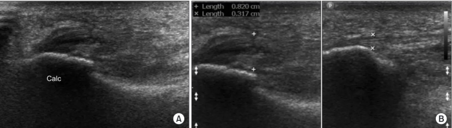
족저근막은 발의 내측부 아치를 지지하는 주된 구조물로서14) 이 에 염증이 발생하는 족저근막염은 발뒤꿈치 통증이 발생했을 때 가장 흔한 원인이다.15) 초음파 검사를 이용하여 족저근막의 두께 가 종골 부착부에서 4 mm 이상이면 족저근막염을 진단할 수 있
는데16,17) Sabir 등16)은 자기공명영상 검사와 비교해서 80%의 민감
도와 88.5%의 특이도를 보고하였다.
Figure 1. Ultrasonography images showing the sprained ankle ligaments. (A) An ultrasonography image shows a small defect (arrow) in the middle portion of the swollen anterior talofibular ligament, which is consistent with a mild to moderate sprain. (B) This ultrasonography image clearly shows a complete rupture of the anterior talofibular ligament. (C) Ultrasonography of the anterior talofibular ligament was performed in ankle plantar flexion and slight inversion, in which position the ligament was tensioned. (D) On the other hand, ultrasonography of the calcaneofibular ligament was performed in ankle dorsiflexion and slight inversion. (E) Calcaneofibular ligament (arrow) is well visualized in ankle dorsiflexion and slight inversion.
Moderate effusion is observed around the calcaneofibular ligament on this image. (F) The integrity of anterior inferior tibiofibular ligament (arrow) can be confirmed by ultrasonography, when a syndesmosis injury is suspected. LM, lateral malleolus; Calc, calcaneus; TIB, tibia; FIB, fibula.
족저근막은 초음파의 장축 영상에서 정상적으로 3-4 mm 이하 의 섬유성 밴드 모양으로 보이며, 족저근막염이 있으면 종골 부 착부에서 방추형(fusiform)으로 두꺼워지고 전반적인 저반사를 보인다(Fig. 2).17,18) Walther 등19)에 의하면 족저근막염 환자의 40%
가 도플러 검사에서 근막 자체와 그 주위에 충혈(hyperemia)이 관 찰된다고 하였다.
3. 족저근막 파열(plantar fascia tear)
족저근막의 파열은 드물게 발생하며 부분 파열 혹은 완전 파열의 형태로 나타날 수 있다.20) 족저근막염은 종골 부착부 주변에서 주 로 발생하는 데 비해 족저근막 파열은 족저근막의 원위부, 즉 중 간 부위에서 발생한다(Fig. 3A).21) 족저근막의 원위부, 파열은 주 로 외상과 관련 있는데 운동선수들이 뛰거나 점프하는 동작에 서 발생하며 만성적인 과사용도 발생 요인 중의 하나로 여겨지고 있다.21) 급성 파열이 있으면 부분적으로 섬유구조의 무반사 단속 (anechoic interruption)이 관찰되고, 그 주위에 액체(fluid)가 고이 게 되는데 족지 및 족관절을 신전시키면 족저근막에 장력이 가해 져 더 잘 볼 수 있다(Fig. 3B, 3C). 자연 발생하는 파열은 종골 부착 부에서 발생하는데 대부분 족저근막염에 대한 스테로이드 주사
치료의 합병증과 관계된다.22)
족저근막 파열의 진단은 병력 청취와 이학적 검사만으로도 충 분히 가능하나 정확한 손상의 위치 및 범위 확인을 위해 영상학 적 검사를 시행한다.20) Jeswani 등23)은 족저근막의 부종과 손상에 의한 섬유 구조의 단속을 구별하는 데 초음파가 자기공명영상 검 사보다 유용하다고 하였다. 또한 동적 초음파를 이용하면 손상 부위의 결손을 더 잘 확인할 수 있다.
4. 족장판 파열(plantar plate tear)
족장판은 중족지 관절의 족저부에 위치하는 섬유연골 구조로 관 절의 안정화에 중요하며 스포츠 활동 중에 과도한 부하를 받으면 이 구조물이 약해져서 파열이나 불안정성이 발생할 수 있다.24) 족 장판은 초음파 장축 영상에서 굴곡건과 중족 골두 사이에 끼어있 는 모습으로 가장 잘 관찰되며, 균일하게 반사되는 구조가 중족 골 경부 골간에서부터 족지 기저부까지 연결되어 있다.24,25) 족장 판의 파열은 주로 2번째 중족지 관절에서 발생하고 3번째와 4번 째에서도 발생할 수 있다.24) 파열이 발생하면 저반사 혹은 불균질 한(heterogeneous) 부분이 섬유구조 안에 보이고, 발가락을 족배 굴곡시키면 그 크기가 커지는 것을 관찰할 수 있다(Fig. 4). 파열
A B
Calc
Figure 2. (A) Long-axis ultrasonography image shows moderate swelling of the plantar fascia in the calcaneal insertion site. (B) The thickness of the plantar fascia is more than 8 mm in the left image, while that of the other side is less than 4 mm. Calc, calcaneus.
Calc
A B C
Figure 3. (A) Photograph of the right foot shows a bruise along the plantar fascia. (B) An ultrasonography image shows a partial interruption of the plantar fascia (arrow) near the calcaneal insertion. (C) T2-weighted fat suppression magnetic resonance imaging confirms the partial rupture of the plantar fascia (arrow). Calc, calcaneus.
은 보통 원위부와 중앙부에 위치하지만 내측이나 외측으로 확장 될 수도 있다.24,26) 한편 초음파상에서 파열이 있어도 증상이 없는 것이 대부분이므로 임상적 증상과 연관지어 파열의 진단을 내려 야 한다는 보고도 있다.27)
5. 아킬레스건 손상(Achilles tendon injury)
아킬레스건은 인체에서 가장 크고 강한 건으로 다른 건과는 달리 활액막이 없고 대신 건주위 조직(paratenon)이 얇은 혈관 막(vas- cular membrane)에 의해 둘러싸여 있다.28) 아킬레스건 손상은 스 포츠와 연관성이 높으며 주로 달리기, 축구, 테니스, 베드민턴 운 동 중에 발생한다.29-32) 병변은 종골부착부의 근위 2-6 cm 부위에 주로 발생하는데 족저굴곡된 발이 갑자기 족배굴곡될 때 혹은 족
관절 뒷부분을 직접 타격받을 때 손상이 발생한다.33)
부분 파열은 건 내부에 경계가 명확한 저반사 영역이 보이며 장축 영상에서 섬유 모양(fibrillar pattern)이 부분적으로 소실된
다.34,35) 초음파상으로 건병증과 부분 파열을 구분하는 것은 어렵
A B
2 3
Figure 4. (A) Anteroposterior radiograph of the left foot shows medial subluxation of the 2nd metatarsophalangeal (MTP) joint. (B) An ultrasonography images show the tear of the lateral part of the plantar plate (arrow) in the 2nd MTP joint. 2: the 2nd metatarsal head, 3: the 3rd metatarsal head.
Figure 5. Long-axis ultrasonography image shows complete rupture and gap formation of the Achilles tendon.
A
B
GC
Sol
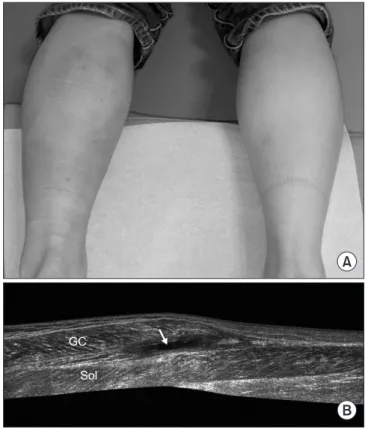
Figure 6. (A) Photograph shows a bruise in the proximal part of left calf. (B) The extended field of view ultrasonography image shows a hematoma (arrow) in the junction of the medial GC and the Sol, which is consistent with a ‘tennis leg’ injury. GC, gastrocnemius muscle; Sol, soleus muscle.
지만 두 경우 모두 비수술적으로 치료가 가능하기 때문에 임상적 으로 문제가 되지 않는 경우가 대부분이다.36,37) 또한 도플러 검사 에서 신생혈관 형성(neovascularization)을 확인할 수 있는데 임상 적 예후보다는 통증의 심한 정도와 연관이 있다고 한다.38) 급성 파열의 경우 대부분 임상적으로 명확하지만 20% 정도에 서는 진단을 놓치는 경우도 발생한다.39) 초음파로 검사할 때 종골 부착부부터 근건 결합부까지 확인해야 하며 완전 파열 시 초음파 에서 섬유구조의 파괴와 2개의 분절로 나누어지는 것을 관찰할 수 있다(Fig. 5). 시간이 지나면 결손 부위는 혈종으로 채워져 이 후 육아 조직(granulation tissue)과 섬유성 반흔으로 변하게 된다.
Hartgerink 등40)은 아킬레스건 손상 부위로 전방의 지방이 들어 오는 것을 발견하였고, 완전 파열의 경우 족척건(plantaris tendon) 이 더 쉽게 관찰되는 것을 발견하였다. 초음파 검사를 시행할 때 족관절을 굴곡, 신전하면서 건의 결손이 좁아지고 넓어지는 것을 관찰하면 완전 파열을 진단하는 데 도움이 된다.
6. 비복근 내측두 파열(gastrocnemius medial head injury)
비복근의 내측두 파열은 Tennis leg라고도 불리우며 외상에 의해 발생한다.41) 이 손상은 다양한 형태로 나타날 수 있는데 내측 비 복근의 손상이 가장 많으나 그 외 하퇴 비복근과 가자미근 사이 의 근막 파열 또는 드물게 가자미근의 손상으로 나타나기도 한
다.42,43) 40세 이후에 주로 발생하며 스포츠 활동이나 일상생활 중
족관절 부위가 과하게 신전되었을 때 발생하므로 달리기나 라켓 볼 등을 하다가 발생하는 경우가 많다(Fig. 6A).30,44) 임상적 증상 만으로도 진단을 내릴 수 있으나 초음파 검사를 시행하면 정확한 손상 부위 및 손상 정도의 확인에 도움을 받을 수 있다. 또한 초 음파 검사는 슬와낭종(Baker
’
s cyst), 심부 정맥 혈전증(deep vein thrombosis)과 감별하기 위한 표준적인 검사법으로도 여겨진다.45) 비복근 내측두의 파열이 있으면 근육 내에 정상적으로 보이 던 고반사와 저반사의 평행선들이 단절되고 비복근의 원위부 부 착부위에 불명확한 반사가 보이게 되며, 간혹 비복근과 가자미 근 사이에 혈종이 관찰되기도 한다(Fig. 6B).30,46) 초음파 단축(short axis) 영상을 이용하면 근육 단면을 한번에 볼 수 있어 부분 혹은 완전 파열 여부를 확인할 수 있다.파열이 발생하고 난 후 치료 과정에서 초음파 검사를 통해 추 시하면 고여있던 액체가 흡수되면서 무반사에서 저반사로 변하 는 과정과 파열 부위가 섬유 반흔 조직으로 대체되어 가는 소견 을 볼 수 있다. 초음파 단축 영상에서 둥근 모양의 고반사가 관찰 되고, 도플러 종축 영상에서 색깔 신호(color signal)가 없다면 심 부 정맥 혈전증을 의심해야 한다.47)
7. 비골건 손상(peroneal tendon injury)
비골건 복합체(peroneal tendon complex)는 단비골근(peroneus
brevis muscles)및 장비골근(peroneus longus muscles)과 비골건들, 활액막 건초(synovial tendon sheath), 상부 및 하부 지대(superior and inferior retinaculum), 비 부골(Os peroneum) 등으로 구성된
다.48-51) 과상부(supramalleolar region)의 근위부에서는 장비골건이
단비골건의 외측에 위치하며, 원위부로 내려오면서 단비골근의 크기가 줄어들고 건으로 바뀌게 되어 과부(malleolar region)에서 는 장비골건이 단비골건의 외측후방으로 주행하고 두 건은 외과 의 후방 구(sulcus)에 위치한다.48)
비골건 손상은 직접적인 외상은 드물고, 대부분 불안정성으로 인한 미세 손상의 누적으로 인해 건초염이 발생한 상태에서 족관 절 염좌 때와 같이 과도한 장력이 가해져 발생한다.52-55)
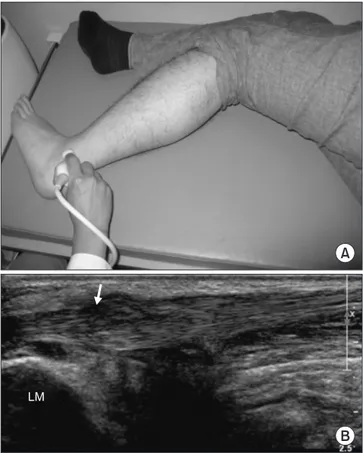
초음파는 비골건의 손상을 진단하는 데 매우 효과적이다. 초음 파 검사를 할 때는 하퇴부 밑을 받친 상태로 측와위를 취한 후 고 관절을 약간 굴곡 및 내회전시키고 슬관절을 30도 정도 굴곡시 키면 비골건의 장축 및 단축 영상을 확인할 수 있다(Fig. 7A). 추 가적으로 장비골건의 원위부는 해부학적으로 족저부에 있으므 로 복와위를 취해서 검사를 할 수도 있다. Grant 등53)에 의하면 25 명의 비골건 손상 환자에 대해서 동적 초음파 검사와 수술 소견
A
B
LM
Figure 7. (A) Ultrasonography of peroneal tendon was performed in lateral decubitus position with hip and knee flexion. (B) A long-axis ultrasonography image shows a partial tear (arrow) of the peroneus brevis tendon. LM, lateral malleolus.
을 비교하고 초음파의 민감도, 특이도, 정확도가 각각 100%, 85%, 90%로서 동적 초음파 검사가 비골건의 손상 유무를 확인하는 데 있어 매우 중요하다고 주장하였다.
비골건의 부분 파열은 초음파 영상에서 얇아지거나 혹은 두꺼 워져 보이며 정상적인 섬유구조가 소실되고 내부에 불규칙한 저 반사 소견으로 나타나게 된다(Fig. 7B).
8. 비골건 불안정성(peroneal tendon instability)
비골건의 불안정성은 두 가지 형태로 나타난다.56) 첫 번째는 하 나 혹은 두 개의 건 모두가 외과의 앞쪽으로 탈구 혹은 아탈구되 는 것으로 신체검사상 저항을 준 상태로 족관절을 외번시킬 때 건의 이상 움직임을 촉지할 수 있다. Oden57)은 비골건의 불안정 성을 해부학적 병변에 따라서 4가지로 분류하였는데 1형은 지대 (retinaculum)의 견열에 의한 낭(pouch)의 생성, 2형은 지대의 비골 부착부 손상(tear), 3형은 지대의 비골 부착부 견열 골절, 4형은 지
대의 후방부 손상을 말하며 1형과 3형이 가장 흔하다고 하였다.
비골건 불안정성의 또 다른 형태로 일시적으로 건들의 앞뒤 관 계가 바뀌는 불안정성이 있는데 이는 건초내 아탈구(retrofibular intrasheath subluxation)로 알려져 있고, 비골건이 비골 터널(pe- roneal tunnel) 밖으로 빠지지 않아서 신체 검사만으로는 진단할 수 없다.58,59) Raikin 등58)은 건초내 아탈구를 단비골건의 장축 파열 (longitudinal split tear) 여부에 따라서 A형, B형으로 분류하였는데 A형은 단비골건이 정상 상태이고, B형은 장비골건이 단비골건의 갈라진(split) 부위로 아탈구되는 상태를 말한다.
초음파를 이용하면 동적인 검사를 통해 비골건 탈구를 쉽게 확 인할 수 있으며 이는 컴퓨터 단층촬영이나 자기공명영상 검사로 얻을 수 없는 초음파만의 확실한 장점이다.3,56) 특히 단축 영상으 로 확인하는 것이 비골건과 비골 사이의 위치 관계를 평가하는 데 도움이 된다(Fig. 8). 평가하는 동안 젤(gel)을 충분히 사용하여 변환기에 과도한 압력을 가하지 않아야 건의 탈구를 쉽게 발견할 수 있다. 건들의 위치를 확인하고 정상적인 위치에 있다면 건초 내 아탈구 혹은 탈구를 의심하고 족관절을 움직이면서 검사를 계 속한다(Fig. 9). 일단 불안정성이 확인되면 단 비골건의 장축 파열 을 확인해야 하며 그 외 건초 두께의 증가와 삼출물의 생성을 확 인한다.
결 론
초음파 검사는 자기공명영상 검사에 비해서 가격이 저렴하고 접 근성이 좋은 장점이 있으며 동적 검사를 이용하면 인대 파열이나 건의 탈구와 같은 스포츠 손상의 진단에도 유리하다. 이러한 초 음파의 장점을 활용하면 족부와 족관절 스포츠 손상의 정확한 진 단에 많은 도움이 될 것이다.
Figure 9. Short-axis dynamic ultrasonography image shows the instability of the peroneus brevis tendon within the peroneal sheath.
The patient was requested to do active eversion of the ankle joint while undergoing the ultrasonography. PB; peroneus brevis tendon; PL, peroneus longus tendon; LM, lateral malleolus.
A B
LM
PL PB
LM
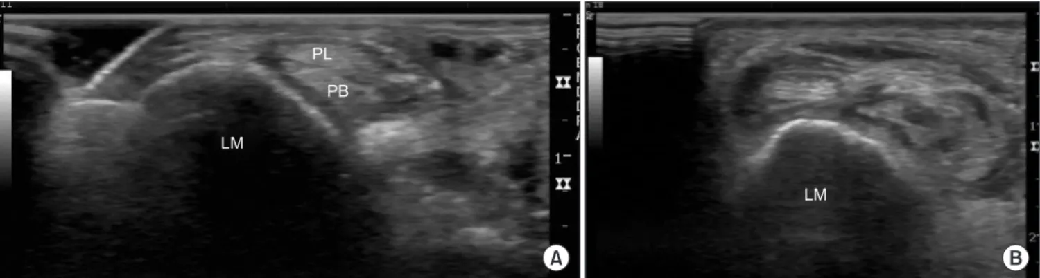
Figure 8. Short-axis ultrasonography images of the retromalleolar area show the normal (A) and the dislocated (B) peroneal tendons. LM, lateral malleolus; PL, peroneus longus tendon; PB, peroneus brevis tendon.
CONFLICTS OF INTEREST
The authors have nothing to disclose.
REFERENCES
1. Bianchi S, Martinoli C, Gaignot C, De Gautard R, Meyer JM.
Ultrasound of the ankle: anatomy of the tendons, bursae, and ligaments. Semin Musculoskelet Radiol. 2005;9:243-59.
2. Jacobson JA, van Holsbeeck MT. Musculoskeletal ultrasonog- raphy. Orthop Clin North Am. 1998;29:135-67.
3. Khoury V, Cardinal E, Bureau NJ. Musculoskeletal sonogra- phy: a dynamic tool for usual and unusual disorders. AJR Am J Roentgenol. 2007;188:W63-73.
4. Morvan G, Busson J, Wybier M, Mathieu P. Ultrasound of the ankle. Eur J Ultrasound. 2001;14:73-82.
5. Fong DT, Hong Y, Chan LK, Yung PS, Chan KM. A system- atic review on ankle injury and ankle sprain in sports. Sports Med. 2007;37:73-94.
6. Ferran NA, Maffulli N. Epidemiology of sprains of the lateral ankle ligament complex. Foot Ankle Clin. 2006;11:659-62.
7. Garrick JG. The frequency of injury, mechanism of inju- ry, and epidemiology of ankle sprains. Am J Sports Med.
1977;5:241-2.
8. Peetrons P, Creteur V, Bacq C. Sonography of ankle liga- ments. J Clin Ultrasound. 2004;32:491-9.
9. Brasseur JL, Luzzati A, Lazennec JY, Guérin-Surville H, Rog- er B, Grenier P. Ultrasono-anatomy of the ankle ligaments.
Surg Radiol Anat. 1994;16:87-91.
10. Campbell DG, Menz A, Isaacs J. Dynamic ankle ultrasonog- raphy. A new imaging technique for acute ankle ligament injuries. Am J Sports Med. 1994;22:855-8.
11. Bozić R, Weiser J. [Epidemiologic data of rupture of the fib- ular ligament of the upper ankle joint]. Aktuelle Traumatol.
1991;21:118-20. German.
12. Copercini M, Bonvin F, Martinoli C, Bianchi S. Sonographic diagnosis of talar lateral process fracture. J Ultrasound Med.
2003;22:635-40.
13. Friedrich JM, Schnarkowski P, Rübenacker S, Wallner B. Ul- trasonography of capsular morphology in normal and trau- matic ankle joints. J Clin Ultrasound. 1993;21:179-87.
14. Hedrick MR. The plantar aponeurosis. Foot Ankle Int.
1996;17:646-9.
15. DeMaio M, Paine R, Mangine RE, Drez D Jr. Plantar fasciitis.
Orthopedics. 1993;16:1153-63.
16. Sabir N, Demirlenk S, Yagci B, Karabulut N, Cubukcu S.
Clinical utility of sonography in diagnosing plantar fasciitis. J Ultrasound Med. 2005;24:1041-8.
17. Kane D, Greaney T, Shanahan M, et al. The role of ultraso- nography in the diagnosis and management of idiopathic plantar fasciitis. Rheumatology (Oxford). 2001;40:1002-8.
18. Gibbon WW. Plantar fasciitis: US imaging. Radiology.
1992;182:285.
19. Walther M, Radke S, Kirschner S, Ettl V, Gohlke F. Power Doppler findings in plantar fasciitis. Ultrasound Med Biol.
2004;30:435-40.
20. Draghi F, Gitto S, Bortolotto C, Draghi AG, Ori Belometti G.
Imaging of plantar fascia disorders: findings on plain radiog- raphy, ultrasound and magnetic resonance imaging. Insights Imaging. 2017;8:69-78.
21. McNally EG, Shetty S. Plantar fascia: imaging diagnosis and guided treatment. Semin Musculoskelet Radiol. 2010;14:334- 43.
22. Sellman JR. Plantar fascia rupture associated with corticoste- roid injection. Foot Ankle Int. 1994;15:376-81.
23. Jeswani T, Morlese J, McNally EG. Getting to the heel of the problem: plantar fascia lesions. Clin Radiol. 2009;64:931-9.
24. Gregg J, Silberstein M, Schneider T, Marks P. Sonographic and MRI evaluation of the plantar plate: a prospective study.
Eur Radiol. 2006;16:2661-9.
25. Ford LA, Collins KB, Christensen JC. Stabilization of the subluxed second metatarsophalangeal joint: flexor tendon transfer versus primary repair of the plantar plate. J Foot An- kle Surg. 1998;37:217-22.
26. Gregg JM, Silberstein M, Schneider T, Kerr JB, Marks P. So- nography of plantar plates in cadavers: correlation with MRI and histology. AJR Am J Roentgenol. 2006;186:948-55.
27. Quinn TJ, Jacobson JA, Craig JG, van Holsbeeck MT. So- nography of Morton's neuromas. AJR Am J Roentgenol.
2000;174:1723-8.
28. Maffulli N, Sharma P, Luscombe KL. Achilles tendinopathy:
aetiology and management. J R Soc Med. 2004;97:472-6.
29. Kannus P, Natri A. Etiology and pathophysiology of tendon ruptures in sports. Scand J Med Sci Sports. 1997;7:107-12.
30. Bianchi S, Martinoli C, Abdelwahab IF, Derchi LE, Damiani S.
Sonographic evaluation of tears of the gastrocnemius medial head ("tennis leg"). J Ultrasound Med. 1998;17:157-62.
31. Kvist M. Achilles tendon injuries in athletes. Sports Med.
1994;18:173-201.
32. Schönbauer HR. [Diseases of the Achilles tendon]. Wien Klin Wochenschr Suppl. 1986;168:1-47. German.
33. Bencardino J, Rosenberg ZS, Delfaut E. MR imaging in sports injuries of the foot and ankle. Magn Reson Imaging Clin N Am. 1999;7:131-49, ix.
34. Fornage BD. Achilles tendon: US examination. Radiology.
1986;159:759-64.
35. Blei CL, Nirschl RP, Grant EG. Achilles tendon: US diagno- sis of pathologic conditions. Work in progress. Radiology.
1986;159:765-7.
36. Paavola M, Paakkala T, Kannus P, Järvinen M. Ultrasonog- raphy in the differential diagnosis of Achilles tendon injuries and related disorders. A comparison between pre-opera- tive ultrasonography and surgical findings. Acta Radiol.
1998;39:612-9.
37. Aström M, Gentz CF, Nilsson P, Rausing A, Sjöberg S, Westlin N. Imaging in chronic achilles tendinopathy: a com- parison of ultrasonography, magnetic resonance imaging and surgical findings in 27 histologically verified cases. Skeletal Radiol. 1996;25:615-20.
38. Zanetti M, Metzdorf A, Kundert HP, et al. Achilles tendons:
clinical relevance of neovascularization diagnosed with pow- er Doppler US. Radiology. 2003;227:556-60.
39. Scheller AD, Kasser JR, Quigley TB. Tendon injuries about the ankle. Clin Sports Med. 1983;2:631-41.
40. Hartgerink P, Fessell DP, Jacobson JA, van Holsbeeck MT.
Full- versus partial-thickness Achilles tendon tears: sono- graphic accuracy and characterization in 26 cases with surgi- cal correlation. Radiology. 2001;220:406-12.
41. Miller WA. Rupture of the musculotendinous juncture of the medial head of the gastrocnemius muscle. Am J Sports Med.
1977;5:191-3.
42. Kwak HS, Han YM, Lee SY, Kim KN, Chung GH. Diagno- sis and follow-up US evaluation of ruptures of the medial head of the gastrocnemius ("tennis leg"). Korean J Radiol.
2006;7:193-8.
43. Severance HW Jr, Bassett FH 3rd. Rupture of the plantar- is--does it exist? J Bone Joint Surg Am. 1982;64:1387-8.
44. Jarolem KL, Wolinsky PR, Savenor A, Ben-Yishay A. Tennis leg leading to acute compartment syndrome. Orthopedics.
1994;17:721-3.
45. Slawski DP. Deep venous thrombosis complicating rupture of the medial head of the gastrocnemius muscle. J Orthop Trau-
ma. 1994;8:263-4.
46. Delgado GJ, Chung CB, Lektrakul N, et al. Tennis leg: clinical US study of 141 patients and anatomic investigation of four cadavers with MR imaging and US. Radiology. 2002;224:112- 9.
47. Flecca D, Tomei A, Ravazzolo N, Martinelli M, Giovagnorio F.
US evaluation and diagnosis of rupture of the medial head of the gastrocnemius (tennis leg). J Ultrasound. 2007;10:194-8.
48. Bianchi S, Delmi M, Molini L. Ultrasound of peroneal ten- dons. Semin Musculoskelet Radiol. 2010;14:292-306.
49. Saupe N, Mengiardi B, Pfirrmann CW, Vienne P, Seifert B, Zanetti M. Anatomic variants associated with peroneal tendon disorders: MR imaging findings in volunteers with asymptomatic ankles. Radiology. 2007;242:509-17.
50. Numkarunarunrote N, Malik A, Aguiar RO, Trudell DJ, Resnick D. Retinacula of the foot and ankle: MRI with anatomic correlation in cadavers. AJR Am J Roentgenol.
2007;188:W348-54.
51. Hyer CF, Dawson JM, Philbin TM, Berlet GC, Lee TH. The peroneal tubercle: description, classification, and relevance to peroneus longus tendon pathology. Foot Ankle Int.
2005;26:947-50.
52. Smania L, Craig JG, von Holsbeeck M. Ultrasonographic findings in peroneus longus tendon rupture. J Ultrasound Med. 2007;26:243-6.
53. Grant TH, Kelikian AS, Jereb SE, McCarthy RJ. Ultrasound diagnosis of peroneal tendon tears. A surgical correlation. J Bone Joint Surg Am. 2005;87:1788-94.
54. Dombek MF, Lamm BM, Saltrick K, Mendicino RW, Cat- anzariti AR. Peroneal tendon tears: a retrospective review. J Foot Ankle Surg. 2003;42:250-8.
55. Rademaker J, Rosenberg ZS, Delfaut EM, Cheung YY, Sch- weitzer ME. Tear of the peroneus longus tendon: MR imag- ing features in nine patients. Radiology. 2000;214:700-4.
56. Neustadter J, Raikin SM, Nazarian LN. Dynamic sonographic evaluation of peroneal tendon subluxation. AJR Am J Roent- genol. 2004;183:985-8.
57. Oden RR. Tendon injuries about the ankle resulting from skiing. Clin Orthop Relat Res. 1987;216:63-9.
58. Raikin SM, Elias I, Nazarian LN. Intrasheath subluxation of the peroneal tendons. J Bone Joint Surg Am. 2008;90:992-9.
59. McConkey JP, Favero KJ. Subluxation of the peroneal ten- dons within the peroneal tendon sheath. A case report. Am J Sports Med. 1987;15:511-3.
족부 및 족관절 스포츠 손상에서 초음파의 활용
문영석 • 김종빈 • 안재훈
가톨릭대학교 의과대학 서울성모병원 정형외과학교실
족관절과 족부의 스포츠 손상은 임상에서 흔히 만날 수 있다. 초음파는 이러한 손상의 진단에 매우 유용한데 이는 자기공명영상 검사 에 비해 가격이 저렴하고 접근성이 뛰어나며 동적인 검사가 가능하다는 것 때문이다. 본 종설에서는 족부와 족관절에서 발생할 수 있 는 스포츠 손상 중 대표적인 질환의 초음파 소견에 집중하였다.
색인단어: 족부, 족관절, 스포츠 손상, 초음파
접수일 2018년 5월 16일 수정일 2018년 8월 24일 게재확정일 2018년 10월 14일 책임저자 안재훈
06591, 서울시 서초구 반포대로 222, 가톨릭대학교 의과대학 서울성모병원 정형외과학교실
TEL 02-2258-2837, FAX 02-535-9834, E-mail jahn@catholic.ac.kr, ORCID https://orcid.org/0000-0002-4253-3943
Copyright © 2019 by The Korean Orthopaedic Association
“This is an Open Access article distributed under the terms of the Creative Commons Attribution Non-Commercial License (http://creativecommons.org/licenses/by-nc/4.0/) which permits unrestricted non-commercial use, distribution, and reproduction in any medium, provided the original work is properly cited.”