Vol. 21, No. 2, 2009 209
Received October 17, 2008, Accepted for publication December 8, 2008
Reprint request to: Kwang Hyun Cho, M.D., Department of Derma- tology, Seoul National University College of Medicine, 28, Yeongeon- dong, Jongno-gu, Seoul 110-744, Korea. Tel: 82-2-2072-2412, Fax:
82-2-742-7344, E-mail: [email protected] Ann Dermatol Vol. 21, No. 2, 2009
CASE REPORT
A Case of Epstein-Barr Virus-associated Hydroa Vacciniforme
Sun Young Huh, M.D., Mira Choi, M.D., Kwang Hyun Cho, M.D.
Department of Dermatology, Seoul National University College of Medicine, Seoul, Korea
Hydroa vacciniforme (HV) is a photosensitivity disorder characterized by recurrent necrotic vesiculopapules on sun-exposed areas, which heal spontaneously during adolescence. Recently, an association has been reported be- tween latent Epstein-Barr virus (EBV) infection and atypical HV-like eruption and malignant potential. However, latent EBV infection has also been reported in the setting of typical HV. An 11-year-old girl presented with recurrent, scattered, discrete vesicular eruptions with scarring on the face and the extensor surfaces of both forearms. In-situ hybridization was carried out to detect latent EBV infection. Based on the clin- ical and histopathological findings, typical EBV-associated HV was suspected. (Ann Dermatol 21(2) 209∼212, 2009) -Keywords-
Epstein-Barr virus, Hydroa vacciniforme
INTRODUCTION
Hydroa vacciniforme (HV) is a rare photosensitivity dis- order characterized by recurrent necrotic vesiculopapules on sun-exposed areas, which heal with vacciniform scarring. HV usually starts during childhood and resolves spontaneously during adolescence without systemic involvement.
Recently, there have been reports of patients with atypical HV-like eruptions in Asia and Mexico1-7. In contrast with the symptoms seen in typical HV, these patients showed facial swelling, indurated nodules on non-sun-exposed
and sun-exposed areas, high-grade fever, wasting, and hepatosplenomegaly. Several of the reported patients pro- gressed to hematological malignancies and death. Latent Epstein-Barr virus (EBV) infection was detected in most of the patients with this atypical HV-like eruption.
However, Iwatsuki et al8 reported that some patients with atypical HV-like eruptions did not progress to overt malig- nant hematological neoplasia. In addition, the inves- tigators reported six patients with the typical manifes- tations of HV who had a number of EBV-encoded small nuclear RNA (EBER)+ cells in the cellular infiltrates in the dermis. These findings support the possibility that typical HV is also associated with latent EBV infection.
There have been several reports of HV in Korea1-3,9-12. However, to our knowledge, there has been no previous report of typical HV with confirmed latent EBV infection in Korea. We report a rare case of typical HV that oc- curred in association with latent EBV infection in an 11-year-old girl.
CASE REPORT
An 11-year-old Korean girl presented with a 5-year history of recurrent, scattered, discrete vesicular eruptions with scars on the face and the extensor surfaces of both forearms.
Her skin lesions were aggravated by sun exposure and healed spontaneously with crusts and mild scars. There was no family or personal history of cutaneous photosensitivity.
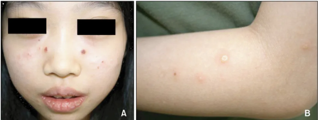
Physical examination showed multiple erythematous pap- ules, vesicles, crusts, and shallow scars on the face, espe- cially on the cheeks (Fig. 1). Except for the skin lesions, there were no remarkable findings on physical examination.
Minimal erythemal doses (MEDs) of visible light and UVB were measured with EktagraphicⓇ (Eastman-Kodak, USA) and UV 800Ⓡ (Waldmann, Germany) respectively. UVA MED measurements and photoprovocation tests were not performed due to patient rejection. The MED was 50
SY Huh, et al
210 Ann Dermatol
Fig. 1. The patient showed multiple, discrete vesicular eruptions with scarring on the face (A) and the extensor surfaces of the forearms (B).
Fig. 2. (A) A skin biopsy obtained from the vesicular lesion on her forearm. Intraepidermal vesiculation with superficial and deep perivascular and periappendageal infiltrations in the upper dermis (H&E, ×200). (B) In situ hybridization. Many cells positive for EBV-encoded small nuclear RNA are present in the infiltrates (×200).
mJ/cm2 for UVB. No response was observed for a 30-minute exposure to visible light. Initial laboratory in- vestigations showed that the erythrocyte sedimentation rate (27 mm/h) and the antistreptolysin O titer (433, pos- itive) were slightly elevated, and the anti-SSB/La antibody was positive. Other laboratory studies were within normal limits, including complete blood cell count with differ- ential cell count, platelet count, liver and renal function tests, antinuclear antibody concentration, lupus eryth- ematosus cell preparation, anti-SSA/Ro antibody, and an- ti-Smith antibody.
Histopathologic examination showed intraepidermal ve- siculation with superficial and deep perivascular lympho- cytic infiltrations in the dermis (Fig. 2A). In situ hybrid- ization was carried out on paraffin-embedded skin biopsy sections, using fluorescein-conjugated oligonucleotide probes for EBV early RNAs (EBERs) (Dakopatts, Glostrup, Denmark) for the detection of latent EBV infection.
Distinct, dark blue precipitates representing EBER tran- scripts were found in the nuclei of the lymphoid cells in the dermal infiltrate (Fig. 2B). Immunohistochemical stain-
ing was performed using monoclonal antibodies to CD3 (pan T-cell marker), CD45RO (memory T-cell marker), CD8 (helper T-cell marker), CD20 (B-cell marker), CD56 (NK-cell marker) (Dakopatts, Glostrup, Denmark), and la- tent membrane protein-1 (LMP-1). Most of the infiltrating cells in the dermis had a positive T-cell phenotype: CD3 and CD45RO positivity with some CD8 positivity. Only a few CD56-positive cells were present in the perivascular area. CD20-positive cells and LMP-1-positive cells were absent (Fig. 3).
Based on the clinical and histopathological findings, typi- cal EBV-associated hydroa vacciniforme was suspected.
Avoidance of sunlight and sunscreen application was recommended. After 1 year of follow-up, the patient has manifested no recurrence and shows no evidence of lym- phoproliferative disorders.
DISCUSSION
Hydroa vacciniforme was first described by Bazin et al13 in 1862. It is clinically characterized by recurrent necrotic
A Case of Epstein-Barr Virus-associated Hydroa Vacciniforme
Vol. 21, No. 2, 2009 211 Fig. 3. Immunohistochemical stains.
The dermal infiltrates are composed mainly of lymphocytes expressing CD3-positive (A) and CD45RO- positive (B) cells (Immunoperoxidase stain, ×100).
vesiculopapules on sun-exposed areas, which heal with vacciniform scarring. It is known to be a disease of early childhood that regresses spontaneously in adolescence and does not impair the general health of the patient14. Iwatsuki et al8 reported 6 patients with clinically and histo- logically typical HV who had EBV-encoded small nuclear RNA (EBER)+ cells in the dermal infiltrate. They suggested the possibility that typical HV and atypical HV are variants within the same disease spectrum of EBV-associated lym- phoproliferative disorders8,15.
However, patients with EBV-associated HV-like eruptions, mainly reported in Asia1-3,16-18, present with atypical skin lesions in non-sun-exposed areas, as well as associated systemic symptoms. They occasionally progress to devel- op hematological malignancies. Such a clinical entity has been regarded as differing from typical HV, although it has several similar clinical and histopathological findings.
The EBV-associated HV-like eruptions reported in Korea1-3,19 have shown features more typical of EBV-associated lym- phoproliferative disorders than of typical HV: skin lesions recurring continuously irrespective of sun exposure, asso- ciated systemic symptoms, with most patients progressing to develop hematological malignancies. Thus, in contrast to the preponderance of typical HV cases seen in Caucasians, most of the cases reported in Asians, includ- ing Koreans, show EBV-associated HV-like eruptions with malignant potential.
In the current case, clinical manifestations were similar to those seen in typical HV, with a benign course for the skin lesions, which cleared up after photoprotection was implemented. Associated latent EBV infection was con-
firmed by the presence of EBV-related RNAs, similar to those seen in the patients described by Iwatsuki et al8. Immunohistochemical staining revealed that the EBER- containing cells predominantly had a T-cell phenotype (CD3, CD45RO). Only a few cells had an NK-cell (CD56) phenotype, and no cells had a B-cell (CD20) phenotype.
No patients expressed LMP-1, similar to the patients de- scribed by Iwatsuki et al8. LMP-1 is a gene product of la- tent EBV infection and is known to have oncogenic activ- ity18. However, detection of this gene product is not suit- able for a screening test for EBV infection, since LMP-1 is not always expressed by EBV-infected cells20.
Previously, our group reported 6 patients with EBV-asso- ciated lymphoproliferative lesions presenting as HV-like eruption, with 3 different clinical courses19. The varying clinical courses in patients with EBV-associated HV-like eruptions are considered to be associated with the number of EBER+ cells in the skin lesions, subtype of EBV, and immune status of the patient16. Detailed pathophysio- logical mechanisms related to the various clinical manifes- tations occurring in the same disease spectrum remain to be determined.
Six cases of typical HV have been reported in Korea9-12, including a case of HV confirmed by repetitive UVA pho- totesting that showed spontaneous remission with the use of topical sunscreen9. To our knowledge, there has been no previous report of typical HV with confirmed latent EBV infection in Korea. Hence, we report this interesting case, which we believe to represent the first reported case of this entity in Korea.
SY Huh, et al
212 Ann Dermatol
REFERENCES
1. Cho KH, Kim CW, Lee DY, Sohn SJ, Kim DW, Chung JH. An Epstein-Barr virus-associated lymphoproliferative lesion of the skin presenting as recurrent necrotic papulovesicles of the face. Br J Dermatol 1996;134:791-796.
2. Cho KH, Kim CW, Kwon OS, Yang SG, Park KC, Park MH, et al. Epstein-Barr virus-associated lymphoproliferative erup- tion with progression to large granular lymphocytic leu- kaemia. Br J Dermatol 1997;137:426-430.
3. Cho KH, Kim CW, Heo DS, Lee DS, Choi WW, Rim JH, et al. Epstein-Barr virus-associated peripheral T-cell lymphoma in adults with hydroa vacciniforme-like lesions. Clin Exp Dermatol 2001;26:242-247.
4. Magana M, Sangueza P, Gil-Beristain J, Sanchez-Sosa S, Salgado A, Ramon G, et al. Angiocentric cutaneous T-cell lymphoma of childhood (hydroa-like lymphoma): a distinc- tive type of cutaneous T-cell lymphoma. J Am Acad Derma- tol 1998;38:574-579.
5. Ruiz-Maldonado R, Parrilla FM, Orozco-Covarrubias ML, Ridaura C, Tamayo Sanchez L, Duran McKinster C. Edema- tous, scarring vasculitic panniculitis: a new multisystemic disease with malignant potential. J Am Acad Dermatol 1995;32:37-44.
6. Oono T, Arata J, Masuda T, Ohtsuki Y. Coexistence of hy- droa vacciniforme and malignant lymphoma. Arch Dermatol 1986;122:1306-1309.
7. Asada H, Okada N, Tei H, Yamamura T, Hashimoto K, Kondo K, et al. Epstein-Barr virus-associated large granular lymphocyte leukemia with cutaneous infiltration. J Am Acad Dermatol 1994;31:251-255.
8. Iwatsuki K, Xu Z, Takata M, Iguchi M, Ohtsuka M, Akiba H, et al. The association of latent Epstein-Barr virus infection with hydroa vacciniforme. Br J Dermatol 1999;140:715-721.
9. Hann SK, Im S, Park YK, Lee S. Hydroa vacciniforme with unusually severe scar formation: diagnosis by repetitive UVA phototesting. J Am Acad Dermatol 1991;25:401-403.
10. Paik SA, Hahm JH, Kook HI. Three cases of hydroa vacciniforme. Korean J Dermatol 1977;15:341-345.
11. Lim SD. A case of hydroa vacciniforme. Korean J Dermatol 1960;1:65-67.
12. Chang SH, Yoon TY. A case of hydroa vacciniforme with oc- ular involvment. Korean J Dermatol 1993;31:612-615.
13. Bazin E, Baudot E, Guérard L. Leçons théoriques et cliniques sur les affections génériques de la peau. Vol. 1. Paris: Dela- haye, 1862:132.
14. Bickers DR, Demar LK, DeLeo V, Poh-Fitzpatrick MB, Aronberg JM, Harber LC. Hydroa vacciniforme. Arch Derma- tol 1978;114:1193-1196.
15. Iwatsuki K, Satoh M, Yamamoto T, Oono T, Morizane S, Ohtsuka M, et al. Pathogenic link between hydroa vaccini- forme and Epstein-Barr virus-associated hematologic disor- ders. Arch Dermatol 2006;142:587-595.
16. Iwatsuki K, Ohtsuka M, Harada H, Han G, Kaneko F.
Clinicopathologic manifestations of Epstein-Barr virus-asso- ciated cutaneous lymphoproliferative disorders. Arch Der- matol 1997;133:1081-1086.
17. Wu YH, Chen HC, Hsiao PF, Tu MI, Lin YC, Wang TY.
Hydroa vacciniforme-like Epstein-Barr virus-associated mon- oclonal T-lymphoproliferative disorder in a child. Int J Der- matol 2007;46:1081-1086.
18. Iwatsuki K, Xu Z, Ohtsuka M, Kaneko F. Cutaneous lympho- proliferative disorders associated with Epstein-Barr virus in- fection: a clinical overview. J Dermatol Sci 2000;22:181- 195.
19. Cho KH, Lee SH, Kim CW, Jeon YK, Kwon IH, Cho YJ, et al.
Epstein-Barr virus-associated lymphoproliferative lesions pre- senting as a hydroa vacciniforme-like eruption: an analysis of six cases. Br J Dermatol 2004;151:372-380.
20. Rowe M, Lear AL, Croom-Carter D, Davies AH, Rickinson AB. Three pathways of Epstein-Barr virus gene activation from EBNA1-positive latency in B lymphocytes. J Virol 1992;
66:122-131.