ISSN 2234-3806 • eISSN 2234-3814
Ann Lab Med 2013;33:283-287
http://dx.doi.org/10.3343/alm.2013.33.4.283
The First Korean Case of Sphingobacterium
spiritivorum Bacteremia in a Patient with Acute Myeloid Leukemia
Young Rae Koh, M.D.1, Shine Young Kim, M.D.1, Chulhun L Chang, M.D.1, Ho-Jin Shin, M.D.2, Kye-Hyung Kim, M.D.2, and Jongyoun Yi, M.D.1
Departments of Laboratory Medicine1 and Internal Medicine2, Pusan National University School of Medicine, Busan, Korea
Sphingobacterium spiritivorum has been rarely isolated from clinical specimens of immu- nocompromised patients, and there have been no case reports of S. spiritivorum infection in Korea to our knowledge. We report a case of S. spiritivorum bacteremia in a 68-yr-old woman, who was diagnosed with acute myeloid leukemia and subsequently received che- motherapy. One day after chemotherapy ended, her body temperature increased to 38.3°C.
A gram-negative bacillus was isolated in aerobic blood cultures and identified as S. spiri- tivorum by an automated biochemical system. A 16S rRNA sequencing analysis confirmed that the isolate was S. spiritivorum. The patient received antibiotic therapy for 11 days but died of septic shock. This is the first reported case of human S. spiritivorum infection in Korea. Although human infection is rare, S. spiritivorum can be a fatal opportunistic patho- gen in immunocompromised patients.
Key Words: Sphingobacterium spiritivorum, Bacteremia, Immunocompromised patient, 16S rRNA sequencing
Received: November 1, 2012 Revision received: January 14, 2013 Accepted: April 30, 2013
Corresponding author: Jongyoun Yi Department of Laboratory Medicine, Pusan National University Yangsan Hospital, 20 Geumo-ro, Mulgeum-eup, Yangsan 626-770, Korea
Tel: +82-55-360-1870 Fax: +82-55-360-1880 E-mail: socioliberal@yahoo.co.kr
© The Korean Society for Laboratory Medicine.
This is an Open Access article distributed under the terms of the Creative Commons Attribution Non-Commercial License (http://creativecom- mons.org/licenses/by-nc/3.0) which permits unrestricted non-commercial use, distribution, and reproduction in any medium, provided the original work is properly cited.
INTRODUCTION
Sphingobacterium species are non-fermentative, gram-negative rods that are positive for biochemical tests such as catalase and oxidase production, but are negative for that of indole [1]. Sphin- gobacterium species were previously described as unnamed bacteria (part of Centers for Disease Control and Prevention group IIk). Holmes et al. [2, 3] proposed the genus name Flavo- bacterium for the bacteria, while in 1983, Yabuuchi et al. [4] first proposed the name Sphingobacterium for the genus. The genus Sphingobacterium was created to classify organisms that con- tain large amounts of sphingophospholipid compounds in their cell membranes, and have other taxonomic features that distin- guish them from Flavobacterium species [4].
Sphingobacterium species have usually been isolated from soil, plants, foodstuffs, and water sources, but the isolation of
the species from human clinical specimens has been rarely re- ported worldwide [5]. Furthermore, Sphingobacterium spiritivo- rum have been rarely isolated from clinical specimens of immu- nocompromised patients, and there have been no case reports of S. spiritivorum infection in Korea to our knowledge. We report a case of S. spiritivorum bacteremia in a patient, who had un- dergone chemotherapy for acute myeloid leukemia.
CASE REPORT
A 68-yr-old woman was admitted to our hospital for dyspnea that had lasted for 7 days. She had no history of smoking or pul- monary diseases. A physical examination did not find lymph- adenopathy or organomegaly. An initial complete blood cell count showed the following results: hemoglobin, 11.3 g/dL; white blood cell counts, 2.86×10⁹/L (neutrophils, 40%; lymphocytes,
57%; and monocytes, 3%); platelet counts, 47×109/L. A periph- eral blood smear revealed pancytopenia with no leukemic blasts.
A bone marrow (BM) examination was performed to evaluate pancytopenia. The BM aspirate smears and cytochemical stain- ing showed increased myeloblasts (40%) and other myeloid precursors. The patient was diagnosed with acute myeloid leu- kemia, not otherwise specified, based on the 2008 WHO classi- fication system [6].
She received chemotherapy of cytarabine 160 mg and idaru- bicin 20 mg for 3 days, and idarubucin 20 mg for 4 days. One day after the chemotherapy ended, her body temperature in- creased to 38.3°C. Her blood pressure, pulse rate, and respira- tory rate were 90/70 mmHg, 110/min, and 20/min, respectively.
Her C-reactive protein (CRP) level increased to 6.31 mg/dL, and her procalcitonin level was 0.08 ng/mL (reference range: <0.05 ng/mL).
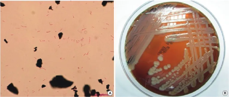
Four sets of blood cultures were collected: 2 sets from periph- eral veins and 2 sets from the central venous catheter. The pa- tient received empirical antibiotic therapy with intravenous ce- fepime (2 g every 12 hr). After 1 day of incubation, gram-nega- tive bacilli grew in an aerobic culture bottle that contained the culture from the central venous catheter. After 2 days of incuba- tion, Gram-negative bacilli also grew in an aerobic culture bottle containing the culture from the peripheral vein (Fig. 1A). The positive culture broth was inoculated onto a blood agar plate (BAP) and a MacConkey agar plate (MAC) and incubated for 48
hr at 37°C with 5% CO2. Non-hemolytic, light yellow-colored col- onies grew on the BAP (Fig. 1B). A few colonies also grew on the MAC. Catalase and oxidase tests were positive, but the in- dole test was negative. The organism was identified as S. spiri- tivorum with 98.0% probability using the Vitek 2 Gram-Negative Identification card (BioMérieux inc., Marcy-l’Etoile, France).
To confirm the identity of the isolate, 16S ribosomal RNA (rRNA) sequencing analysis was performed. InstaGene Matrix (Bio-Rad Laboratories, Hercules, CA, USA) was used to extract the bacterial genomic DNA, and the first 500 base pairs on the 5′ end of the 16S rRNA gene were amplified and sequenced us- ing MicroSeq 500 16S rDNA Bacterial Identification PCR and Sequencing Kits (Applied Biosystems, Foster City, CA, USA).
The sequencing product was analyzed on a 3130 Genetic Ana- lyzer (Applied Biosystems) according to the manufacturer’s in- structions.
The resulting sequence of the patient-derived isolate was compared with sequences stored in GenBank (http://www.ncbi.
nlm.nih.gov/genbank), EMBL (The European Molecular Biology Laboratory, http://www.ebi.ac.uk/embl), RDP-II (The Ribosomal Database Project, http://rdp.cme.msu.edu), and EzTaxon (http://
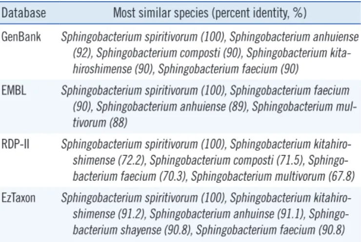
www.eztaxon.org) databases. The percent identity between the isolate from the patient and its closely related Sphingobacterium species of the 4 databases are shown in Table 1. The GenBank and RDP-II databases showed that the 16S rRNA gene se- quence of the isolate from the patient was 100% homologous with that of S. spiritivorum strain NCTC 11386 (Accession num-
Fig. 1. (A) Gram-negative bacilli from smear preparations of the positive blood cultures (Gram stain, ×1,000). (B) Yellow-colored colonies of Sphingobacterium spiritivorum on a blood agar plate.
A B
ber: GenBank, NR044077.1; RDP-II, S000752320). The EMBL and EzTaxon databases showed that the 16S rRNA gene se- quence of the isolate from the patient was 100% homologous with that of S. spiritivorum strain ATCC 33861 (Accession num- ber: EMBL, ACHA02000013; EzTaxon, ACHA01000008). The isolate from the patient showed percent identity of >99% with S.
spiritivorum and >0.8% separation from other species. Thus, the isolate was confirmed to be S. spiritivorum [7].
For phylogenetic analysis, the resulting sequence was com-
pared with those of reference strains of the most closely related Sphingobacterium species present in the GenBank databases.
A phylogenetic tree was constructed by the neighbor-joining method using the Microseq 500 bp 16S rRNA sequences (Fig. 2).
Antimicrobial susceptibility was tested using the AST-N132 card from the Vitek 2 system (BioMérieux). The isolate was sus- ceptible to cefepime, ciprofloxacin, levofloxacin, meropenem, minocycline, and trimethoprim-sulfamethoxazole; had moderate susceptibility to cefotaxime, ceftazidime, imipenem, and ticarcil- lin-clavulanic acid; but was resistant to amikacin, aztreonam, colistin, gentamicin, piperacillin, piperacillin-tazobactam, ticar- cillin, and tobramycin.
The central venous catheter was removed. The antibiotic regi- men was changed from cefepime to ciprofloxacin, because nephrotoxicity was suspected to be due to increased blood urea nitrogen and creatinine levels. After 3 days, the patient’s fever subsided. Subsequent blood culture tests were negative for S.
spiritivorum and any other microorganism. However, on the fifth day of ciprofloxacin therapy, she developed a fever again and her general condition worsened. On the eleventh day, she died of septic shock.
DISCUSSION
Sphingobacterium species are non-fermentative, non-motile, non-spore-forming aerobic gram-negative bacilli. They produce Table 1. Sequence comparison between the isolate from the pa-
tient and its most similar species
Database Most similar species (percent identity, %)
GenBank Sp hingobacterium spiritivorum (100), Sphingobacterium anhuiense (92), Sphingobacterium composti (90), Sphingobacterium kita- hiroshimense (90), Sphingobacterium faecium (90)
EMBL Sp hingobacterium spiritivorum (100), Sphingobacterium faecium (90), Sphingobacterium anhuiense (89), Sphingobacterium mul- tivorum (88)
RDP-II Sp hingobacterium spiritivorum (100), Sphingobacterium kitahiro- shimense (72.2), Sphingobacterium composti (71.5), Sphingo- bacterium faecium (70.3), Sphingobacterium multivorum (67.8) EzTaxon Sp hingobacterium spiritivorum (100), Sphingobacterium kitahiro-
shimense (91.2), Sphingobacterium anhuinse (91.1), Sphingo- bacterium shayense (90.8), Sphingobacterium faecium (90.8) Database: GenBank (http://www.ncbi.nlm.nih.gov/genbank), EMBL (The European Molecular Biology Laboratory, http://www.ebi.ac.uk/embl), RDP-II (The Ribosomal Database Project, http://rdp.cme.msu.edu), and EzTaxon (http://www.eztaxon.org).
Sphingobacterium kitahiroshimense 10C (NR041636.1) Sphingobacterium anhuiense CW 186 (NR044477.1)
Sphingobacterium faecium DSM 11690 (NR025537.1)
Sphingobacterium thalpophilum DSM 11723 (NR042135.1) Sphingobacterium multivorum (NR043196.1)
Sphingobacterium composti T5-12 (NR041363.1) Sphingobacterium shayense HS 39 (FJ816788.1)
Sphingobacterium spiritivorum NCTC 11386 (NR044077.1)
Sphingobacterium antarcticum DSM 15311 (FR733711.1)
0.01
Isolate of patient
Fig. 2. Phylogenetic relationships of the isolate from the present patient and related Sphingobacterium species, constructed by the neigh- bor-joining method by using the Microseq 500 bp 16S rRNA sequences. All names and accession numbers are given as cited in the Gen- Bank database. The tree was drawn with branch length as the evolutionary distances. The scale bar length of 0.01 indicates 1% sequence distance.
catalase, oxidase, and urease [1]. Sphingobacterium species grow on both BAP and MAC [2, 3]. Colonies are yellowish, cir- cular, slightly convex, smooth, opaque, and non-hemolytic on BAP after 2 days of incubation [2, 3]. To date, 15 species, in- cluding S. anhuiense, S. antarcticus, S. bambusae, S. ca- nadense, S. composti, S. daejeonense, S. faecium, S. hepari- num, S. kitahiroshimense, S. multivorum, S. piscium, S. shay- ense, S. siyangense, S. spiritivorum, and S. thalpophilum, have been described in the genus Sphingobacterium [4, 8-17].
Sphingobacterium species are usually isolated from soil, wa- ter, and plant material, and only a few case reports of human infections caused by the species have been published [5]. Pre- viously reported Sphingobacterium species isolated from hu- man clinical specimens were S. multivorum and S. spiritivorum.
To date, 7 cases of S. multivorum infection have been reported worldwide in relation to septicemia [5, 18-20], peritonitis [21], respiratory tract infection [22], and necrotizing fasciitis [23].
Only 3 cases of S. spiritivorum infection have been reported worldwide [24-26]. The present case and previously reported cases are compared in Table 2. In 2002, Marinella [24] first de- scribed a case of cellulitis-associated sepsis caused by an S.
spiritivorum infection. In 2003, Tronel et al. [25] reported a case of S. spiritivorum bacteremia. In 2005, Kronel et al. [26] reported a case of cellulitis-associated sepsis caused by S. spiritivorum from the water reservoir of a steam iron.
In the present case, the patient was in an immunosuppressed condition due to the chemotherapy to treat acute myeloid leuke- mia. The patient was diagnosed with a catheter-related blood- stream infection because the time interval of positive blood cul- ture signs from those between the peripheral vein and central venous catheter cultures was more than 2 hr. The source and transmission route of the S. spiritivorum infection in this case may have been a skin entry site of an intravascular device or a subcutaneous path of the catheter that had been in a close proximity with the natural habitats of this organism.
The 16S rRNA sequencing analysis can be a useful and de- finitive method particularly for the identification of clinically sig- nificant bacterial isolates with ambiguous biochemical profiles or
of rarely encountered bacterial species [27, 28]. We confirmed the identity of the blood isolate, first identified biochemically as S. spiritivorum, by 16S rRNA sequencing analysis.
Sphingobacterium species are generally resistant to amino- glycosides and polymyxin B, but are susceptible to quinolones and trimethoprim-sulfamethoxazole in vitro. Susceptibility to β-lactam antibiotics is known to vary [1]. In 2009, Lambiase et al. [29] reported that 13 S. multivorum and 8 S. spiritivorum iso- lates from sputum samples in 332 patients with cystic fibrosis were resistant to aminoglycosides, but susceptible to quinolones and trimethoprim-sulfamethoxazole. They also found that S.
multivorum isolates were resistant to all β-lactams, whereas the S. spiritivorum isolates were susceptible to ceftazidime, piper- acillin, and carbapenems. The isolate of this case was suscepti- ble to cefepime, meropenem, minocycline, as well as ciprofloxa- cin, levofloxacin, and trimethoprim-sulfamethoxazole.
This is the first reported case of human S. spiritivorum infec- tion in Korea, which shows that S. spiritivorum can be a fatal human opportunistic pathogen in immunocompromised pa- tients, despite human infection being rare.
Authors’ Disclosures of Potential Conflicts of Interest
No potential conflicts of interest relevant to this article were re- ported.
Acknowledgements
This work was supported by a 2-Year Research Grant of Pusan National University.
REFERENCES
1. Versalovic J, Carroll KC, et al. eds. Manual of clinical microbiology. 10th ed. Washington, DC: ASM Press, 2011:723-7.
2. Holmes B, Owen RJ, Weaver RE. Flavobacterium multivorum, a new species isolated from human clinical specimens and previously known as group IIk, biotype 2. Int J Syst Bacteriol 1981;31:21-34.
Table 2. Cases of Sphingobacterium spiritivorum infection
Case No. [Reference] Sex Age Predisposing condition Symptom Source Diagnosis Outcome 1. Marinella (2002) [23] M 72 Parkinson disease Fever, chill, convulsion Blood Cellulitis, sepsis Recovered 2. Tronel et al. (2003) [24] M 84 Refractory anemia Fever, leg rash Blood Foot cellulitis, sepsis Recovered 3. Kämpfer et al. (2005) [25] F 34 NA Fever, dry cough BAL Extrinsic allergic alveolitis Recovered
4. [PR] F 68 Acute myeloid leukemia, chemotherapy Fever Blood Sepsis Died
Abbreviations: M, male; F, female; BAL, bronchoalveolar lavage; PR, present report; NA, not available.
3. Holmes B, Owen RJ, Hollis DG. Flavobacterium spiritivorum, a new species isolated from human clinical specimens. Int J Syst Bacteriol 1982;32:157-65.
4. Yabuuchi E, Kaneko T, Yano I, Moss CW, Miyoshi N. Sphingobacterium gen. nov., Sphingobacterium spiritivorum comb. nov., Sphingobacteri- um multivorum comb. nov., Sphingobacterium mizutae sp. nov., and Flavobacterium indologenes sp. nov.: Glucose-nonfermenting gram- negative rods in CDC groups IIK-2 and IIb. Int J Syst Bacteriol 1983;33: 580-98.
5. Aydo an M, Yumuk Z, Dündar V, Arisoy ES. Sphingobacterium multivo- rum septicemia in an infant: Report of a case and review of the litera- ture. Türk Mikrobiyol Cem Derg 2006;36:44-8.
6. Swerdlow SH, Campo E, Harris NL, Jaffe ES, Pileri SA, Stein H, et al.
WHO Classification of Tumours of Haematopoietic and Lymphoid Tis- sues. Lyon, France: IARC; 2008.
7. Clinical and Laboratory Standards Institute. Interpretive criteria for iden- tification of bacteria and fungi by DNA target sequencing; Approved guideline. CLSI document MM18-A. Wayne, PA: Clinical and Laboratory Standards Institute, 2008.
8. Wei W, Zhou Y, Wang X, Huang X, Lai R. Sphingobacterium anhuiense sp. nov., isolated from forest soil. Int J Syst Evol Microbiol 2008;58:2098- 101.
9. Shivaji S, Ray MK, Rao NS, Saisree L, Jagannadham MV, Kumar GS, et al. Sphingobacterium antarcticus sp. nov., a psychrotrophic bacterium from the soils of Schirmacher oasis, Antarctica. Int J Syst Bacteriol 1992;42:102-06.
10. Duan S, Liu Z, Feng X, Zheng K, Cheng L. Sphingobacterium bambu- sae sp. nov., isolated from soil of bamboo plantation. J Microbiol 2009; 47:693-8.
11. Mehnaz S, Weselowski B, Lazarovits G. Sphingobacterium canadense sp. nov., an isolate from corn roots. Syst Appl Microbiol 2007;30:519-24. 12. Yoo SH, Weon HY, Jang HB, Kim BY, Kwon SW, Go SJ, et al. Sphingo- bacterium composti sp. nov., isolated from cotton-waste composts. Int J Syst Evol Microbiol 2007;57:1590-3.
13. Kim KH, Ten LN, Liu QM, Im WT, Lee ST. Sphingobacterium daejeo- nense sp. nov., isolated from a compost sample. Int J Syst Evol Microbi- ol 2006;56:2031-6.
14. Takeuchi M and Yokota A. Proposals of Sphingobacterium faecium sp.
nov., Sphingobacterium piscium sp. nov., Sphingobacterium heparinum comb. nov., Sphingobacterium thalpophilum comb. nov. and two geno- species of the genus Sphingobacterium and synonymy of Flavobacteri- um yabuuchiae and Sphingobacterium spiritivorum. J Gen Appl Micro- biol 1992;38:465-82.
15. Matsuyama H, Katoh H, Ohkushi T, Satoh A, Kawahara K, Yumoto I.
Sphingobacterium kitahiroshimense sp. nov., isolated from soil. Int J
Syst Evol Microbiol 2008;58:1576-9.
16. He X, Xiao T, Kuang H, Lan X, Tudahong M, Osman G, et al. Sphingo- bacterium shayense sp. nov., isolated from forest soil. Int J Syst Evol Microbiol 2010;60:2377-81.
17. Liu R, Liu H, Zhang CX, Yang SY, Liu XH, Zhang KY, et al. Sphingobac- terium siyangense sp. nov., isolated from farm soil. Int J Syst Evol Mi- crobiol 2008;58:1458-62.
18. Potvliege C, Dejaegher-Bauduin C, Hansen W, Dratwa M, Collart F, Tielemans C, et al. Flavobacterium multivorum septicemia in a hemodi- alyzed patient. J Clin Microbiol 1984;19:568-9.
19. Freney J, Hansen W, Ploton C, Meugnier H, Madier S, Bornstein N, et al. Septicemia caused by Sphingobacterium multivorum. J Clin Micro- biol 1987;25:1126-8.
20. Areekul S, Vongsthongsri U, Mookto T, Chettanadee S, Wilairatana P.
Sphingobacterium multivorum septicemia: a case report. J Med Assoc Thai 1996;79:395-8.
21. Dhawan VK, Rajashekaraiah KR, Metzger WI, Rice TW, Kallick CA.
Spontaneous bacterial peritonitis due to a group IIk-2 strain. J Clin Mi- crobiol 1980;11:492-5.
22. Reina J, Borrell N, Figuerola J. Sphingobacterium multivorum isolated from a patient with cystic fibrosis. Eur J Clin Microbiol Infect Dis 1992; 11:81-2.
23. Grimaldi D, Doloy A, Fichet J, Bourgeois E, Zuber B, Wajsfisz A, et al.
Necrotizing fasciitis and septic shock related to the uncommon gram- negative pathogen Sphingobacterium multivorum. J Clin Microbiol 2012;50:202-3.
24. Marinella MA. Cellulitis and sepsis due to Sphingobacterium. JAMA 2002;288:1985.
25. Tronel H, Plesiat P, Ageron E, Grimont PA. Bacteremia caused by a novel species of Sphingobacterium. Clin Microbiol Infect 2003;9:1242-4. 26. Kämpfer P, Engelhart S, Rolke M, Sennekamp J. Extrinsic allergic alveo- litis (hypersensitivity pneumonitis) caused by Sphingobacterium spiri- tivorum from the water reservoir of a steam iron. J Clin Microbiol 2005; 43:4908-10.
27. Woo PC, Ng KH, Lau SK, Yip KT, Fung AM, Leung KW, et al. Usefulness of the MicroSeq 50016S ribosomal DNA-based bacterial identification system for identification of clinically significant bacterial isolates with ambiguous biochemical profiles. J Clin Microbiol 2003;41:1996-2001. 28. Arosio M, Nozza F, Rizzi M, Ruggeri M, Casella P, Beretta G, et al. Eval-
uation of the MicroSeq 50016S rDNA-based gene sequencing for the diagnosis of culture-negative bacterial meningitis. New Microbiol 2008; 31:343-9.
29. Lambiase A, Rossano F, Del Pezzo M, Raia V, Sepe A, de Gregorio F, et al. Sphingobacterium respiratory tract infection in patients with cystic fi- brosis. BMC Res Notes 2009;2:262.