Bee Venom이 NF-κB의 불활성화에 의한 세포자멸사를 통해 PC-3 세포의 증식에 미치는 영향
오현준ᆞ송호섭
경원대학교 한의과대학 침구학교실
목적 : 이 연구는 봉약침의 봉독과 그 주요성분인 멜리틴이 NF-κB의 활성억제와 세포자멸사 관련 단백 질의 발현 조절을 통하여 세포자멸사를 유도함으로써 전립선 암세포주인 PC-3 세포의 성장을 억제하는지를 확인하고 해당 기전을 살펴보고자 하였다.
방법 : 봉독이나 멜리틴을 처리한 후 PC-3의 성장억제를 관찰하기 위해 WST-1 assay, CCK-8 assay를 시행하였고, 세포자멸사 조절단백질의 변동 관찰에는 western blot analysis를 시행하였고, 세포자멸사와 연 관된 NF-κB의 활성 변화를 관찰하기 위해 EMSA를 시행하였으며, PC-3에서 봉독이나 멜리틴과 NF-κB의 상호작용을 관찰하기 위해 transient transfection assay를 시행하여 세포생존율과 NF-κB의 활성 변동을 측 정하였다.
결과 : PC-3 세포에 봉독이나 멜리틴을 처리한 후, 전립선암세포의 성장, 세포자멸사의 유발, 세포자멸사 관련 단백질의 발현, NF-κB의 활성, NF-κB의 p50, IKKα, IKKβ 치환 후 NF-κB의 활성과 PC-3 세포 증 식에 미치는 영향을 관찰하여 다음과 같은 결과를 얻었다.
1. PC-3 세포에서 봉독이나 멜리틴을 처리한 후 세포자멸사가 유도되어 세포성장이 억제되었고, 세포자 멸사 관련 단백질 중 분리된 PARP, caspase-3, -9는 유의한 증가를, Bcl-2, XIAP, cXIAP2는 유의한 감소를 나타내었다.
1)
Bee Venom Inhibits PC-3 Cell Proliferation Through Induction of Apoptosis Via Inactivation
of NF-κB
Oh Hyun-jun and Song Ho-sueb
Dept. of Acupuncture & Moxibustion, College of Oriental Medicine, Kyungwon University
․This research was supported by the Kyungwon University Research Fund in 2010
․Acceptance : 2010. 3. 5. ․Adjustment : 2010. 5. 26. ․Adoption : 2010. 5. 26.
․Corresponding author : Song Ho-sueb, Kyungwon Gil Oriental Medical Hospital, 117 Yong-dong Jung-gu Incheon Republic of Korea.
Tel. 82-32-770-1214 E-mail : [email protected]
국문초록
Original Article
2. PC-3 세포에서 봉독이나 멜리틴을 처리한 후 NF-κB의 활성은 유의한 감소를 나타내었다.
3. PC-3 세포에서 NF-κB의 p50, IKKα, IKKβ를 치환하여 작용기를 없애고 봉독이나 멜리틴을 처리하였 을 경우에도 NF-κB의 활성이 유의한 감소를 나타내었다.
결론 : 이상의 결과는 봉독이나 멜리틴이 NF-κB의 활성 억제를 통하여 인간 전립선암세포주인 PC-3의 세포자멸사를 유발함으로써 증식억제 효과가 있음을 입증한 것으로, 전립선암의 예방과 치료에 대한 효과적 인 치료제 개발에 도움이 될 것으로 기대된다.
핵심 단어 : 봉독, 멜리틴, 전립선암, PC-3 cell, 세포자멸사, NF-κB, cleavaged PARP, Bcl-2, XIAP, cIAP2, transfection
Ⅰ. Introduction
Prostate cancer is the most commonly diagnosed malignancy in men in many industrialized countries1). The management of prostate cancer remains a com- plex and intricate problem, because metastatic hormone- refractory prostate cancer remains incurable with currently available therapies including androgen de- privation with luteinizing hormone-releasing hormone agonists, nonsteroidal androgen receptor antagonists, androgen withdrawal, corticosteroids, chemotherapy, radiation therapy, radiopharmaceuticals, immunologic approaches, gene therapy and novel molecular targets, etc2,3). However surgical technique has been developed and clinical outcome following radio- therapy has been also improved up to now, not a few patients yet meet a recurrence of prostate cancer frequently by the transition of androgen sensitive to androgen refractory state4-7). Therefore, to execute suppression of androgen-independent prostate cancer, novel therapeutic approaches must be developed.
Apoptosis is generally mediated through multiple pathways, involving a complex array of biochemical regulators and molecular interactions8), one of which is NF-κB and its signal pathway. Recent several studies have reported that NF-κB is constitutively activated in human prostate cancer cells including PC-3 cells and promote their survival9,10) implying prostate cancer cell proliferation can be regulated
by induction of apoptosis via NF-κB inactivation.
The Biochemical and morphological character- istics of apoptosis includes caspase activation, release of cytochrome c, nuclear condensation, protease activation and DNA fragmentation8). Apoptosis has emerged as a significantly relative mechanism for the inhibition of cancer growth11,12). Especially in prostate cancer, delaying androgen refractory state through induction of apoptosis via down regulating activity of NF-κB and modulating apoptosis regulatory proteins has been regarded to be related with anti-proliferation of the cancer9,10).
Lee et al13). partly demonstrated that Bee venom (BV) can inactivate NF-κB and suppress the expres- sion of inflammation related genes such as iNOS, COX-2 and cPLA2 and thereby prevent anti- apoptotic ability of NF-κB causing human androgen sensitive prostate cancer LNCaP cells undergo apoptosis, in vitro, suggesting BV should be one of the agents capable of preventing androgen depend- ent prostate cancer from changing into androgen refractory one through promoting apoptosis via inactivation of NF-κB and modulation of apoptosis regulatory proteins, thereby inhibiting proliferation of prostate cancer.
To reconfirm the hypotheses in androgen in- dependent prostate cancer, based on the above findings, I therefor conducted a mechanism study, determining whether BV exerts inhibitory effect on human androgen insensitive prostate cancer PC-3 cell proliferation via induction of apoptotic cell death
through inactivation of NF-κB and modulation of other apoptosis related genes and whether pro- mising interaction between SVT and p50 of NF-κB in PC-3 cells from the previous report14) can be intactly applied to BV interacting with signal molecules such as p50 in PC-3 cells.
Ⅱ. Materials and methods
1. Materials
Dried BV was purchased from You-Miel BV Ltd(Hwasoon, Jeonnam, Korea). The composition of the BV was as follow: 45~50% melittin, 2.5~3%
apamin, 2~3% MCD peptide, 12% PLA2, 1% lyso- PLA, 1~1.5% histidine, 4~5% 6pp lipids, 0.5%
secarpin, 0.1% tertiapin, 0.1% procamine, 1.5~2%
hyaluronidase, 2~3% amine, 4~5% carbohydrate and 19~27% other, including protease inhibitor, gluco- sidase, invertase, acid phosphomonoesterase, dopa- mine, norepinephirne and unknown amino acids, with >99.5% purity. Melittin(MT) was purchased from Sigma Chemical Co(St Louis, MO, USA). Goat polyclonal antibody to COX-2(1 : 500) and TNF(1 : 500), mouse polyclonal antibody to iNOS(1 : 500) and all of the secondary antibodies such as Akt, phosphorylated Akt, Bax, Bcl-2, PARP, caspase-3, -9, cleaved caspase-3, -9, cIAP2, XIAP, MMP-2, -13, used in Western blot analysis were purchased from Santa Cruz Biotechnology(Santa Cruz, CA, USA).
T4 polynucleotide kinase was obtained from Pro- mega(Madison, WI, USA). Poly(dI-dC), horseradish peroxidase-labeled donkey anti-rabbit secondary anti- body and ECL detection reagent were obtained from Amersham Pharmacia Biotech(Piscataway, NJ, USA). Reagents for sodium dodecyl sulfate (SDS)–
polyacrylamide gel electrophoresis were purchased from Bio-Rad(Hercules, CA, USA). All other rea- gents were purchased from Sigma unless other- wise stated.
2. Cell culture
The PC-3 human prostate cancer cell was ob- tained from ATCC(American type culture coll- ection, Rockville, MD, USA). Prostate cells were cultured in RPMI-1640 medium(Life Technologies Inc., Gaithersberg, MD, USA) supplement with 10%
fetal calf serum(FCS ; Collaborative Biomedical Products, Bedford, MA, USA) and antibiotics, peni- cillin/streptomycin(100unit/ml, Bioproducts, Walker- sville, MD, USA) cell cultures were maintained at 37℃ in a humidified atmosphere of 5% CO2.
3. Cell viability assay 1) WST-1 assay
Cells were plated at a density of 1×10⁵ cells per well in 96-well plate and subconfluent cells were exposed to different doses of MT(0.5~2.5㎍) or BV(1~10㎍) for 24, 48, 72hr. After treatment, cell viability was measured by WST-1 assay(Dojindo Laboratory, Kumamoto, Japan) according to the manufacturer’s instructions. WST-1 solution was added to cells in 96-well plates, cells were in- cubated at 37.5℃ for 1hr and the optical density of each well was read at 450nm.
2) CCK-8 assay
Cells were plated at a density of 1×10⁴ cells per well in 96-well plates and subconfluent cells were exposed to different doses of MT(0.5~2.5㎍) or BV(1~10㎍) for 24, 48 or 72hr. After treatment, cell viability was measured by Cell Counting Kit-8 (CCK-8) system(Dojindo Laboratory, Kumamoto, Japan) according to the manufacturer’s instructions.
CCK-8 solution was added to cells in 96-well plates, cells were incubated at 37°C for 1hr and the optical density of each well was read at 450nm.
4. Western blot analysis
Cells were homogenized with lysis buffer [50 mM Tris pH 8.0, 150mM NaCl, 0.02% sodium azide, 0.2% SDS, 1mM PMFS, 10μl/ml aprotinin, 1%
igapel 630(Sigma-Aldrich, St Louis, MO, USA), 10 mM NaF, 0.5mM EDTA, 0.1mM EGTA and 0.5%
sodium deoxycholate] and centrifuged at 23,000g for 1hr. Equal amount of proteins(80μg) were separated on a SDS/12%-polyacrylamide gel and transferred to a nitrocellulose membrane(Hybond ECL, Amer- sham Pharmacia Biotech Inc., Piscataway, NJ, USA).
Blots were blocked for 2hr at room temperature with 5%(w/v) non-fat dried milk in Tris-buffered saline [10mM Tris(pH 8.0) and 150mM NaCl]
solution containing 0.05% tween-20. The membrane was incubated for 5hr at room temperature with specific antibodies Bax, Bcl-2, cleaved PARP, cleaved caspase-3, -9, cIAP2, XIAP(Santa Cruz, CA, USA).
The blot was then incubated with the corres- ponding conjugated antirabbit immunoglobulin g-hor- seradish peroxidase(Santa Cruz, CA, USA). Im- munoreactive proteins were detected with the ECL western blotting detection system. The relative density of the protein bands was scanned by densi- tometry using MyImage(SLB, Seoul, Korea) and quantified by Labworks 4.0 software(UVP Inc, Upland, California, USA).
5. Preparation of Nuclear extracts and Electromobility shift assays
It was performed according to the manufacturer’s recommendations(Promega, Madison, WI, USA).
Briefly, 1×10⁶cells/ml was washed twice with 1×
PBS, followed by the addition of 1ml of PBS and the cells were scraped into a cold Eppendorf tube.
Cells were spun down at 15,000g for 1min and the resulting supernatant was removed. Solution A(50mM HEPES, pH 7.4, 10mM KCl, 1mM EDTA, 1mM EGTA, 1mM dithiothreitol, 0.1μg/ml phenylmethyl- sulfonyl fluoride, 1μg/ml pepstatin A, 1μg/ml leu- peptin, 10μg/ml soybean trypsin inhibitor, 10μg/ml aprotinin and 0.5% Nonidet P-40) was added to the pellet in a 2 : 1 ratio(v/v) and allowed to incubate on ice for 10min. Solution C(solution A + 10% gly- cerol and 400mM KCl) was added to the pellet in a 2 : 1 ratio(v/v) and vortexed on ice for 20min. The cells were centrifuged at 15,000g for 7min and the
resulting nuclear extract supernatant was collected in a chilled Eppendorf tube. Consensus oligonucleo- tides were end-labeled using T4 polynucleotide kinase and [g-32P] ATP for 10min at 37℃. Gel shift reactions were assembled and allowed to incubate at room temperature for 10min followed by the addition of 1μl(50,000~200,000cpm) of 32P- labeled oligonucleotide and another 20min of incub- ation at room temperature. For supershift assays, nuclear extracts from cells treated with MT(0.5~
2.5㎍) or BV(1-10㎍) were incubated with specific antibodies against Rel-A NF-κB isoforms for 1hr before EMSA. For competition assays, nuclear extracts from cells treated with MT(0.5~2.5㎍) or BV(1~10㎍) were incubated with unlabelled NF-κB oligonuclaotide(50X, 100X and 200X) or labeled SP-1(100X) and AP-1(100X) for 30min before EMSA. Subsequently 1μl of gel loading buffer was added to each reaction and loaded onto a 6%
nondenaturing gel and electrophoresed until the dye was three-fourths of the way down the gel. The gel was dried at 80℃ for 1hr and exposed to film overnight at 70℃. The relative density of the DNA-protein binding bands was scanned by densi- tometry using MyImage(SLB, Seoul, Korea) and quantified by Labworks 4.0 software(UVP Inc, Upland, California, USA).
6. Assay of luciferase activity
PC-3 cells were cotreated with MT(0.5~2.5㎍) or BV(1-10㎍) and TNF-α. Luciferase activity was measured using a luciferase assay kit(Promega) and WinGlow software, according to the manufacturer’s instructions(Berthold, Wildbad, Germany).
7. Transient transfection assay
A fusion gene containing pCMV promoter and mutant of NF-kB signal molecules such as p50, IKKα and IKKβ was used. PC-3 cells were transfected with these mutant genes constructs.
Cell viability was measured after stimulating the cells for 24hr with MT(1~2.5㎍/㎖) for 24hr.
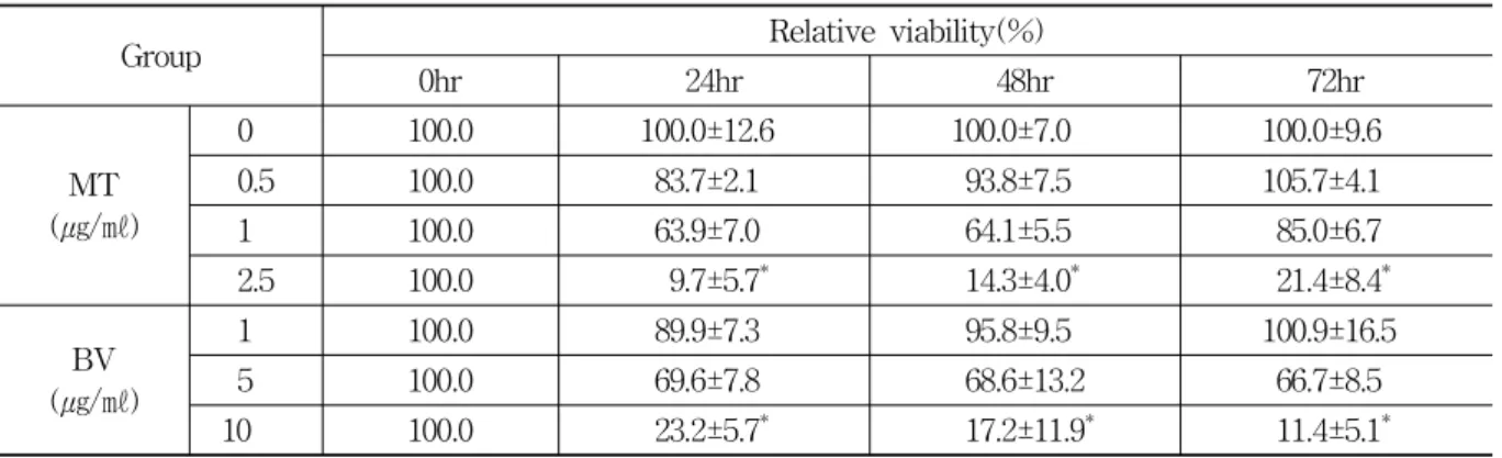
Group Relative viability(%)
0hr 24hr 48hr 72hr
MT (㎍/㎖)
0.0 100.0 100.0±12.6 100.0±7.0 100.0±9.6
0.5 100.0 83.7±2.1 93.8±7.5 105.7±4.1
1.0 100.0 63.9±7.0 64.1±5.5 85.0±6.7
2.5 100.0 9.7±5.7* 14.3±4.0* 21.4±8.4*
BV (㎍/㎖)
1.0 100.0 89.9±7.3 95.8±9.5 100.9±16.5
5.0 100.0 69.6±7.8 68.6±13.2 66.7±8.5
10.0 100.0 23.2±5.7* 17.2±11.9* 11.4±5.1*
Values are the mean±SEM of 3 independent experiments performed in triplicate.
MT and BV represent Melittin and Bee venom respectively. * : representsp<0.05 compared with control.
Table 1. PC-3 Cell Growth After Treatment of MT or BV
Fig. 1. Morphological changes of PC-3 cells by MT or BV
Morphological changes were observed under microscope(magnification, 200 × ).
The figures are representative of three experiments, with triplicate of each experiment.
8. Statistical analysis
Data were analyzed using one-way ANOVA followed by Tuckey test as a post-hoc test.
Differences were considered significant at
p
<0.05.Ⅲ. Results
1. Inhibition of PC-3 cell growth
Morphological alteration of the cells was demon- strated in Fig. 1. Once the cells were exposed to MT(0.5-2.5㎍/㎖) or BV(1-10㎍/㎖), the cells were not grown and died in a dose dependent manner.
To evaluate an effect of MT or BV on the cell growth of PC-3 cells, I analyzed cell viability using
WST-1 assay and direct cell counting. MT or BV inhibited prostate cancer cell growth in a dose dependent manner, the percentage of control significantly decreased by 2.5㎍/㎖ of MT at 24, 48 and 72hr was 9.7±5.7, 14.3±4.0 and 21.4±8.4%, which of control significantly decreased by 10㎍/㎖
of BV at 24, 48 and 72hr was 23.2±5.7, 17.2±11.9 and 11.4±5.1%(Table 1, Fig. 1).
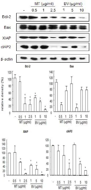
2. Expression of apoptosis regulatory proteins
Anti- and pro-apoptotic effectors modulate apo- ptosis known as programmed cell death which involve a large number of proteins16). The change of mitoondrial transmembrane potential induced by transcation of Bax protein plays a major role in promotion of apoptosis. As mitochondrial membrane
Fig. 2. Effect of MT or BV on expression of apoptosis related proteins in PC-3 cells
Equal amounts(50µg) of whole cell lysates were subjected to electrophoresis and analyzed by apoptosis regulatory molecules(Bax, Bcl-2, XIAP, cIAP2). The relative density was analyzed by densitometry. Similar patterns of protein expression were obtained from three experiments. Values are mean±SEM of two experiments, with triplicate of each experiment.
* : representsp<0.05 significant compared from control.
potential(MMP) is decreased, increased permeabili- zation of this membrane allows matrix condensation and release of cytochrome c to the intermembrane space and activation of subsequent downstream ca- spases such as caspase-3, -9. Activated caspase-3
Fig. 3. Effect of MT or BV on expression of pro-apototic gene in PC-3 cells
Equal amounts(50µg) of whole cell lysates were subjected to electrophoresis and analyzed by apoptosis regulatory molecules(cleaved PARP, cleaved caspase 3, cleaved caspase 9). The relative density was analyzed by densitometry. Similar patterns of protein expression were obtained from three experiments. Values are mean±SEM of two experiments, with triplicate of each experiment.
* : represents p<0.05 significant compared from control.
cleaves intra-cellular proteins such as PARP which are vital to cell survival and growth and PARP cleavage has been used as an important marker of apoptosis.
Among those anti-apoptotic protein, the human
Group Apoptotic gene exression (relative density, %)
Cleaved-PARP Cleaved caspase 3 Cleaved caspase 9
MT (㎍/㎖)
0.0 100.0±1.0* 100.0±1.0* 100.0±1.0*
0.5 127.12±10.1* 156.8±16.2* 183.9±13.5*
1.0 84.8±14.0* 89.4±31.4* 150.6±42.5*
2.5 133.5±11.0* 121.42±14.4* 173.2±62.5*
BV (㎍/㎖)
1.0 88.5±10.4* 95.3±12.1* 124.5±31.1*
5.0 48.6±14.8* 127.4±13.5* 226.4±43.1*
10 42.2±11.8* 148.5±28.1* 220.0±31.7*
Values are the mean±SEM of 3 independent experiments performed in triplicate.
BV and MT represents Bee venom and melittin respectively. * : represents p<0.05 significant compared with contol.
Table 3. Pro-Apoptotic gene Expressions in Prostate Cancer PC-3 Cells
Group Apoptotic gene exression(relative density, %)
Bcl-2 Bax XIAP cIAP2
MT (㎍/㎖)
0.0 100.0±1.0* 100.0±1.0 100.0±1.0* 100.0±1.0*
0.5 89.4±14.5* 23.5±6.7 103.2±30.9* 82.7±20.6*
1.0 23.5±17.7* 72.0±15.6 58.2±3.4* 65.0±32.7*
2.5 21.4±13.5* 96.0±23.4 6.7±2.8* 21.4±19.6*
BV (㎍/㎖)
1.0 41.4±6.9* 13.0±13.1 44.2±4.3* 100.9±5.7*
5.0 29.7±4.9* 74.2±9.7 26.6±17.3* 42.7±8.2*
10.0 6.3±2.0* 74.5±5.4 2.3±0.7* 18.4±1.3*
Values are the mean±SEM of 3 independent experiments performed in triplicate.
BV and MT represents Bee venom and melittin respectively. * : representsp<0.05 significant compared with contol.
Table 2. Apoptotosis Related gene Expressions in Prostate Cancer PC-3 Cells
inhibitor of apoptosis(IAP) family includes cellular IAP1(cIAP1), cIAP2, X-linked IAP(XIAP). cIAP2 is inducible by a variety of NF-κB-inducing stimuli in multiple cell lines, whereas induction of cIAP1 and XIAP appears to be cell type- and stimulusepen- dent. Therefore, to gain insight into mechanisms controlling apoptosis in PC-3 cells, we observed the effect of MT and BV on anti-apoptotic and pro- poptotic proteins. Fig. 2, 3 reveal a western blot analysis of Bax, Bcl-2, XIAP, cIAP2, cleaved caspase-3, cleavaged PARP, cleaved caspase-9 expressions in PC-3 cells treated with a different dose of MT or BV. Compared with control, ex- ession of pro-apoptotic proteins such as cleaved caspase-3 and cleaved caspase-9 in the PC-3 cells treated by 0.5, 1 and 2.5μg/㎖ of MT or 1, 5 and 10 μg/㎖ of BV was increased in a dose dependent manner, whereas that of cleavaged PARP was
increased only by 0.5 and 2.5μg/㎖ of MT and moreover Bax was not increased in the above concentration of MT or BV.
Meanwhile, anti-apoptosis related proteins including Bcl-2, XIAP, cIAP2 were decreased by 0.5, 1 and 2.5μg of MT or 1, 5 and 10μg/㎖ of BV dose dependently, compared with control(Fig. 2, 3).
From the above findings, we confirmed the ability of MT or BV to exert influence upon apo- ptosis related proteins inducing Bax, caspase-3, -9, cleaved caspase-3, cleavaged PARP, cleaved caspase- 9 up-regulation and Bcl-2, XIAP, cIAP2 down regulation.
Observing the expression of the proteins con- cerned more concretely, Bcl-2 significantly deeased by 1 and 2.5μg/㎖ of MT or 1, 5 and 10μg/㎖ of BV was 23.5±17.7 and 21.4±13.5% or 41.4±6.9, 29.7±
4.9 and 6.3±2.0%. XIAP significantly decreased by 1
and 2.5μg/㎖ of MT or 1, 5 and 10μg/㎖ of BV was 58.2±3.4 and 6.7±2.8% or 44.2±4.3, 26.6±7.3 and 2.3±0.7%. cIAP2 significantly decreased by 2.5μg/㎖
of MT or 5 and 10μg/㎖ of BV was 21.4±19.6% or 42.7±8.2 and 18.4±1.3%.
Cleavaged PARP significantly increased by 0.5 and 2.5μg/㎖ of MT was 127.12±10.1 and 133.5±
11%.
Cleaved caspase-3 significantly increased by 0.5 and 2.5μg/㎖ of MT or 5 and 10μg/㎖ of BV was 156.8±16.2 and 121.42±14.4% or 127.4±13.5 and 148.5±
28.1%. Cleaved caspase-9 significantly increased by 0.5, 1 and 2.5μg/㎖ of MT or 1, 5 and 10μg/㎖ of BV was 183.9±13.5, 150.6±42.5 and 173.2±62.5% or 124.5±31.1, 226.4±43.1 and 220.0±31.7%(Table 2, 3).
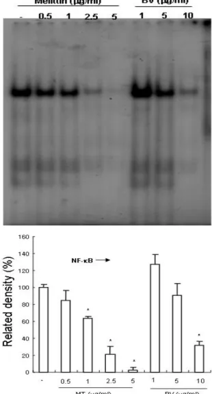
3. Inhibition of NF-κB
It was demonstrated that MT or BV negatively regulates NF-κB and suppresses expression of inflammation related genes including iNOS and COX-2 via inhibition of NF-κB. In addition, NF-κB is known to inhibitory transcription factor of apo- ptosis. To investigate the hypothesis whether MT or BV can inactivate NF-κB and thereby prevent anti-apoptotic ability of NF-κB causing prostate cancer PC-3 cells go apoptosis, I assessed NF-κB activity in the cells treated for different con- centration with MT or BV for 24hr by EMSA.
Group Related density(%)
MT (㎍/㎖)
0 100.0±3.0*
0.5 85.0±12.0*
1 63.0±3.0*
2.5 21.0±10.0*
5 2.0±4.0*
BV (㎍/㎖)
1 127.0±12.0*
5 91.0±14.0*
10 32.0±5.0*
Values are the mean±SEM of 3 independent exper- iments performed in triplicate. BV and MT represents Bee venom and melittin respectively.
* : representsp<0.05 significant compared with contol.
Table 4. Effect of MT or BV on NF-κB Activity in PC-3 Cells
NF-κB was highly activated in this cell, however the activation of NF-κB was gradually decreased by the culture in the presence of MT or BV in the cells(Fig. 4). In NF-kB activity, the density of control significantly decreased by MT 1, 2.5 and 5μg/
㎖ or BV 10μg/㎖ was 63±3 and 21±10 and 2±4%
or 32±5%(Table 4). I also conducted a luci- ferase assay in order to determine the role of MT or BV in NF-κB dependent gene transcriptilucion.
Fig. 4. Inhibition of NF-κB in prostate cancer PC-3 cells by MT or BV
Activation of NF-κB was determined by electrophoretic mobility shift assay(EMSA). Nuclear extracts from PC-3 cells with MT(0.5, 1, 2.5, 5μg/㎖) or BV(1, 5, 10μg/㎖) were incubated in binding reactions of 32P-labeled oligon- ucleotide containing the B sequence. NF-κB DNA binding activity was determined by EMSA.
* : representsp<0.05 significant compared from control.
PC-3 cells were transfected with this promoter- reporter gene construct and transcriptional activities were measured after TNF-α stimulation with or without MT or BV. As shown in Fig. 5, cotreat- ment of the transfected cells with MT significantly inhibited the luciferase activity induced by TNF-α in PC-3 cells. The luciferase activity of control
Fig. 5. Effects of MT or BV on TNF-α induced NF-κB dependent luciferase activity in PC-3 cells
PC-3 cells were transfected with pNF-κB-Luc plasmid (5NF-κB) and activated with TNF-α in absence or presence of MT(0.5, 1, 2.5μg) or BV(1, 5, 10μg) for 2hr and the luciferase activity was determined. Values are the mean±SEM of 3 independent experiments performed in triplicate. The level of induction was calculated relative to the luciferase activity in unstimulated transfected cells.
* : represents p<0.05 significant compared from control.
Group Luciferase activity normal 1842.5±142.5* control 3713.7±193.1* MT
(㎍/㎖)
0.5 2987.5±151.3* 1 2240.4±104.1* 2.5 1870.0±137.9* BV
(㎍/㎖)
1 3713.7±193.1* 5 2948.0±126.2* 10 2263.8±96.9*
Values are the mean±SEM of 3 independent exper- iments performed in triplicate. BV and MT represents Bee venom and melittin respectively.
* : representsp<0.05 significant compared with contol.
Control was treated by TNF-α.
Table 5. Effect of MT or BV on Iuciferase Activity in PC-3 Cells
significantly decreased by MT 1 and 2.5㎍/㎖ was 2240.4±104.1 and 1870.0±137.9(Table 5).
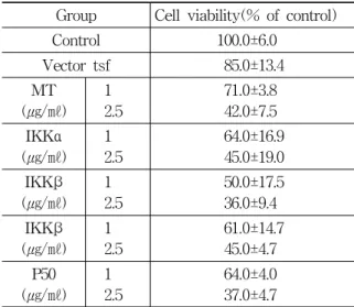
4. Mechanism of anti-apoptotic NF-κB inactivation
To confirm whether MT also inhibits NF-κB
Fig. 6. Effect of MT on cell viability in the re- presentative NF-κB signal molecule mutant plasmid transfected PC-3 cells
A fusion gene containing pCMV promoter and mutants of NF-κB signal molecules such as p50(C62S), IKKα (C178A), IKKβ(C179A), IKKβ(K44A) was used. PC-3 cells were transfected with these mutant genes constructs. Cell viability and NF-κB activity were measured after stimu- lating the cells with MT 1 and 2.5㎍/㎖ for 24hr.
Group Cell viability(% of control)
Control 100.0±6.00
Vector tsf 85.0±13.4
MT (㎍/㎖)
1 2.5
71.0±3.80 42.0±7.50 IKKα
(㎍/㎖) 1 2.5
64.0±16.9 45.0±19.0 IKKβ
(㎍/㎖) 1 2.5
50.0±17.5 36.0±9.40 IKKβ
(㎍/㎖) 1
2.5 61.0±14.7
45.0±4.70 P50
(㎍/㎖) 1 2.5
64.0±4.00 37.0±4.70
Values are the mean±SEM of 3 independent experi- ments performed in triplicate. BV and MT represents Bee venom and melittin respectively.
Table 6. Effect of MT on Cell Viability in the NF-κB Signal Molecule Transfected PC-3 Cells
through strong binding to cysteine residues with sulfhydryl group in PC-3 cells, transient trans- fection assay using a fusion gene containing pCMV promoter and mutants of NF-κB signal molecules such as p50(C62S), IKKα(C178A), IKKβ(C179A), IKKβ (K44A). PC-3 cells were transfected with these mutant genes constructs. Cell viability was mea- sured after stimulating the cells with MT(1, 2.5μg/㎖) for 24hr. Contrary to the above, present study re- vealed that MT generally inhibited proliferation dose dependently in the PC-3 cells transfected with above mutants without conspicuous reverse effect, although cell viability in IKKα and IKKβ mutant group was merely increased by 2.5μg/㎖ of MT(Fig. 6, Table 6).
Ⅳ. Discussion
Prostate cancer is the most frequently diagnosed solid tumor in American men which is responsible for 40,000 deaths per year and becomes the second leading cause of cancer-related death among men in the United States1,16). At early stage, the pro- liferation of prostate cancer cells is mainly de- pendent upon androgen and androgen ablation is generally used as a treatment17). However, androgen- independent growth occurs after all18), leading to metastasis formation in over 85% of patients with advanced prostate cancer. No effective treatments are available to manage the metastatic tumors which are responsible for mortality. The molecular mech- anisms responsible for the transition to androgen- independence are not yet clear and novel therapeutic strategies are needed to overcome the intractable advanced prostate cancer.
Most of the present available cytotoxic anti- cancer drugs mediate their effect via induction of apoptosis in cancer cells19,20) and induction of apo- ptosis is suggested as one of the major mech- anisms for the selective therapy of various cancers such as prostate cancer19-22).
Apoptosis is a mechanism by which cells ex- ecute an endogenous program of cell death in
response to various stimuli including hemothera- peutic agents, ionizing radiation, TNF-α, fas ligand, dexamethasone and other agents of stress or injury23).
The apoptosis program is mediated through multiple pathways, involving a complex array of biochemical regulators and molecular interactions.
The well defined set of biochemical and morpho- logical changes that characterize apoptosis include caspase activation, release of cytochrome c, nuclear condensation, protease activation and DNA frag- mentation24,25).
In case of advanced prostate cancer, cancer cells become resistant to apoptosis and do not respond to cytotoxic chemotherapeutic agents21). Therefore, the agents that induce apoptotic cell death in androgen refractory cancer could be useful in controlling this malignancy22).
In the present study, to ascertain the effect of BV, as an alternative to manage androgen inde- pendent cancer through induction of apoptosis, on proliferation of androgen independent PC-3 cells, apoptosis regulatory proteins, activity of NF-κB and signal molecules of NF-κB signal pathway, as well as findings from Lee et al13).’s report, low concentration of MT(2.5㎍/㎖) or BV(10㎍/㎖) inhi- bited prostate cancer cell growth time and dose- dependently, demonstrating morphologic apoptotic ch- aracteristics of the cells.
One of the most significant events in apoptosis is mitochondrial dysfunction. Loss of mitochondrial transmembrane potential(MTP) elicits the release of cytochrome c from mitochondria to cytosol26). After release, it promotes caspase-9 and -3 subsequently activating downstream caspase-327). Activated caspase- 3 cleaves intracellular protein PARP that is vital to cell survival and growth and is referred to be an important marker of apoptosis28). It has been evi- dent that the proapoptotic protein Bax plays an essential role in the onset of MTP changes and induces cytochrome c release, which, as a repre- sentative pro-apoptotic protein, generally predo- minates over the anti-apoptotic protein Bcl-229), thus Bax/Bcl-2 ratio increases during apoptosis28).
The human inhibitor of apoptosis(IAP) family in- cluding cellular IAP1(cIAP1), cIAP2, X-linked IAP (XIAP), neuronal apoptosis inhibitor protein and survivin also down regulates apoptosis as an anti- apoptotic protein30,31).
According to the result revealed about the above Anti- and pro-apoptotic effectors modulating apo- ptosis, Bax was not changed significantly by BV or MT in androgen refractory PC-3 cells inconsistent with Lee et al’s finding that Bax was dose dependently and significantly increased by MT or BV. However, in this study, Bcl-2 was significantly down regulated by MT or BV, thus Bax/Bcl-2 ratio remained increased. Pro-apoptotic cleaved caspase-3 and cleaved caspase-9 in the PC-3 cells significantly and dose-dependently increased by MT or BV, while PARP cleaved by caspase-3 subsequently increased only by MT. Anti-apoptosis related proteins including Bcl-2, XIAP, cIAP2 were all significantly decreased by MT or BV, compared with control.
From these findings, we confirmed the ability of MT or BV to exert influence upon apoptosis related proteins inducing cleaved caspase-3, cleavaged PARP, cleaved caspase-9 up-regulation and Bcl-2, XIAP, cIAP2 down regulation, suggesting MT or BV inhibit growth of androgen refractory prostate cancer PC-3 cells through induction of apoptosis via apoptosis regulatory proteins.
Recently agents inhibiting NF-κB have shown to induce apoptotic cell death, thereby inhibiting cancer cell growth9,10,32). Therefore, it was implied that agents capable of suppressing NF-κB in its signal pathway may be potentially available in the prevention and management of prostate cancer growth via induction of apoptotic prostate cancer cell death. In this study, to investigate the hypothesis whether MT or BV can inactivate NF-κ B and thereby prevent anti-apoptotic ability of NF- κB causing prostate cancer PC-3 cells undergo apoptosis, MT or BV down regulated anti-apoptotic NF-κB and its transcriptional activity significantly.
As for the mechanism how natural toxins acts with signal molecules in the NF-κB signal pathway, Son
et al14) revealed that at the molecular level, SVT inhibited constitutively activated NF-κB signaling by impairing IκBα phosphorylation with inhibition of p50 translocation and it bound with sulfhydryl group of cysteine residue in NF-κB, IKKα and IKK β resulting in down regulation of NF-κB activity19). Moreover, abolished SVT-induced apoptotic cell death was found in the cells transfected with mutant p50, IKKα and β in which the cysteine residue was replaced with other amino acids19). Inconsistent with the above report, Lee et al13). demonstrated MT or BV inhibited androgen dependent LNCaP cells proliferation and NF-κB activation. However, the reverse effect was not actually shown in the cells transfected with p50 (C62S) mutant, suggesting that BV may act with the other signal molecules in the NF-κB signal pathway and recommending further mechanism study. Thus, following Lee et al’s suggestion, to confirm whether MT inhibits NF-κB through strong binding to cysteine residues with sulfhydryl group of signal molecules of NF-κB signal pathway in androgen insensitive PC-3 cells, I performed transient transfection assay using a fusion gene containing pCMV promoter and mutants of NF-κB signal molecules such as p50(C62S), IKKα(C178A), IKKβ(C179A), IKKβ(K44A). PC-3 cells were trans- fected with these mutant genes constructs and their cell viability was measured. As a result, although cell viability in IKKα and IKKβ mutant group was merely increased by 2.5μg/㎖ of MT, cell viability was generally decreased by MT dose dependently in the PC-3 cells transfected with above mutants, implyng conspicuous reverse effect in the mutant NF-κB signal molecules’ groups was not observed in the PC-3 cells, similar with the previous re- port13).
Therefore, further study is needed to elucidate the mechanism, but the noteworthy findings in this study is the identification of anti-proliferative efficacy of BV through inactivation of NF-κB and modulation of other apoptosis related proteins in human androgen refractory prostate cancer PC-3 cells, similar with the previous report14) in hormone
dependent LNCaP cells. It suggests that BV should be one of the agents capable of inhibiting growth of prostate cancer having aptitude of transition from androgen dependent state to androgen inde- pendent state, with increasing morbidity and mor- tality, because it exerts influence upon androgen dependent and androgen refractory prostate cancer as well.
Ⅴ. References
1 1. H Gronberg. Prostate cancer epidemiology.
Lancet. 2003 ; 361 : 859-64.
2. Walsh PC, Partin AW, Epstein JI. Cancer control and quality of life following anatomical radical retropubic prostatectomy. J Uro. l994 ; 152 : 1831-6.
3. Dawson N, Moul J, Higano C. Hormone refrac- tory prostate cancer : Current issues and treat- ment options. Glenview IL : Physicians &
Scientists Publishing Co Inc. 1999 ; 45.
4. Stamey TA, McNeal JE. Campbell’s urology, 6th ed. Philadelphia : WB Saunders. 1992 : 345.
5. Kozlowski JM, Ellis WJ, Grayhack JT. Advanced prostatic carcinoma. Early versus late endocrine therapy. Urol Clin North Am. 1991 ; 18 : 15-24.
6. Isaacs JT, Lundmo PI, Berges R, Martikainen P, Kyprianou N, English HF. Androgen regulation of programmed death of normal and malignant prostatic cells. J Androl. 1992 ; 13 : 457-64.
7. Ismail M, Gomella LG. Current treatment of ad- vanced prostate cancer. Tech Urol. 1997 ; 3 : 16-24.
8. Green DR. Apoptotic pathways : Paper wraps stone blunts scissors. Cell. 2000 ; 102 : 1-4.
9. Raj GV, Sekula JA, Guo R, Madden JF, Daaka Y. Lysophosphatidic acid promotes survival of androgen-insensitive prostate cancer PC3 cells via activation of NF-kappaB. Prostate. 2004 ; 61 : 10513.
10. Syrovets T, Gschwend JE, Buchele B et al.
Inhibition of IkappaB kinase activity by acetyl-
boswellic acids promotes apoptosis in androgen- independent PC-3 prostate cancer cells
in vitro
andin vivo
. J Biol Chem. 2005 ; 280 : 6170-80.11. Cattaneo-Pangrazzi RMC, Schott H, Wunderli- Allenspach H, Rothen-Rutishauser B, Guenthert M, Schwendener RA. Cell cycle arrest and p53-independent induction of apoptosis
in vitro
by the new anticancer drugs 5-FdUrd-P- FdCydOct and dCyd-Pam-P-FdUrd in DU-145 human prostate cancer cells. J Cancer Res Clin Oncol. 2000 ; 126 : 247-56.12. Lowe SW, Lin AW. Apoptosis in cancer. Car- cinogenesis 2000 ; 21 : 485-95.
13. HS Lee, HS Song. Bee venom inhibits LNCaP cell proliferation through induction of apoptosis via inactivation of NF-κB. Journal of acupunc- ture and moxibustion 2007 ; 25(2) : 59-74.
14. DJ Son, MH Park, SJ Chae et al. Inhibitory effect of snake venom toxin from Vipera lebetina turanica on hormone-refractory human prostate cancer cell growth : induction of apoptosis through inactivation of nuclear factor κB. Mol Cancer Ther. 2007 ; 6(2) : 675-83.
15. Wolf BB, Green DR. Suicidal tendencies : apo- ptotic cell death by caspase family proteinases.
J Biol Chem. 1999 ; 274 : 20049-52.
16. American Cancer Society. Cancer facts and fig- ures 2001. Am Cancer Soc. 2001 ; 42.
17. DG Bostwick, J Qian, Effect of androgen depriv- ation therapy on prostatic intraepithelial neo- plasia. Urology. 2001 ; 58 : 91-3.
18. BJ Feldman, D Feldman, The development of androgen-independent prostate cancer. Nat Rev Cancer. 2001 ; 1 : 34-45.
19. Lowe SW, Lin AW. Apoptosis in cancer. Car- cinogenesis. 2000 ; 21 : 48595.
20. Guseva NV, Taghiyev AF, Rokhlin OW, Cohen MB. Death receptor-induced cell death in prostate cancer. J Cell Biochem. 2004 ; 91 : 7099.
21. Gurumurthy S, Vasudevan KM, Rangnekar VM.
Regulation of apoptosis in prostate cancer. Can- cer Metastasis Rev. 2001 ; 20 : 22543.
22. Kantoff PW. New agents in the therapy of hormone-refractory patients with prostate
cancer. Semin Oncol 1995 ; 22(1 Suppl 1) : 324.
23. Isaacs JT. Apoptosis: Translating theory to therapy for prostate cancer. J Natl Cancer Inst.
2000 ; 92 : 1367-9.
24. Ripple GH, Wilding G. Drug development in prostate cancer. Semin Oncol. 1999 ; 26 : 217- 26.
25. Green DR. Apoptotic pathways : Paper wraps stone blunts scissors. Cell. 2000 ; 102 : 1-4.
26. Thornberry NA and Lazbrik Y. Caspases : en- emies within. Science. 1998 ; 281 : 1312-6.
27. Darmon AJ, Nicholson DW and Bleackley RC.
Activation of the apoptotic protease CPP32 by cytotoxic T cell derived granzyme B Nature.
1995 ; 377 : 446-8.
28. Sedlak TW, Oltvai ZN, Yang E et al. Multiple
Bcl2 family members demonstrate selective dimerization with Bax. Proc Natl Acad Sci USA. 1995 ; 92 : 7834-8.
29. Green DR and Reed JC. Mitochondria and Apo- ptosis. Science. 1998 ; 281 : 1309-12.
30. LaCasse EC, Baird S, Korneluk RG, MacKenzie AE. The inhibitors of apoptosis (IAPs) and their emerging role in cancer. Oncogene. 1998 ; 17 : 3247-59.
31. Deveraux QL, Reed JC. IAP family proteins- suppressors of apoptosis. Genes Dev. 1999 ; 13 : 239-52.
32. Li Y, Sarkar FH. Inhibition of nuclear factor kappaB activation in PC3 cells by genistein is mediated via Akt signaling pathway. Clin Cancer Res. 2002 ; 8 : 2369-77.