Background: The purpose of this study was to investigate the efficacy of muscle energy techniques (MET) of upper trapezius and sternocleidomastoid muscles on Bell’s palsy.
Methods: In this retrospective study, we screened the medical records of patients with Bell’s palsy who had received inpatient and outpatient treatment at the Department of Acupuncture & Moxibustion, Korean Medicine Hospital Dong-Eui University between November 28, 2016 and April 30, 2017. A total of 34 out of 93 Bell’s palsy patients met the inclusion criteria. The 34 patients were divided into two groups: Group A patients had undergone Korean–Western combination treatment and MET of upper trapezius and sternocleidomastoid muscles; Group B patients had undergone Korean–Western combination treatment only.
MET of upper trapezius and sternocleidomastoid muscles had been performed three times a week during the inpatient period, and two to three times a week during the outpatient period. Yanagihara scores had been assessed at the first visit, and 1, 2, 3, and 4 weeks after the first visit.
Results: Group A Yanagihara scores were significantly improved during each interval from the first visit to 4 weeks later. Group B Yanagihara scores were also significantly improved except during the first week. During every period, the improvements observed in Yanagihara score were significantly higher in Group A than in Group B.
Conclusion: These results suggest that MET of upper trapezius and sternocleidomastoid muscles may be effective treatment for Bell’s palsy.
©2017 Korean Acupuncture & Moxibustion Medicine Society. This is an open access article under the CC BY- NC-ND license (http://creativecommons.org/licenses/by-nc-nd/4.0/).
Article history:
Submitted: June 27, 2017 Revised: August 9, 2017 Accepted: August 22, 2017 Keywords:
Bell’s palsy, muscle energy technique, sternocleidomastoid muscle, upper trapezius Original Article
Effects of Muscle Energy Technique of Upper Trapezius and Sternocleidomastoid Muscles on Bell’s Palsy
Jong-Hyeon Park
1, Yoon-Joo Lee
2, Hye-Min Ryu
1, Seung-Jeong Lee
1, Eun-Jin Park
1, Choon-Ho Song
1, Cheol-Hong Kim
1, Hyun-Min Yoon
1,*
1 Department of Acupuncture & Moxibustion Meridian & Acupoint, College of Korean Medicine, Dong-Eui University, Busan, Korea 2 Department of Acupuncture & Moxibustion Meridian & Acupoint, College of Korean Medicine, Dong-Eui University, Ulsan, Korea
ARTICLE INFO ABSTRACT
Journal of Acupuncture Research
Journal homepage: https://www.e-jar.org
*
Corresponding author.Department of Acupuncture & Moxibustion Meridian & Acupoint, College of Korean Medicine, Dong-Eui University, 62 Yangjeong-ro, Busanjin-gu, Busan 47227, Korea E-mail: [email protected]
https://doi.org/10.13045/jar.2017.02131 pISSN 2586-288X eISSN 2586-2898
©2017 Korean Acupuncture & Moxibustion Medicine Society, Published by E-Tree Publishing. This is an open access article under the CC BY-NC-ND license (http://creativecommons.org/licenses/by-nc-nd/4.0/).
Introduction
Facial nerves are mixed nerves consisting of motor, sensory, and parasympathetic nerves [1]. Facial nerves are also involved in facial muscle movement, tear and saliva secretion, tympanum protection, and taste sensation by the tongue [2].
Infra-nuclear facial nerve palsy without clear causes is referred to as idiopathic Bell’s palsy [3] and represents the most common facial nerve disorder, with a prevalence of 11–40 cases per 100,000 persons. The incidence of Bell’s palsy does not vary among different sex and age groups, but has been reported to be higher among patients with diabetes [2].
Vascular ischemia, viruses, genetic factors, and autoimmunity have all been highlighted as possible causes of Bell’s palsy. Thus far,
vascular ischemia appears to be the most likely cause of Bell’s palsy, and cold exposure, emotional shock, and anxiety are known to cause circulatory disturbance [4]. According to Korean medicine, Bell’s palsy occurs when the harmful energy of a cold invades the meridian system in a weak state, as well as when stasis stagnates the meridian system, obstructs qi and blood, and blocks the flow of nutrients into muscles and tendons [3].
The major clinical symptoms of Bell’s palsy include loss of
wrinkles in the forehead due to facial nerve paralysis on one side
of the face, epiphora due to an inability to close the eyes, ocular
discomfort, and lip distortion toward the contralateral side. Minor
symptoms include impaired taste at the tip of the tongue, sound
sensitivity, and ringing in the ears [5]. Approximately 60% of
patients with Bell’s palsy experience pain in the ipsilateral mastoid
as a preceding symptom, and severe paralysis occurs within the next 48 hours [6].
The muscle energy technique (MET) is a form of chuna manual treatment on fascia and is based on active muscle relaxation techniques that use the intrinsic energy of the muscles to relax and stretch stiff muscles [7]. Compared to other manipulative therapies, MET has a wider range of applications for treating the nervous and musculoskeletal systems, proceeds smoothly, and allows patients to actively participate in the treatment process [8]. The efficacy of MET has been proven both experimentally and clinically [8].
While manual therapy has been undergoing a change from techniques using fast and weak twisting movements to softer techniques that focus more on soft tissues [8], there have not been any clinical reports on the treatment of Bell’s palsy using MET.
Therefore, in this study, we retrospectively compared patients with Bell’s palsy who received Korean–Western combination treatment and MET of upper trapezius and sternocleidomastoid (SCM) muscles with those who received Korean–Western combination treatment only.
Materials and Methods Patients
We reviewed the medical records of patients who visited the Department of Acupuncture & Moxibustion, Korean Medicine Hospital Dong-Eui University who were diagnosed with Bell’s palsy, and who had received inpatient and outpatient treatment between November 28, 2016 and April 30, 2017. Approval for the study was received from the institutional review board of Korean Medicine Hospital Dong-Eui University (IRB no. 2017-02).
The inclusion criteria were: (1) patients had undergone more than 4 weeks of treatment, including 7 days of inpatient treatment, following the initial diagnosis of Bell’s palsy; (2) patients were aged 20–70 years; (3) patients had presented to hospital within 10 days of the onset of Bell’s palsy; (4) patients had no previous history of stroke or cancer; and (5) patients had never received insulin injections for diabetes.
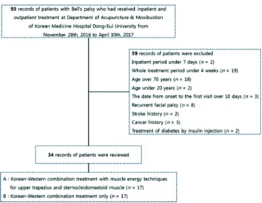
Of the 93 patients with Bell’s palsy who were treated during the study period, 59 were excluded as they did not meet all of the inclusion criteria. Of the 34 patients who were included in this study, 17 received Korean–Western combination treatment and MET of upper trapezius and SCM muscles (Group A), and 17 received Korean–Western combination treatment only (Group B) (Fig. 1).
Treatments Acupuncture
Disposable stainless steel needles (0.20 mm in diameter, 30.00 mm in length; Dong Bang Medical Co. Ltd., Daecheon, Chungcheongnam-do, Korea) were used to apply acupuncture twice daily during inpatient treatment, and two to three times a week during outpatient treatment. Acupuncture was applied on Chanjuk (BL2), Yangbaek (GB14), Sajukgong (TE23), Seungeub (ST1), Yeonghyang (LI20), Hagwan (ST7), Gwollyo (SI18), Sugu (GV26), Seungjang (CV24), Jichang (ST4), Hyeopgeo (ST6), and Yepung (TE17) on the ipsilateral side, and the stomach–
tonification point on the contralateral side for 20 minutes.
Pharmacopuncture
The 10% sweet bee venom used in this study was produced at an extramural herbal dispensary of the Korean Pharmacopuncture Research Institute. A 1.0 mL disposable insulin syringe (29 G
Fig. 1. Flowchart showing how patients were selected for inclusion.
× 1/2 inch; Shin Chang Medical Co. Ltd., Gumi, Korea) was used to inject the solution in Chanjuk (BL2), Yangbaek (GB14), Sajukgong (TE23), Seungeub (ST1), Yeonghyang (LI20), Hagwan (ST7), Gwollyo (SI18), Sugu (GV26), Seungjang (CV24), Jichang (ST4), Hyeopgeo (ST6), and Yepung (TE17) on the ipsilateral side.
Separate injections were carried out to administer 0.05–0.10 mL of the solution per acupoint; total volume of solution injected per treatment was 0.5 mL. Pharmacopuncture therapy was performed once daily during inpatient treatment, and two to three times a week during outpatient treatment.
Herbal medicine
Depending on patients’ symptoms, Igigeopung Powder, Bohyeol Decoction, Ikgi Decoction, and Geopung Decoction were administered (Table 1).
Western examination and treatment
All patients were diagnosed with Bell’s palsy based on neurological test results. A nerve conduction velocity (NCV) test was performed 7–10 days after the onset of Bell’s palsy to measure the severity of paralysis. Patients were orally administered high-dose steroids for the first 5 days. The dose was gradually reduced over the course of the next 5 days. Patients who refused pharmacotherapy or who were at high risk of side effects were excluded from steroid administration. Antiviral agents, drugs that improved blood circulation, and vitamins were also administered.
To prevent corneal damage, an artificial solution and eye ointment were applied. Patients received facial massage and electrotherapy at a physical therapy center once daily (except during holidays) throughout the period of hospitalization.
MET of upper trapezius and SCM muscles
An acupuncture specialist performed MET on patients three times a week during the inpatient period and two to three times a week during the outpatient period. The precise protocols used are described below.
MET of the upper trapezius muscle: With the patient in the
supine position, the therapist supported the back of the patient’s
seconds later, and the area where the contact hand was placed was diagonally pushed toward the foot on the same side to stretch the muscles while the patient was in a relaxed state. This process was repeated three to four times [7].
Methods Data collection
Initial diagnosis, treatment progress, and NCV test results from patients’ medical records were reviewed.
Facial paralysis assessment
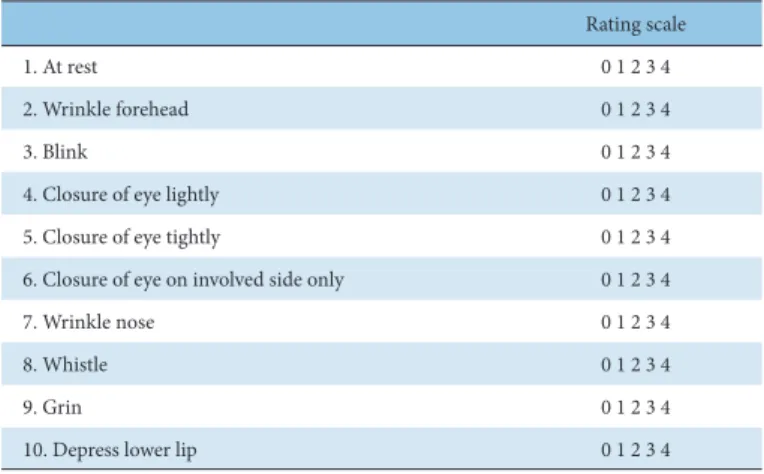
We used the Yanagihara unweighted grading system (Y-system, Table 2) to assess the severity of facial paralysis. Facial paralysis measurements were taken on the first visit (P0), and 1 (P1), 2 (P2), 3 (P3), and 4 (P4) weeks after the first visit. The Y-system is an assessment method that classifies facial function into 10 different domains without weighting any values. Using the Y-system, facial paralysis can be easily and quickly assessed and quantified without the use of special equipment, while avoiding subjective errors.
For these reasons, the Y-system is the most commonly used of all regional scales [9].
Statistical analyses
All statistical analyses were performed with SPSS version 18.0 for Windows (SPSS Inc., Chicago, IL, USA). Results are expressed as means ± standard deviations. A Kolmogorov-Smirnov test was used to assess data for normality.
To test the homogeneity of categorical data for Group A and Group B, the Chi-square test was used for data with a normal distribution, and Fisher’s exact test was used for data that did not follow a normal distribution. Student’s t-test was used to test the homogeneity of continuous data that were normally distributed, and the Mann–Whitney U test was used for continuous data that were not normally distributed. The level of statistical significance was set at p < 0.05.
The significance of improvements in patient condition at different treatment intervals (P0 to P1, P1 to P2, P2 to P3, P3 to P4) within each group was tested with a paired t-test following a test for normality (p < 0.001). The significance of the differences in improvement in patient condition in each treatment period (P0 to P1, P0 to P2, P0 to P3, P0 to P4) between Group A and Group B was tested using Student’s t-test following a test of normality (p <
0.05).
head with his contact hand, and placed his stabilizing hand on the patient’s clavicle, which is where the insertion site of the upper trapezius muscle is located. The contact hand was used to bend the patient’s neck forward, then rotate and bend it toward the ipsilateral side to check for an elastic barrier, and then to place it at a mid-point between the initial position and the fully rotated position. The patient was then asked to breathe in, hold their breath, and perform isometric contraction (20% of maximal force) in the direction that would allow the muscle’s point of origin to get closer to its insertion, while the therapist pressed on the patient in the opposite direction. The patient was then asked to breathe out 6–7 seconds later, and the muscles—including those with an elastic barrier—were stretched while the patient was in a relaxed state.
This process was repeated three to four times [7].
MET of the SCM muscle: The patient lay in the supine position with a cushion under the shoulder to keep the head slightly bent backward onto the bed [8]. The therapist placed his contact hand on the ipsilateral mastoid on the temporal region, which is the insertion site of the SCM muscle, and his stabilizing hand on the sternum, which is the point of origin. The patient was asked to rotate his head to the contralateral side and lift his head while holding his breath, while the therapist pressed on the patient in the opposite direction. The patient was then asked to breathe out 6–7
Igigeopung Powder
Root or rhizome of Saposhnikovia Radix·Root of Paeoniae Radix Alba (baked with alcohol)·Root of Angelicae Dahuricae Radix·Unripe pericarp of Citrii Unshiu Immaturi Pericarpium·Rhizome of Cnidii Rhizoma·Tuberous root of Gastrodiae Rhizoma·Root of Angelicae Pubescentis Radix·Rhizome of Notopterygii Rhizoma·Root of Platycodi Radix·Root of Glycyrrhizae Radix·Floral axis of Schizonepetae Spica·Fruit of Ponciri Fructus Pericarpium·Old peel of Citri Reticulatae Pericarpium·Tuberous root of Arisaematis Rhizoma·Root of Linderae Radix 5 g, Tuberous root of Typhonii Tuber·Bombycis Corpus cum Batryticatus 4 g
Bohyeol Decoction
Honey roasted root of Astragali Radix 8 g, Fruit of Jujubae Fructus 7 g, Sclerotium of Poria (Hoelen)·Root of Atractylodis Rhizoma Alba·Root of Angelicae Gigantis Radix·Roasted root of Glycyrrhizae Radix·Old peel of Citri Reticulatae Pericarpium·Root of Ginseng Radix·Rhizome of Zingiberis Rhizoma Recens·Root of Rehmanniae Radix Preparat 4 g, Bombycis Corpus cum Batryticatus·Root or rhizome of Saposhnikovia Radix·Tuberous root of Pinelliae Rhizoma·Rhizome of Notopterygii Rhizoma·Root of Gentianae Macrophyllae Radix·Tuberous root of Arisaematis Rhizoma 3 g, The whole of Scorpio·Alcohol-washed rhizome of Cimicifugae Rhizoma·Alcohol-washed root of Bupleuri Radix 1 g
DecoctionIkgi
Sclerotium of Poria (Hoelen)·Baked root of Atractylodis Rhizoma Alba·Root of Paeoniae Radix Alba·Rhizome of Cnidii Rhizoma·Root of Angelicae Gigantis Radix·Roasted root of Glycyrrhizae Radix·Honey roasted root of Astragali Radix·Old peel of Citri Reticulatae Pericarpium·Root of Ginseng Radix·Root of Rehmanniae Radix Preparat 6 g, Bombycis Corpus cum Batryticatus·Root or rhizome of Saposhnikovia Radix·Tuberous root of Pinelliae Rhizoma·Rhizome of Notopterygii Rhizoma·Root of Gentianae Macrophyllae Radix·Tuberous root of Arisaematis Rhizoma·Rhizome of Cimicifugae Rhizoma·Root of Bupleuri Radix 4 g, The whole of Scorpio 2 g
Geopung Decoction
Sclerotium of Poria (Hoelen)·Root of Polygoni Multiflori Radix 8 g, Rhizome of Cnidii Rhizoma·Pericarp of Aurantii Pericarpium·Rhizome of Cyperi Rhizoma·Root of Linderae Radix 6 g, Tuberous root of Pinelliae Rhizoma (treated with ginger juice)·Rhizome of Atractylodis Rhizoma·Root of Angelicae Gigantis Radix·Branch of Cinnamomi Ramulus·Root of Paeoniae Radix Rubra·Root of Ginseng Radix·Root of Achyranthis Radix 4 g, Root of Aucklandiae Radix 3 g·Root of Glycyrrhizae Radix 2 g
Table 1. Herbal Medicine Prescriptions Table 2. The Yanagihara Unweighted Grading System
Rating scale
1. At rest 0 1 2 3 4
2. Wrinkle forehead 0 1 2 3 4
3. Blink 0 1 2 3 4
4. Closure of eye lightly 0 1 2 3 4
5. Closure of eye tightly 0 1 2 3 4
6. Closure of eye on involved side only 0 1 2 3 4
7. Wrinkle nose 0 1 2 3 4
8. Whistle 0 1 2 3 4
9. Grin 0 1 2 3 4
10. Depress lower lip 0 1 2 3 4
Results
General characteristics
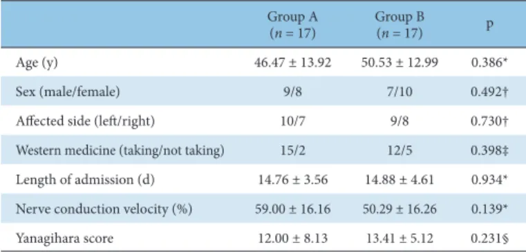
The data extracted from the medical records of the 34 patients who were included in this study included age, sex, affected side, whether or not they took Western medicine, length of admission, NCV, and Yanagihara score. There were no statistically significant differences in any of these characteristics between Groups A and B (Table 3).
Causes of paralysis
Several causes of facial nerve paralysis were recorded: overwork, stress, exposure to cold, common cold, and idiopathic (Table 4).
Multiple responses were allowed. Overwork and stress were the most common causes.
Symptoms at onset
The symptoms reported by patients at disease onset are shown in Table 5. Multiple responses were allowed. Almost two-thirds of patients experienced postauricular pain and lacrimation.
Yanagihara score
The Yanagihara scores for Groups A and B at P0, P1, P2, P3, and P4 are shown in Table 6. The treatment intervals between each of these time points were defined as follows: from the first visit to 1 week later (P01), from 1 week to 2 weeks later (P12), from 2 weeks to 3 weeks later (P23), and from 3 weeks to 4 weeks later (P34).
Within-group changes
Significant improvements in Yanagihara score were observed at all time intervals in Group A. In Group B however, significant improvements in Yanagihara score were observed at all time intervals except for P01 (Table 7).
Between-group comparison
Changes in Yanagihara score at each time period were calculated as differences in the score compared to that at the first visit.
Significantly greater improvements were observed in Group A than in Group B for all time periods (Table 8).
Table 3. General Characteristics of Patients
Data are presented as mean ± SD.
*Student’s t-test.
†Chi-square test.
‡Fisher’s exact test.
§Mann–Whitney U test.
Group A
(n = 17) Group B
(n = 17) p
Age (y) 46.47 ± 13.92 50.53 ± 12.99 0.386*
Sex (male/female) 9/8 7/10 0.492†
Affected side (left/right) 10/7 9/8 0.730†
Western medicine (taking/not taking) 15/2 12/5 0.398‡
Length of admission (d) 14.76 ± 3.56 14.88 ± 4.61 0.934*
Nerve conduction velocity (%) 59.00 ± 16.16 50.29 ± 16.26 0.139*
Yanagihara score 12.00 ± 8.13 13.41 ± 5.12 0.231§
Table 6. Yanagihara Score
Data are presented as mean ± SD.
P0, the first visit; P1, 1 week after the first visit; P2, 2 weeks after the first visit; P3, 3 weeks after the first visit; P4, 4 weeks after the first visit.
Group A Group B
P0 12.00 ± 8.13 13.41 ± 5.12
P1 17.76 ± 6.34 13.59 ± 7.14
P2 25.59 ± 6.58 20.18 ± 7.69
P3 31.06 ± 6.52 26.59 ± 8.27
P4 35.12 ± 5.31 30.18 ± 8.14
Table 4. Causes of Paralysis*
*Multiple responses were allowed.
Cause Group A (n) Group B (n) Total (n)
Overwork 7 8 15
Stress 7 8 15
Exposure to cold 6 5 11
Common cold 3 2 5
Unknown 4 5 9
Table 5. Symptoms at Onset*
*Multiple responses were allowed.
Symptom Group A (n) Group B (n) Total (n)
Postauricular pain 9 13 22
Lacrimation 10 11 21
Scheroma 9 9 18
Hyperacusis 2 3 5
Hypoacusis 0 1 1
Dysgeusia 5 5 10
Tinnitus 3 3 6
Dizziness 1 1 2
Other 2 1 3
Discussion
The motor nerve fibers from the motor nucleus of the facial nerve assist in controlling facial expressions and making the movements that are necessary for speech and mastication.
Secretomotor fibers of the facial nerve, which originate from the superior salivary nucleus, regulate the secretory activities of the submandibular glands, sublingual glands, lachrymal glands, olfactory mucosa, and palatine mucosa. Special visceral afferent fibers of the facial nerve, which originate from the nucleus tractus solitarius, occupy two-thirds of the anterior region of the tongue and palate. They are responsible for taste sensation in the anterior two-thirds of the tongue and sensation in the anterior two-thirds of the soft palate [1].
Facial nerve paralysis can be classified as central or peripheral [3], and differentiating between the two types of paralysis is important.
As the frontalis muscle is controlled by both hemispheres of
the cerebrum, facial nerve paralysis due to upper motor neuron
lesions affects the bottom two-thirds of the face only. In contrast,
peripheral facial nerve lesions not only paralyze the muscles
involved in the closing of the mouth and eyes but also affect the frontalis muscle, thus obscuring wrinkles on the forehead [10]. Peripheral facial nerve paralysis can be classified as nuclear paralysis or infra-nuclear paralysis. Infra-nuclear facial nerve paralysis without a clear cause is referred to as Bell’s palsy [3].
Bell’s palsy suddenly occurs without any evident causes or injuries. The most likely cause of Bell’s palsy is vascular ischemia.
It has been hypothesized that constriction of the arterial vessels interferes with blood flow into the facial nerves, thus causing the nerves within the facial canal to swell and become pressurized, consequently leading to paralysis [4].
Postauricular pain is the most common early symptom of Bell’s palsy [11]. When the patient tries to firmly close the eye on the paralyzed side of the face, the pupil of the eye moves abnormally upward relative to the pupil of the other eye; this symptom is referred to as Bell’s phenomenon [10]. Patients with Bell’s palsy may not be able to wrinkle their forehead or whistle, and food may collect within the mouth on the ipsilateral side such that fluids leak from the mouth. If the chorda tympani nerve becomes affected, taste sensation may become impaired in two-thirds of the tongue tip. If the stapedius muscle becomes affected, sound sensitivity may also occur.
Aside from acupuncture and herbal medicine, Korean medical treatments include electroacupuncture [12], pharmacopuncture [13], embedding therapy [14], acupotomy [15], and chuna manual therapy [16,17]. These therapies have been clinically practiced, and their efficacy has been clinically proven. In Western medicine, the administration of steroids and antiviral agents, physical therapy, and surgery are used [5].
MET stretches shortened muscles to relieve tenderness and pain, and to recover the original state of the muscles. This not only removes trigger points within the muscles, but also relieves pain in the ligament or periosteum at the attachment point. In a study by Lewit and Simons, MET was effective for 330 out of 351 muscles, muscle relaxation was observed in 63% of all patients in the second
examination 3 months later, and pain relief was achieved in 23% of muscles [18].
With regard to the mechanism underlying MET, during isometric exercise, in which the forces applied by the therapist and the patient onto the agonistic muscle match, reciprocal inhibition occurs in the antagonistic muscle, and post-isometric relaxation occurs in the agonistic muscle due to physiological and neurological reactions. Thereby, contracted muscles and tissues can be stretched and relaxed.
MET resembles chuna manual therapy for the meridian muscle in Korean medicine [19], which applies stimuli onto the surface of the patient’s body to control the meridian system, to prevent and treat diseases. Therapeutic effects are achieved through correction of particular parts of the body such as joints and the skeletal structure using manual force [20].
The effectiveness of chuna manual therapy for treating Bell’s palsy has been reported in a clinical study by Park et al [16], in which the danmuji anchu traction technique was performed, and in a clinical study by Jeong et al [17], in which JS supine cervical distraction, supine cervical manipulation, and cranial base release were performed. These chuna manual therapy techniques for Bell’s palsy fall under the areas of chuna joint distraction therapy, chuna spine and joint manipulation therapy, and chuna craniosacral therapy. However, clinical research on fascia chuna therapy for Bell’s palsy is currently lacking.
The Department of Acupuncture & Moxibustion, Korean Medicine Hospital Dong-Eui University has been performing MET of upper trapezius and SCM muscles associated with facial nerve disorders to treat Bell’s palsy. Significantly better treatment outcomes have been observed in patients who receive Korean–
Western combination treatment and MET compared to patients who receive Korean–Western combination treatment only. In this retrospective analysis of the medical records of 34 patients who were diagnosed with Bell’s palsy and who met the criteria for inclusion into the study, overwork and stress were the most common causes of paralysis, followed by cold exposure, unknown causes, and the common cold. Postauricular pain was the most common early symptom at onset, followed by lacrimation, scheroma, dysgeusia, tinnitus, hyperacusis, dizziness, and hypoacusis.
In Group A (patients who had received Korean–Western combination treatment and MET of upper trapezius and SCM muscles), Yanagihara scores were significantly improved at all time intervals (P01, P12, P23, P34). Yanagihara scores were also significantly improved in Group B (patients who had received Korean–Western combination treatment only) at all time intervals except for P01. For each of the time periods P01, P02, P03 and P04, the improvements observed in Yanagihara score were significantly greater in Group A than in Group B. Based on these results, the treatment of Bell’s palsy with MET of upper trapezius and SCM muscles may be beneficial. The theoretical background on the application of MET on these two muscles is given below.
First, the effects of treatment on the SCM muscle on Bell’s palsy can be considered from an anatomical perspective. To begin with, its effects on the autonomic nervous system can be considered.
The SCM muscle is anatomically closely associated with the stellate ganglion, which is a central ganglion through which sympathetic nerves that travel to the head, neck, and chest pass [21], and carotid arteries and the SCM muscle are ventrally distributed [22].
The Gisa (ST11) of the stomach meridian is an acupuncture point located on the inner side of the SCM muscle and corresponds anatomically to the stellate ganglion. Trigger points of the SCM muscle cause symptoms of autonomic nerve disorders such as headache, dizziness, and nausea [23], possibly by contracting to
Table 8. Between-Group Comparison of Changes in Yanagihara Score*
Data are presented as mean ± SD.
*Student’s t-test.
P01, interval from the first visit to 1 week later; P02, interval from the first visit to 2 weeks later; P03, interval from the first visit to 3 weeks later; P04, interval from the first visit to 4 weeks later.
Group A Group B t p
P01 5.76 ± 4.89 0.18 ± 6.14 2.936 0.006
P02 13.59 ± 6.37 6.76 ± 6.08 3.197 0.003
P03 19.06 ± 7.47 13.18 ± 7.25 2.330 0.026
P04 23.12 ± 7.72 16.76 ± 6.33 2.624 0.013
Table 7. Within-Group Changes in Yanagihara Score*
*Paired t-test.
P01, interval from the first visit to 1 week later; P12, interval from 1 week to 2 weeks later; P23, interval from 2 weeks to 3 weeks later; P34, interval from 3 weeks to 4 weeks later.
t p t p
P01 4.858 < 0.001 0.119 0.907
P12 10.250 < 0.001 5.378 < 0.001
P23 6.344 < 0.001 5.684 < 0.001
P34 4.976 < 0.001 4.999 < 0.001
Group A Group B