Brief Report
Vol. 29, No. 2, 2017 229
Received December 14, 2015, Revised February 23, 2016, Accepted for publication April 4, 2016
Corresponding author: Takeshi Namiki, Department of Dermatology, Graduate School of Medical and Dental Sciences, Tokyo Medical and Dental University, 1-5-45, Yushima, Bunkyo-ku, Tokyo 113-8510, Japan. Tel: 81-3-5803-5286, Fax: 81-3-5803-0143, E-mail: tnamderm@tmd.ac.jp
This is an Open Access article distributed under the terms of the Creative Commons Attribution Non-Commercial License (http://creativecommons.org/
licenses/by-nc/4.0) which permits unrestricted non-commercial use, distribution, and reproduction in any medium, provided the original work is properly cited.
Copyright © The Korean Dermatological Association and The Korean Society for Investigative Dermatology
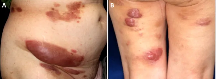
Fig. 1. (A) Multiple erythematous tumors and plaques on the patient’s trunk. The largest tumor was observed at the right lower abdomen.
(B) Multiple erythematous tumors on the lower extremities.
https://doi.org/10.5021/ad.2017.29.2.229
A Case of Primary Cutaneous Gamma-Delta T-Cell Lymphoma with Pautrier Microabscess
Kohei Kato, Takeshi Namiki, Makiko Ueno, Madoka Iikawa, Shown Tokoro, Aya Nishizawa, Kouhei Yamamoto
1, Keiko Miura
1, Hiroo Yokozeki
Departments of Dermatology and 1Pathology, Graduate School of Medical and Dental Sciences, Tokyo Medical and Dental University, Tokyo, Japan
Dear Editor:
Primary cutaneous gamma-delta T-cell lymphoma (PC- GD-TCL) accounts for less than 1% of primary cutaneous T-cell lymphomas (CTCLs). PCGD-TCL is characterized by a highly aggressive clinical course, while mycosis fungoides (MF) has a relatively favorable prognosis1,2. PCGD-TCL is recognized for its complexity and hetero- geneity3. Here we present a case of PCGD-TCL with Pautrier microabscess.
A 69-year-old female presented with erythematous tumors and plaques on her trunk and extremities. Initially, eryth-
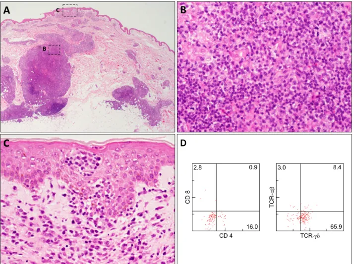
emas and plaques had been noticed at her lower ex- tremities and had spread to her trunk and upper ex- tremities in the past year and a half. Physical examination revealed multiple erythematous tumors and plaques on her trunk and extremities (Fig. 1). Histopathological ex- amination from an erythematous plaque showed focal in- filtrates of atypical lymphocytes in the dermis (Fig. 2A, B).
Epidermotropic infiltrates of atypical lymphocytes and ag- gregations of atypical lymphocytes as Pautrier’s micro- abscess in the epidermis were recognized (Fig. 2C). Flow cytometric analysis and immunohistochemistry both showed
Brief Report
230 Ann Dermatol
Fig. 2. (A) Histology of a biopsy specimen of an erythematous plaque showing focal infiltrates of atypical lymphocytes in the dermis.
The dotted boxes indicate the locations shown at higher magnification in Fig. 1B and 1C (H&E, ×20). (B) Infiltrates of atypical lymphocytes intermingled with histiocytes in the dermis (H&E, ×400). (C) Aggregations of atypical lymphocytes as Pautrier’s mi- croabscess in the epidermis (H&E, ×400). (D) Flow cytometry analyses of a biopsy specimen from the skin using a CD45/SSC gating procedure. CD45/SSC gated cells with various surface antigens of CD4, CD8, T-cell receptor (TCR)-αβ, TCR-γδ shown in two dot plots. (E) Immunohistochemistry of a biopsy specimen from the skin showing a positive reaction forCγM1 (×400). (F) Immunohistochemistry of a biopsy specimen from the skin showing a negative reaction for βF1 (×400). (G) Immunohistochemistry from a lymph node showing a positive reaction for CγM1 (×400).
positive reactions for CD2, CD3 and CD5, a partially pos- itive reaction for CD4, and negative reactions for CD7, CD8, CD20 and CD56 in atypical lymphocytes (Fig. 2D).
A positive reaction for CγM1 and negative reactions for T-cell restricted intracellular antigen-1 and granzyme B were demonstrated by immunohistochemistry (Fig. 2E).
Atypical lymphocytes in the epidermis were positive for C γM1 and negative for βF1 (Fig. 2F). Positron emission tomography revealed multiple accumulation sites with flu- deoxyglucose in the skin and inguinal lymph nodes. His- topathological examination of a lymph node showed me- dium to large sized atypical lymphocytes. Flow cytometric analysis and immunohistochemistry of the lymph node both showed identical results with the skin in addition to a
positive reaction for CγM1 by immunohistochemistry (Fig. 2G). Southern blots showed TCR gene rearrangements by probes of Jγ. A diagnosis of PCGD-TCL was made.
Treatments with radiation and cyclophosphamide-hydroxy- daunorubicin-oncovin-prednisone (CHOP) had diminished most tumors and plaques, but the tumors recurred. CHOP therapy excluding oncovin due to peripheral neuropathies was effective, and most tumors and plaques have been un- der control for two years since the start of the initial therapy.
Although PCGD-TCL and MF should be distinct clinical entities in terms of their histogenesis, an accurate diag- nosis of PCGD-TCL is sometimes challenging due to the aberrant expression of surface markers in neoplastic cells
Brief Report
Vol. 29, No. 2, 2017 231 Fig. 2. Continued.
and technical complexities such as insufficient antibodies that work in paraffin-embedded sections. A previous study of 146 CTCLs showed that 5 PCGD-TCLs and 2 MFs were included in 12 CTCLs with T-cell receptor-γ expression3. Another study of 53 PCGD-TCLs using the same antibody showed that the clinical and histopathological features of 6 cases were similar to those of MF4. Further, PCGD-TCL following MF has also been reported5. Those studies and reports may suggest the heterogeneity of PCGD-TCL. In our case, erythematous plaques were observed in addition to tumors and an epidermotropism as Pautrier micro- abscess was recognized. Those clinical and histopatho- logical findings may confuse an accurate diagnosis.
Detailed analyses of such cases will lead to an accurate di- agnosis and the elucidation of the heterogeneity of PCGD-TCL.
ACKNOWLEDGMENT
We thank C. Miyagishi (Pathology Staff Members) for her
technical assistance.
CONFLICTS OF INTEREST
The authors have nothing to disclose.
REFERENCES
1. Toro JR, Liewehr DJ, Pabby N, Sorbara L, Raffeld M, Steinberg SM, et al. Gamma-delta T-cell phenotype is associated with significantly decreased survival in cutaneous T-cell lymphoma. Blood 2003;101:3407-3412.
2. Tripodo C, Iannitto E, Florena AM, Pucillo CE, Piccaluga PP, Franco V, et al. Gamma-delta T-cell lymphomas. Nat Rev Clin Oncol 2009;6:707-717.
3. Rodríguez-Pinilla SM, Ortiz-Romero PL, Monsalvez V, Tomás IE, Almagro M, Sevilla A, et al. TCR-γ expression in primary cutaneous T-cell lymphomas. Am J Surg Pathol 2013;37:375-384.
4. Guitart J, Weisenburger DD, Subtil A, Kim E, Wood G,
Brief Report
232 Ann Dermatol
Received February 2, 2016, Revised March 31, 2016, Accepted for publication April 4, 2016
Corresponding author: Kee Suck Suh, Department of Dermatology, Kosin University College of Medicine, 262 Gamcheon-ro, Seo-gu, Busan 49267, Korea. Tel:
82-51-990-6145, Fax: 82-51-990-3041, E-mail: ksderm77@unitel.co.kr
This is an Open Access article distributed under the terms of the Creative Commons Attribution Non-Commercial License (http://creativecommons.org/
licenses/by-nc/4.0) which permits unrestricted non-commercial use, distribution, and reproduction in any medium, provided the original work is properly cited.
Copyright © The Korean Dermatological Association and The Korean Society for Investigative Dermatology Duvic M, et al. Cutaneous γδ T-cell lymphomas: a spectrum
of presentations with overlap with other cytotoxic lymphomas.
Am J Surg Pathol 2012;36:1656-1665.
5. Suga H, Sugaya M, Miyagaki T, Ohmatsu H, Fujita H, Sato S.
Primary cutaneous γδ T-cell lymphoma following mycosis fungoides. Int J Dermatol 2014;53:e82-e84.
https://doi.org/10.5021/ad.2017.29.2.232
Granulocyte Colony-Stimulating Factor-Induced Psoriasiform Dermatitis Improved by Narrowband Ultraviolet B
Min Soo Jang, Jong Bin Park, Joon Hee Kim, Myeong Hyeon Yang, Kang Hoon Lee, Sang Hwa Han, Kee Suck Suh
Department of Dermatology, Kosin University College of Medicine, Busan, Korea
Dear Editor:
Granulocyte colony-stimulating factor (G-CSF) is a hema- topoietic growth factor that stimulates the proliferation and differentiation of hematopoietic cells in a bone mar- row, and activates neutrophils. There are possible systemic reactions associated with G-CSF, including Sweet’s syn- drome, bullous pyoderma gangrenosum, leukocytoclastic vasculitis, and panniculitis1. Furthermore, G-CSF may in- duce psoriasiform dermatitis, or exacerbate pre-existing psoriasis2.
A 73-year-old man presented with one-month history of pruritic erythematous papules and patches on the whole body. He was diagnosed with gastric cancer and myelo- dysplasia 3 years ago. Cancer treatment included chemo- therapy using decitabine. G-CSF therapy was initiated when the peripheral blood neutrophil level decreased be- low 1,000/mm3. After two treatments with G-CSF (1 and 3 weeks prior to presentation), the skin lesion developed. At 4 weeks after discontinuation of G-CSF, however, the le- sions partially improved. And further G-CSF therapy ex- acerbated the lesions. The patient had no history of der-
matologic diseases. The physical examination revealed er- ythematous variable sized scaly papules and patches on the whole body (Fig. 1A). At the time of skin lesion onset, a blood cell count showed a white blood cell count of 1,330/mm3 (normal: 4,700∼9,600/mm3), neutrophil count of 150/mm3 (normal: 1,880∼6,640/mm3). Histopathologic findings showed epidermal acanthosis, confluent para- keratosis, neutrophils in the stratum corneum (Munro’s microabscess), and dilated capillaries in the dermal papil- lae (Fig. 1C). Based on the clinical course, drug history, and findings of physical and histologic examination, the diagnosis of G-CSF-induced psoriasiform dermatitis was made. As no significant improvement was seen after stop- ping G-CSF, he was treated with 19 cycles of narrowband ultraviolet B (NBUVB) (3 times weekly), which resulted in significant improvement (Fig. 1B). The lesions improved gradually without aggravation and side effect during NBUVB treatment. In follow-up period, he died of sepsis after oph- thalmologic surgery for orbital mucormycosis.
Psoriasiform dermatitis associated with G-CSF is known to be induced by increase in peripheral blood neutrophils,