INTRODUCTION
Hepatocellular carcinoma (HCC) is the fifth most common cancer globally (1). HCC capsules that develop during hepato- carcinogenesis are characteristic of nodular HCC (2). These capsules consist of peritumoral fibrosis and prominent sinu- soids, and are observed in about 70% of nodular HCCs (3, 4).
Therefore, the presence and identification of capsules are impor- tant for the non-invasive diagnosis of HCC. The United Net- work for Organ Sharing (UNOS)-Organ procurement trans- plant network (OPTN) system and the Liver Imaging Reporting
and Data System (LI-RADS) include an encapsulated appear- ance as a feature of HCC (5, 6). Furthermore, the existence of an intact capsule is associated with better survival rates and lower recurrence rates after resection or ablative therapy (7, 8).
Contrast-enhanced MRI using dynamic contrast enhanced T1-weighted gradient echo sequences is the most reliable tech- nique for detecting HCC capsules (4, 9). Extracellular and hep- atobiliary gadolinium-based contrast agents (gadoxetic acid;
Primovist or Eovist; Bayer Healthcare, Berlin, Germany) are commonly used for liver MRI. However, since the pharmacoki- netic and pharmacodynamics of these two contrast agents dif-
Value of Image Subtraction for the Identification of Hepatocellular Carcinoma Capsule on Gadoxetic Acid-Enhanced MRI
가도세틱산-조영증강 MRI에서 간세포암 피막 발견에 대한 영상차감기법의 진단적 가치
Hyunjung Kim, MD
1, Jhii-Hyun Ahn, MD
1*, Jin Sil Moon, MD
2, Seung-Whan Cha, MD
1Department of 1Radiology, 2Center of Biomedical Data Science, Yonsei University Wonju College of Medicine, Wonju Severance Christian Hospital, Wonju, Korea
Purpose: To evaluate value of image subtraction for identifying hepatocellular car- cinoma (HCC) capsule on gadoxetic acid-enhanced MR images.
Materials and Methods: This study involved 108 patients at risk of HCC preopera- tively examined using gadoxetic acid-enhanced MRI with hepatic resection between May 2015 and February 2017. We evaluated qualities of subtraction images and presence of capsular appearance on portal venous or transitional phases conven- tional and subtraction images. We assessed effect of capsular appearance on sub- traction images on HCC.
Results: After excluding 1 patient who had treated by transarterial chemoemboliza- tion prior to surgery and 33 patients with unsatisfactory subtraction image quali- ties, 82 focal hepatic lesions (73 HCC, 5 non-HCC malignancies, and 4 benign) from 74 patients were analyzed. Regarding detection of capsules, sensitivity, accuracy, and area under the receiver operating characteristic curve (AUC) on subtraction im- ages were significantly higher than those on conventional images (95.4%, 89.0%, and 0.80, respectively; p < 0.001), though specificities were same (64.7%). For diag- nosis of HCC, sensitivity, accuracy, and AUC on subtraction images were significant- ly higher than on conventional images (82.2%, 79.3%, and 0.69, respectively; p = 0.011), though specificities were identical (55.6%).
Conclusion: Portal venous or transitional phase gadoxetic acid-enhanced MRI sub- traction images could improve detection of HCC capsule.
Index terms
Hepatocellular Carcinoma Magnetic Resonance Imaging Subtraction Technique
Received November 16, 2017 Revised November 17, 2017 Accepted November 17, 2018
*Corresponding author: Jhii-Hyun Ahn, MD Department of Radiology, Yonsei University Wonju College of Medicine, Wonju Severance Christian Hospital, 20 Ilsan-ro, Wonju 26426, Korea.
Tel. 82-33-741-1474 Fax. 82-33-732-8281 E-mail: radajh@yonsei.ac.kr
This is an Open Access article distributed under the terms of the Creative Commons Attribution Non-Commercial License (https://creativecommons.org/licenses/by-nc/4.0) which permits unrestricted non-commercial use, distri- bution, and reproduction in any medium, provided the original work is properly cited.
J Korean Soc Radiol 2018;79(6):340-347 https://doi.org/10.3348/jksr.2018.79.6.340
fer, they have different effects on capsule appearances. Some in- vestigators have reported capsular appearance of HCC is less frequently observed and less discernable on gadoxetic acid-en- hanced MR images due to relatively high backgrounds of he- patic parenchymal enhancement (10-12).
Subtraction of unenhanced images from gadolinium-en- hanced images has been reported to enable better characteriza- tion of the enhancement patterns of focal liver lesions and to maximize lesion visualization (13-15). In the present study, we evaluated the usefulness of subtraction images generated from portal or transitional phase (TP) images with respect to the de- tection of HCC capsule on gadoxetic acid-enhanced MR images.
MATERIALS AND METHODS
Patients
Our Institutional Review Board approved this retrospective study and waived the requirement for informed consent due to its retrospective nature (IRB No. CR316128). From May 2015 to February 2017, a total of 108 consecutive patients at risk of HCC (91 with liver cirrhosis and 17 with chronic viral hepati- tis) underwent preoperative gadoxetic acid-enhanced liver MRI and hepatic resection within 3 months. One patient who had been treated by transarterial chemoembolization prior to sur- gery was excluded. The remaining 107 patients (71 men, 36 women; mean age, 64 years; range, 45–88 years) had 122 focal hepatic lesions, that is; 108 HCCs, 6 cholangiocarcinomas, 2 combined HCC and cholangiocarcinoma, 1 metastatic adeno- carcinoma, 1 high grade dysplastic nodule, 1 regenerative nod- ule, 2 focal nodular hyperplasia-like nodules, and one intrahe- patic splenosis. Eleven patients had multiple HCCs (9 had two HCCs, one had three HCCs, and one had four HCCs) and one patient had one HCC with one combined HCC and cholangio- carcinoma.
MRI Technique
MRI was performed using a 3.0-Tesla system (Magnetom Skyra; Siemens, Erlangen, Germany) equipped with an abdom- inal 64-channel surface coil. Localizer images were obtained in the supine position, and spectrally fat-suppressed breath-hold T2-weighted turbo spin echo images [repetition time (TR) = 920 ms, echo time (TE) = 106 ms, flip angle = 120°, echo train
length = 94, slice thickness = 5 mm] were obtained in the axial plane. After obtaining a double-echo chemical shift gradient- echo sequence [TR = 4.46 ms, first-echo TE = 1.42 ms (op- posed-phase), second-echo TE = 2.85 ms (in-phase), flip angle
= 9°], dynamic studies were performed using a three-dimen- sional gradient echo sequence (VIBE; Siemens) with ultrafast image reconstruction using parallel imaging algorithms (GRAPPA factor = 2) in the axial plane (TR = 4.2 ms, TE = 2.0 ms, flip angle = 13°, matrix = 352 × 192, field of view = 345 × 380 mm, slice thickness = 2.2 mm, slice spacing = 0 mm, slices
= 96) during a 20-second breath-holding period. For dynamic studies, gadoxetic acid disodium (0.1 mL/kg) (Primovist; Bayer Healthcare, Berlin, Germany) was administered at 2 mL/s with a power injector as a rapid bolus immediately followed by a 30 mL saline flush. Images were acquired before and after the in- travenous injection of gadoxetic acid disodium. The acquisition delay for the arterial phase was usually 20–30 seconds and was determined using a bolus-tracking technique or a test-bolus in- jection technique. Dynamic images were obtained 30, 60 (por- tal venous phase, PVP), 180 (TP) and 300 seconds after injec- tion. Hepatobiliary phase images were obtained 20 minutes after contrast injection. Unenhanced images were electronically subtracted from arterial, portal venous, and TPs by an MRI technologist using the system’s commercially available software.
All scans were forwarded to a picture archiving and communi- cation system.
Image Analysis
One board-certified abdominal radiologist with 4 years of experience, who was not involved in the subsequent image analysis, prepared a list indicating the size and locations of his- topathology-proven lesions to be analyzed without indicating any imaging features.
Two board-certified abdominal radiologists (with 11 and 18 years of experience of liver MRI, respectively) blinded to final diagnoses, independently analyzed the MR images. During the image interpretation, the readers referred to the list indicating the anatomic locations and sizes of target lesions to ensure cor- rect identification. After completing the independent review, two readers resolved discordances by discussion and achieved consensus in all cases. Interobserver agreement was assessed using kappa statistics.
The two readers also analyzed the image qualities of subtrac- tion images using the following 5-point scale (16): 1) denoted overall nondiagnostic image quality, 2) denoted severe subtrac- tion artifacts (maximum thickness of subtraction bands of > 5 mm or more than two–thirds of either liver margins or vascular markings ill-defined), 3) denoted moderate subtraction arti- facts (maximum thickness of subtraction bands of 2.5 mm, or between one–third and two–thirds of either the liver margins or vascular markings ill defined), 4) denoted good overall im- age quality with minimal artifacts (maximum thickness of sub- traction bands < 2 mm, or less than one-third of either the liver margins or vascular markings ill-defined), and 5) denoted per- fect overall subtraction quality without any artifacts. Grades 4 and 5 were considered to indicate satisfactory image quality for subtraction images. Lesions with unsatisfactory image qualities were excluded from further evaluation.
The readers determined the presence or absence of a capsular appearance (a peripheral rim of smooth hyperenhancement) on PVP and/or TP, and LI-RADS categories: (LR-1; definitely benign, LR-2; probably benign, LR-3; indeterminate, LR-4;
probably HCC, LR-5; definitely HCC, LR-5V; tumor in vein, and LR-M; probably malignant, but not specific for HCC) for each of the 82 focal hepatic lesions. Two weeks later (to avoid a recall bias), the readers determined the presence or absence of a capsule on subtraction images of PVP and/or TP, and LI-RADS categories of each of the hepatic lesions.
Statistical Analysis
The detection rates of an capsular appearance by visual as- sessment of PVP and/or TP and using subtraction images of PVP and/or TP were compared using receiver operating char- acteristic (ROC) curve analysis.
To assess the clinical effects of subtraction images for the di- agnosis of HCC using the LI-RADS diagnostic algorithm, we compared the sensitivities, specificities, and accuracies of LR-5 or LR-5V for HCC between the following two situations: 1) when conventionally defined capsular appearance was included as a major feature, and 2) when capsular appearance defined on subtracted images was included as a major feature. Pathological results were used as reference standards.
Interobserver agreement between imaging findings (before consensus) was determined using weighted kappa statistics.
Levels of agreement were defined as follows: poor, ĸ ≤ 0.20; fair, ĸ = 0.21–0.40; moderate, ĸ = 0.41–0.60; good, ĸ = 0.61–0.80;
excellent, ĸ = 0.81–1.00. Two-sided p values of less than 0.05 were considered to indicate statistical significance. The analysis was performed using SAS 9.4 (SAS Institute Inc., Cary, NC, USA), MedCalc software (MedCalc Software, Ostend, Bel- gium), and R 3.1.0 (R Foundation for Statistical Computing, Vienna, Austria).
RESULTS
Image Quality of Subtraction Images
Thirty-three of the 107 patients (30.8%) showed a misregis- tration artifact (Grade 1, one patient; Grade 2, five patients; and Grade 3, 27 patients), and the images concerned were consid- ered unsatisfactory.
Patient and Lesion Characteristics
After excluding the 33 patients with unsatisfactory subtrac- tion images, we finally analyzed 74 patients with 82 focal hepat- ic lesions (73 HCCs, 2 cholangiocarcinomas, 2 combined HCC and cholangiocarcinoma, 1 metastatic adenocarcinoma, 1 high grade dysplastic nodule, 1 regenerative nodule, and 2 focal nodular hyperplasia-like nodules). The baseline characteristics of the 74 patients and 82 focal hepatic lesions are summarized in Table 1.
Pathologically, 63 (86.3%) of the 73 HCCs and 2 (40%) of the 5 other malignancies (1 combined HCC and cholangiocarcino- ma, 1 cholangiocarcinoma) had a fibrous capsule. None of 4 benign lesions had a capsule.
Image Analysis
Imaging evaluations of capsules revealed good agreement be- tween the two reviewers on conventional (ĸ = 0.68) and subtrac- tion images (ĸ = 0.72). Diagnostic performances of hepatocellu- lar capsules on conventional images and subtraction images are summarized in Table 2. The sensitivity and accuracy of detec- tion of HCC on subtraction images were higher than those on conventional images, but specificity did not differ. In addition, area under ROC curve (AUC) was significantly higher for sub- traction images than conventional images (p < 0.001) (Fig. 1).
When focal hepatic lesions were categorized using subtrac-
tion images, the AUC of LR-5 or LR-5V for diagnosing HCC were significantly higher than the AUC of conventional images (0.65 and 0.69, respectively) (p = 0.011) (Table 3).
DISCUSSION
In the present study, it was found subtraction images detected the capsular appearance of HCC more sensitively than conven-
tional images (95.4% and 73.9%, respectively) without loss of specificity (64.7% and 64.7%, respectively), and that the diag- nostic performances of LR-5 and LR-5V, including capsular ap- pearance as a major feature, using subtraction images was sig- nificantly higher than that of conventional images (p = 0.011).
Histologically, HCC capsules are composed of two layers; an inner layer rich in fibrous tissue and an outer, water-rich layer with portal venules (or sinusoids) and newly-formed bile ducts (3, 4). However, imaging does not always show a true fibrous capsule; instead, a pseudocapsule consisting of peritumoral si- nusoids and/or fibrosis may be evident (4, 17). This capsular appearance, be it a true fibrous capsule or pseudocapsule, is one of the specific findings of HCC, because benign nodules and non-HCC malignancies usually do not manifest a capsular ap- pearance (18-22).
Fibrous capsules and pseudocapsules have similar appear- ances on dynamic MR images, and appear as enhancing rims on PVP or TP images due to retention of contrast agent within prominent peritumoral sinusoids and/or fibrosis (9). Approxi- mately 50% of gadoxetic acid is uptaken by hepatocytes, and this uptake starts with first pass through the liver; in fact, it has been reported to become visible as early as 90 sec after injection (23). Furthermore, this rapid uptake by hepatocytes also ex- plains the rapid removal of gadoxetic acid from capsule sinu- soids, and therefore, capsules are faint and not well identified on PVP or TP images.
However, according to our results, the conspicuity of a cap- sule or pseudocapsule is improved on subtraction images gen- erated from PVP or TP images. One possible explanation for this result might be that subtraction images more prominently represent subtle differences in contrast enhancement than con- ventional images, which in turn suggests subtraction images better depict capsular appearance than conventional images.
Table 1. Clinical Characteristics of 74 Patients at Risk of HCC
Characteristic Value
Patient
Total 74 (100)
Age (years) 62 [45–86]
Sex
Male 56 (75.7)
Female 18 (24.3)
Risk factor for HCC
Hepatitis B viral liver cirrhosis 36 (48.6) Hepatitis C viral liver cirrhosis 6 (8.1)
Alcoholic liver cirrhosis 13 (17.6)
Chronic hepatitis B 11 (14.9)
Chronic hepatitis C 2 (2.7)
Other* 6 (8.1)
Tumor
Total 82
Pathologic diagnosis
HCC 73 (89)
Other malignancies 5 (6.1)
Benign 4 (4.9)
Tumor size (cm)
HCC 2.8 [0.8–11.8]
Other malignancies 4.7 [1.5–10.5]
Benign 2.1 [0.7–4.3]
Data are n (%) or median [range] values of patients or focal hepatic lesions.
*IIIncludes cirrhosis with nonalcoholic steatohepatitis (n = 3) or cryptogenic cirrhosis (n = 3).
HCC = hepatocellular carcinoma
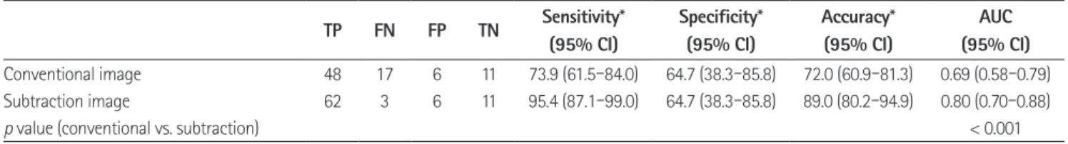
Table 2. Comparison of the Diagnostic Performances of Conventional and Subtraction Images for the Detection of HCC on Gadoxetic Acid- Enhanced MR Images
TP FN FP TN Sensitivity*
(95% CI)
Specificity*
(95% CI)
Accuracy*
(95% CI)
AUC (95% CI) Conventional image 48 17 6 11 73.9 (61.5–84.0) 64.7 (38.3–85.8) 72.0 (60.9–81.3) 0.69 (0.58–0.79) Subtraction image 62 3 6 11 95.4 (87.1–99.0) 64.7 (38.3–85.8) 89.0 (80.2–94.9) 0.80 (0.70–0.88)
p value (conventional vs. subtraction) < 0.001
*Data are % (95% CI) values.
AUC = area under the receiver operating characteristic curve, CI = confidence interval, FN = false negative, FP = false positive, HCC = hepatocellular carci- noma, TN = true negative, TP = true positive
Although some investigators have found that the capsular ap- pearance does not increase diagnostic accuracy for HCC (20), others have asserted that a visualization of a capsule is valuable, as it permits diagnosis of HCC without a definite washout ap- pearance (6, 24). Furthermore, capsular appearance is included as a major imaging feature in the LI-RADS diagnostic algorithm and plays an important role in the diagnosis of HCC. This algo- rithm considers a hyperintense capsular appearance during PVP or TP as a major diagnostic feature of non-invasive HCC (6), and diagnostic performances of LR-5 and LR-5V using capsular appearance on subtraction images for HCC, are higher than that of conventional imaging. Our results imply that subtraction im- ages can perform an important compensatory role in terms of
evaluating capsular appearance when hepatobiliary contrast agents are used. According to recent literature, in patients at risk of developing HCC, a combination of arterial phase hyperen- hancement plus washout or a capsular appearance has near 100% specificity for HCC (10). However, in the present study, the specificities of LR-5 and LR-5V for HCC were poor (55.6%), which appeared to be related to several false positive results at- tributed to the presence of a pseudocapsule.
Previous studies have reported the success of the subtraction technique depends on the degree of misregistration artifact be- tween non-enhanced and enhanced source images (13, 14).
The assessment of subtraction image quality is important be- cause false positive diagnoses can be made for capsules when Table 3. Comparison of the Diagnostic Performances of LR-5 or LR-5V for HCC
TP FN FP TN Sensitivity*
(95% CI)
Specificity*
(95% CI)
Accuracy*
(95% CI)
AUC (95% CI) Conventional image 54 19 4 5 74.0 (62.4-83.5) 55.6 (21.2-86.3) 72.0 (60.9-81.3) 0.65 (0.53-0.75) Subtraction image 60 13 4 5 82.2 (71.5-90.2) 55.6 (21.2-86.3) 79.3 (68.9-87.4) 0.69 (0.58-0.79)
p value (conventional vs. subtraction) 0.011
*Data are % (95% CI) values.
AUC = area under the receiver operating characteristic curve, CI = confidence interval, FN = false negative, FP = false positive, HCC = hepatocellular carci- noma, TN = true negative, TP = true positive
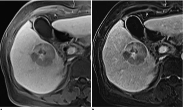
Fig. 1. Case of a 73-year-old woman with hepatocellular carcinoma encased by a histopathologically confirmed fibrous capsule in the right liver.
A. A conventional capsular appearance is not well visualized in the conventional TP image.
B. However, a smooth hyperintense rim is readily detected on the subtracted TP image.
TP = transitional phase
A B
misregistration occurs. In our study, subtraction image quality was unsatisfactory for 33 of the 107 patients (30.8%) showed (Grade 1 in one patient; Grade 2 in five patients; and Grade 3 in 27 patients). Although image quality was unsatisfactory in many patients, the accuracy of capsule detection was high when subtraction images were of Grade 4 quality or better. There were 6 false positive cases for capsular appearance on subtrac- tion images, and all of these cases produced a false positive re- sult on conventional images. Therefore, these 6 false positive cases were considered to be due to pseudocapsules rather than to misregistration of subtraction images. Nevertheless, un- avoidable slice misregistration is the main pitfall of subtraction techniques. Reliable error-correction processing methods for subtraction imaging are needed (14).
This study has several limitations that warrant consideration.
First, the sample size was too small to permit definitive conclu- sions, although all lesions including were confirmed histopath- ologically. The study was powered to detect a > 10% difference in capsule detection rates with an α of 0.05. Based on the actual sample size, the study has a statistical power of 76.0%. Further studies on larger populations are needed to confirm the value of subtraction images for HCC capsule detection. Second, the retrospective nature of the present study inherently introduces the possibility of patient selection bias. Finally, a 2-week inter- val may have been insufficient to avoid recall bias.
In summary, we conclude subtraction images from portal or TP images may be helpful for detecting HCC tumor capsules on gadoxetic acid-enhanced MR images in at-risk patients.
REFERENCES
1. El-Serag HB, Davila JA, Petersen NJ, McGlynn KA. The con- tinuing increase in the incidence of hepatocellular carcino- ma in the United States: an update. Ann Intern Med 2003;
139:817-823.
2. Kojiro M. Histopathology of liver cancers. Best Pract Res Clin Gastroenterol 2005;19:39-62
3. Kadoya M, Matsui O, Takashima T, Nonomura A. Hepato- cellular carcinoma: correlation of MR imaging and histo- pathologic findings. Radiology 1992;183:819-825 4. Ishigami K, Yoshimitsu K, Nishihara Y, Irie H, Asayama Y, Ta-
jima T, et al. Hepatocellular carcinoma with a pseudocap-
sule on gadolinium-enhanced MR images: correlation with histopathologic findings. Radiology 2009;250:435-443 5. Organ Procurement and Transplantation Network. OPTN/
UNOS policy 9.3.G.iv. Available at: http://optn.transplant.
hrsa.gov/ContentDocuments/OPTN_Policies.pdf- nameddest=Policy_09. Published Jan 1, 2015. Accessed Mar 16, 2016
6. Liver Reporting and Data System, version 2013.1. Ameri- can College of Radiology Available at: http://www.acr.org/
Quality-Safety/Resources/LIRADS/. Accessed Mar 16, 2016 7. Miraglia R, Pietrosi G, Maruzzelli L, Petridis I, Caruso S,
Marrone G, et al. Predictive factors of tumor response to trans-catheter treatment in cirrhotic patients with hepa- tocellular carcinoma: a multivariate analysis of pre-treat- ment findings. World J Gastroenterol 2007;13:6022-6026 8. Ng IO, Lai EC, Ng MM, Fan ST. Tumor encapsulation in he-
patocellular carcinoma. A pathologic study of 189 cases.
Cancer 1992;70:45-49
9. Grazioli L, Olivetti L, Fugazzola C, Benetti A, Stanga C, Dettori E, et al. The pseudocapsule in hepatocellular carci- noma: correlation between dynamic MR imaging and pa- thology. Eur Radiol 1999;9:62-67
10. Choi JY, Lee JM, Sirlin CB. CT and MR imaging diagnosis and staging of hepatocellular carcinoma: part II. Extracel- lular agents, hepatobiliary agents, and ancillary imaging features. Radiology 2014;273:30-50
11. Dioguardi Burgio M, Picone D, Cabibbo G, Midiri M, Lagal- la R, Brancatelli G. MR-imaging features of hepatocellular carcinoma capsule appearance in cirrhotic liver: compari- son of gadoxetic acid and gadobenate dimeglumine. Ab- dom Radiol (NY) 2016;41:1546-1554
12. Hope TA, Fowler KJ, Sirlin CB, Costa EA, Yee J, Yeh BM, et al. Hepatobiliary agents and their role in LI-RADS. Abdom Imaging 2015;40:613-625
13. Yu JS, Kim YH, Rofsky NM. Dynamic subtraction magnetic resonance imaging of cirrhotic liver: assessment of high signal intensity lesions on nonenhanced T1-weighted im- ages. J Comput Assist Tomogr 2005;29:51-58
14. Yu JS, Rofsky NM. Dynamic subtraction MR imaging of the liver: advantages and pitfalls. AJR Am J Roentgenol 2003;180:1351-1357
15. Seçil M, Obuz F, Altay C, Gencel O, Ig˘ci E, Sag˘ol O, et al.
The role of dynamic subtraction MRI in detection of hepa- tocellular carcinoma. Diagn Interv Radiol 2008;14:200-204 16. Sundarakumar DK, Wilson GJ, Osman SF, Zaidi SF, Maki JH.
Evaluation of image registration in subtracted 3D dynamic contrast-enhanced MRI of treated hepatocellular carcino- ma. AJR Am J Roentgenol 2015;204:287-296
17. Cho ES, Choi JY. MRI features of hepatocellular carcinoma re- lated to biologic behavior. Korean J Radiol 2015;16:449-464 18. Asayama Y, Nishie A, Ishigami K, Ushijima Y, Takayama Y,
Fujita N, et al. Distinguishing intrahepatic cholangiocarci- noma from poorly differentiated hepatocellular carcinoma using precontrast and gadoxetic acid-enhanced MRI. Di- agn Interv Radiol 2015;21:96-104
19. Khan AS, Hussain HK, Johnson TD, Weadock WJ, Pelletier SJ, Marrero JA. Value of delayed hypointensity and de- layed enhancing rim in magnetic resonance imaging diag- nosis of small hepatocellular carcinoma in the cirrhotic liver. J Magn Reson Imaging 2010;32:360-366
20. Park HJ, Jang KM, Kang TW, Song KD, Kim SH, Kim YK, et al. Identification of imaging predictors discriminating dif- ferent primary liver tumours in patients with chronic liver
disease on gadoxetic acid-enhanced MRI: a classification tree analysis. Eur Radiol 2016;26:3102-3111
21. Rimola J, Forner A, Tremosini S, Reig M, Vilana R, Bianchi L, et al. Non-invasive diagnosis of hepatocellular carcinoma
≤ 2 cm in cirrhosis. Diagnostic accuracy assessing fat, cap- sule and signal intensity at dynamic MRI. J Hepatol 2012;56:1317-1323
22. Suh YJ, Kim MJ, Choi JY, Park YN, Park MS, Kim KW. Dif- ferentiation of hepatic hyperintense lesions seen on ga- doxetic acid-enhanced hepatobiliary phase MRI. AJR Am J Roentgenol 2011;197:W44-W52
23. Vogl TJ, Kümmel S, Hammerstingl R, Schellenbeck M, Schumacher G, Balzer T, et al. Liver tumors: comparison of MR imaging with Gd-EOB-DTPA and Gd-DTPA. Radiology 1996;200:59-67
24. An C, Rhee H, Han K, Choi JY, Park YN, Park MS, et al. Added value of smooth hypointense rim in the hepatobiliary phase of gadoxetic acid-enhanced MRI in identifying tumour capsule and diagnosing hepatocellular carcinoma. Eur Radi- ol 2017;27:2610-2618
가도세틱산-조영증강 MRI에서 간세포암 피막 발견에 대한 영상차감기법의 진단적 가치
김현중
1· 안지현
1* · 문진실
2· 차승환
1목적: 가도세틱산-조영증강 MRI에서 간세포암 피막 발견에 대한 영상차감기법의 진단적 가치를 알아보고자 하였다.
대상과 방법: 2015년 5월부터 2017년 2월까지 가도세틱산-조영증강 MRI를 시행 받고 수술을 시행한 hepatocellular carcinoma (이하 HCC) 고위험군 108명을 대상으로 하였다. 차감영상의 질 및 간문맥기와 이행기의 일반영상과 차감영상 에서 피막 여부에 대해 평가하였고, 차감영상에서의 피막 여부가 Liver Imaging Reporting and Data System에 따른 간세 포암 진단에 미치는 영향을 평가하였다.
결과: 수술 전 경동맥화학색전술을 시행 받았거나 차감영상의 질이 불만족스러운 34명의 환자를 제외한 74명의 환자에 서 82개의 간 병변(간세포암 73개, 그 외 악성종양 5개, 양성종양 4개)에 대해 분석하였다. 피막의 발견에 대한 차감영상 의 민감도, 정확도, 그리고 곡선하면적은 일반영상과 비교하여 통계적으로 유의하게 높았고(각 95.4%, 89.0%, 0.80;
p < 0.001), 특이도는 동일하였다(64.7%). HCC의 진단에 대해서도 차감영상이 일반영상과 비교하여 민감도, 정확도, 그 리고 곡선하면적이 통계적으로 유의하게 높았으며(각 82.2%, 79.3%, 0.69; p = 0.011), 특이도는 동일하였다(55.6%).
결론: 가도세틱산-조영증강 MRI에서 간문맥기 또는 이행기로부터의 차감영상은 간세포암 피막의 발견에 도움이 된다.
연세대학교 원주의과대학 원주세브란스 기독병원 1영상의학과, 2생의학데이터센터