http://dx.doi.org/10.3938/NPSM.69.136
Magnetothermal Properties of Triethylene Glycol-Coated Cobalt-Zinc-Ferrite Nanoparticles for Magnetic Hyperthermia Applications
Ashfaq Ahmad · Hongsub Bae · Ilsu Rhee
∗Department of Physics, Kyungpook National University, Daegu 41566, Korea (Received 8 November 2018 : accepted 14 December 2018)
Cobalt zinc ferrite (Co0.6Zn0.4Fe2O) nanoparticles were synthesized using a one-step hydrother- mal method. Triethylene glycol (TREG) was used as a biocompatible coating material. The TREG- coated nanoparticles were spherical, with an average diameter of 9.82 nm. The Fourier-transform infrared spectra confirmed the presence of a TREG-coating on the surfaces of the nanoparticles.
The particles had a saturation magnetization of 84 emu/g and a coercivity of 32 Oe. The heat- ing capability of the particles was evaluated using an induction system, and the heating curves for aqueous samples with various particle concentrations were obtained. An alternating magnetic field with an amplitude of 4.5 kA/m and a frequency of 216 kHz was applied to the samples. The optimal particle concentration in water for achieving the hyperthermia target temperature of 42
◦C was 4.58 mg/mL. The specific absorption rate (SAR) of the 2-mg/mL sample was 83 W/g.
The linear relationship between the SAR and the square of the field strength was confirmed for a representative 10-mg/mL sample. These findings show that TREG-coated particles are suitable for magnetic hyperthermia applications owing to their superparamagnetic properties and high SAR values.
PACS numbers: 75.20.-g, 61.46.-w, 61.46.Df
Keywords: Zinc-doped cobalt-ferrite nanoparticle, Triethylene glycol coating, High specific absorption rate
I. INTRODUCTION
Magnetic nanoparticles, in particular, ferrite nanopar- ticles, have been extensively studied owing to their po- tential for application in various fields including high- density information storage systems [1], gas sensors [2], magnetocaloric refrigeration [3], magnetic resonance imaging [4,5], medical diagnostics [6], drug delivery, and magnetic hyperthermia [7,8].
Cancer cells are more susceptible to heat than healthy cells owing to their inefficient release of heat; this prop- erty of the cancer cells is used in magnetic hyperthermia [9]. Magnetic nanoparticles generate heat in the presence of a radio frequency (RF) field due to their loss mecha- nisms. Magnetic hyperthermia is a therapeutic concept where magnetic nanoparticles are used as heat generators
∗E-mail: [email protected]
to kill cancer cells. Specific absorption rate (SAR) is de- fined as the amount of heat generated by a unit mass of magnetic components in the presence of a RF magnetic field [10,11]. Magnetic particles with a high SAR value are desirable for magnetic hyperthermia, as the amount of dosage required to increase the temperature to the therapeutic limit can be significantly reduced. The SAR value depends on several factors such as the particle size, shape, chemical composition, magnetic properties, parti- cle concentration, RF-field strength and frequency, etc.
[12,13]. For a controllable hyperthermia, particles with a low Curie temperature are desirable [14].
Cobalt ferrite (CoFe2O4) is a hard magnetic material with a moderate saturation magnetization and large co- ercive force. The magnetic properties of cobalt ferrite can be tuned by substitution with nonmagnetic zinc ions.
The substitution with zinc ions in the tetrahedral sites of
This is an Open Access article distributed under the terms of the Creative Commons Attribution Non-Commercial License (http://creativecommons.org/licenses/by-nc/3.0) which permits unrestricted non-commercial use, distribution, and reproduction in any medium, provided the original work is properly cited.
SAR of the nanoparticles was 83 W/g for a low par- ticle concentration of 5 mg/mL, which revealed that the triethylene glycol (TREG)-coated cobalt zinc ferrite nanoparticles are suitable for magnetic hyperthermia ap- plications.
II. EXPERIMENTAL METHODS
All the chemicals for the synthesis of the particles were purchased from Sigma Aldrich, and used without fur- ther modification. Deionized water was used through- out the synthesis. TREG-coated zinc doped cobalt fer- rite nanoparticles were synthesized using a one-step hy- drothermal technique with a 100-mL teflon stainless- steel autoclave. The nanoparticles were coated with TREG during their synthesis. For the synthesis of the TREG-stabilized Co0.6Zn0.4Fe2O4 particles, 190.34 mg of CoCl2·6H2O, 163.56 mg of ZnCl2·6H2O, and 1 g of FeCl3·6H2O were dissolved in 20-mL deionized water in a 100-mL beaker through magnetic stirring, followed by the addition of 3 mL of TREG. Using a 0.5-M NaOH so- lution, pH was adjusted to 11. The color of the mixture turned dark-brown after the addition of the NaOH solu- tion. The mixture was continuously stirred at ambient temperature using a magnetic stirrer under an argon at- mosphere, introduced to obtain a homogeneous mixture.
Then, the homogenized mixture was transferred into the teflon-lined stainless-steel autoclave with a 40% filling, and kept in a vacuum oven at 160◦C for 12h. Then, the solution was allowed to naturally cool down to room tem- perature in the vacuum oven. The nanoparticles were separated from the solution using a magnet to discard
Fig. 1. (Color online) (a) TEM image and (b) particle size distribution of the TREG-coated cobalt zinc ferrite nanoparticles.
the unreacted residue in the solution after the synthe- sis. This cleaning process was performed several times by redispersing the nanoparticles in water, and then us- ing a magnet to separate the product from the solution until a complete removal of the unreacted products. The particles were redispersed in water for a further charac- terization.
The crystallinity and inverse spinel structure of the sample were analyzed using X-ray diffraction (XRD;
X’pert PRO, PANalytical). The particle shape and size distribution were revealed using transmission electron microscopy (HT 7700, Hitachi Ltd). The stoichiome- try of the nanoparticles was analyzed using inductively coupled plasma spectroscopy (ICP; IRISAP, Thermo Jarrell Ash). A vibrating sample magnetometer (VSM;
MPMS, Quantum Design) was used to measure the mag- netic properties of the particles. A Fourier-transform
Fig. 2. (Color online) XRD patterns of TREG-coated cobalt zinc ferrite nanoparticles. The indices of the crys- tal planes in the figure match with those of inverse spinel ferrite.
infrared spectrometer (FT-IR; Nicolet 380, Thermo Sci- entific USA) was used to confirm the TREG-coating on the surface of the particles. Magnetic hyperthermia mea- surements were performed using an induction heating system (Osung High Tech, OSH-120-B). The tempera- ture of the aqueous solution of particles was measured with a CALEX infrared thermometer (PyroUSB CF, Calex Electronics Limited).
III. RESULTS AND DISCUSSION
X-ray diffraction patterns of the TREG-coated zinc- doped cobalt ferrite nanoparticles are shown in Fig. 1.
The plane indices observed in the graph matched with those of the characteristic inverse spinel structure of fer- rites [JCPDS no. 22-1086].
A TEM image of the coated particles is shown in Fig. 2.
A particle-size distribution histogram is obtained by an- alyzing one hundred representative particles observed in the TEM image. The particles were spherical, with an average diameter of 9.82 nm.
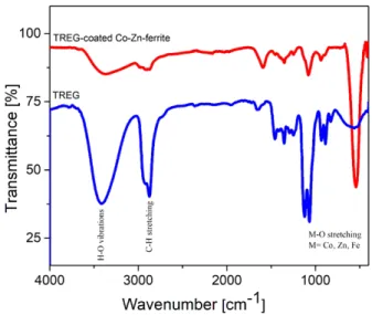
The status of the TREG-coating on the surface of the particles was checked using FT-IR spectra, recorded in the range of 400∼4000 cm−1, as shown in Fig. 3. The spectra exhibit absorption bands, which are in a good agreement with the characteristic FT-IR spectra of pure
Fig. 3. (Color online) FTIR spectra of pure TREG and TREG-coated cobalt zinc ferrite nanoparticles. The ab- sorption bands correspond to the stretching and vibra- tion modes of specific chemical bonds.
TREG and TREG-stabilized cobalt zinc ferrite parti- cles [19]. The C-H stretching at 2800∼2980 cm−1 and C-O stretching at 1060∼1080 cm−1 revealed the pres- ence of TREG molecules, the coating agent on the par- ticles’ surfaces [19]. The broad band around 3400 cm−1 is attributed to the stretching vibration of O-H in the hydroxyl groups of the TREG and water molecules at the particles’ surfaces. The bands at 2874 cm−1 and 2980 cm−1 correspond to the characteristic symmetric and asymmetric C-H stretching vibrations of the (-CH2) methylene groups. The two bands at 1060∼1080 cm−1 correspond to the C-O stretching vibration, which con- firms the presence of organic molecules on the surfaces of the particles. The two bands in the range of 400∼600 cm−1 emerge owing to the metal-oxygen stretching vi- bration at the octahedral and tetrahedral lattice sites.
The bands at 542 cm−1 and 415 cm−1 correspond to the stretching vibrations of Fe-O and Zn-O, respectively.
These bands confirm the TREG coating on the surfaces of the particles [20].
The magnetization of the TREG-coated particles as a function of the applied field at room temperature is shown in Fig. 4. The saturation magnetization of the particles is 84 emu/g, while the coercive force is 32 Oe.
The value of the saturation magnetization is larger than previously reported values [15]. The low coercivity and high saturation magnetization of the particles indicate that the TREG-coated particles are a good candidate
Fig. 4. (Color online) Magnetic moment of the nanopar- ticles as a function of the applied magnetic field. The particles exhibit a negligible coercive force, as shown in the inset.
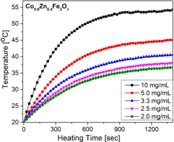
Fig. 5. (Color online) Heating curves of the aqueous so- lutions of the particles with various concentrations. The temperature initially rapidly increases, and then it satu- rates after approximately 900 s.
for magnetic hyperthermia applications [21]. The co- ercive force is small compared with that of the cobalt ferrite nanoparticles [22], most likely owing to the de- crease in magneto-crystalline anisotropy caused by the zinc doping. Similar results have been reported in Ref.
[23].
Calorimetric measurements were performed to evalu- ate the heating efficiency of nanoparticles in the presence of an AC magnetic field. An induction heating system and an IR temperature sensor were employed to obtain the heating curves as a function of the time, as shown in Fig. 5 for the aqueous samples with five different parti- cles concentrations. The samples were measured in the
Fig. 6. (Color online) Concentration dependence of the SAR and saturation temperature. The optimal concen- tration of particles in an aqueous solution for a target temperature of 42◦C is 4.58 mg/mL.
presence of an alternating field with a strength of 4.5 kA/m and frequency of 216 kHz. The temperature of the samples initially rapidly increased, and then it sat- urated within approximately 900 s, when the heat gen- eration of the particles was balanced with the heat loss to the environment. The saturation temperature of the 5-mg/mL sample was 45◦C, higher than the target tem- perature for hyperthermia applications of 42 ◦C. The target temperature can be achieved by controlling the particle concentration in the aqueous solution. The de- pendences of the saturation temperature and SAR as a function of the particles concentration are shown in Fig. 6. With a particles concentration of 4.58 mg/mL, the target temperature of 42◦C can be achieved.
SAR is defined as the generated heat per unit mass of the magnetic constituents in the particles; it can be expressed in W/g. When the particles are dispersed in an aqueous solution, the SAR is [24]:
SAR =∑ cW
mW
mCo+F e
dT
dt, (1)
where, cW is the specific heat of water (4.18 J·g−1·K−1),
dT
dt is the initial temperature increase of the heating curve, and mco+F e is the mass of the magnetic compo- nents (cobalt and iron) in the sample which is determined using inductively coupled plasma spectroscopy.
The heating efficiency of nanoparticles is mainly deter- mined by two types of loss mechanisms, hysteresis loss in multi-domain nanoparticles and relaxation losses in
Fig. 7. (Color online) Field-strength dependence of the heating curves for the 10-mg/mL sample. The saturation temperature increases with the field strength.
superparamagnetic nanoparticles. In our case, the coer- cive force is very small; therefore, we can ignore the heat contribution of the hysteresis loss during temperature in- crease. The relaxation losses are divided into Néel loss, caused by the rotation of the magnetic moment in the presence of an alternating magnetic field, and Brownian loss, caused by the physical rotation of the nanoparticles in the aqueous media. Therefore, both saturation mag- netization and particle size are important factors that determine the SAR [25].
The concentration dependence of the SAR is shown in Fig. 6. The 2-mg/mL sample showed the largest SAR value of 83 W/g; the SAR decreased with the increase of the particles concentration. This behavior can be ex- plained through the dipolar interactions [24,26]. The separation between particles decreases with the increase of the particles concentration, which leads to enhanced dipolar interactions between particles, which in turn re- duces the Néel loss [27].
The effect of the field strength on the heating of the particles was also evaluated. The heating curves for field strengths in the range of 2.4∼4.5 kA/m are shown in Fig. 7. The frequency was fixed to 216 kHz. The power loss in the magnetic particles as a function of the applied field strength is [28]:
P = 1
2µ0χ′′ωH02, (2)
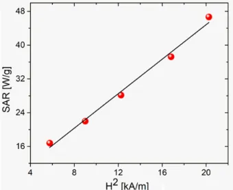
Fig. 8. (Color online) Dependence of the SAR as a func- tion of the square of the field strength. These data are consistent with Eq. (2).
where, H0 and ω are the strength and angular frequency of the alternating magnetic field, while χ′′ is the mag- netic susceptibility of the particles, which depends on the Néel and Brownian relaxation times of the particles.
The SAR is proportional with the power loss expressed by Eq. (2). Fig. 8 shows that there is a linear relationship between the SAR and H02, as expected.
IV. CONCLUSION
Zinc-doped cobalt ferrite nanoparticles were fabricated using a one-step hydrothermal synthesis method and characterized for magnetic hyperthermia applications.
This hydrothermal route is cost effective and highly re- producible. X-ray diffraction measurements confirmed the inverse spinel structure of the particles that were spherical with an average diameter of 9.82 nm. The presence of a TREG coating on the surfaces of the par- ticles was confirmed from the FT-IR spectra. The par- ticles were superparamagnetic with a negligible coercive force. In addition, the particles exhibited a high SAR in an aqueous solution owing to their high saturation magnetization. The optimal concentration of particles for achieving the target hyperthermia temperature of 42
◦C was 4.58 mg/mL. The linear relationship between the SAR and the square of field strength was also confirmed.
These findings show that the TREG-coated cobalt zinc ferrite nanoparticles are suitable for magnetic hyperther- mia applications.
[4] T. Ahmad, H. Bae, Y. Iqbal, I. Rhee and S. Hong et al., J. Magn. Magn. Mater. 381, 151 (2015).
[5] J.-H. Lee, Y.-M. Huh, Y.-W. Jun, J.-W. Seo and J.-T. Jang et al., Nat. Med. 13, 95 (2007).
[6] D. H. Kim, H. Zeng, T. C. Ng and C. S. Brazel, J.
Magn. Magn. Mater. 321, 3899 (2009).
[7] Q. A. Pankhurst, J. Connolly, S. K. Jones and J.
Dobson, J. Phys. D: Appl. Phys. 36, R167 (2003).
[8] A. Ahmad, H. Bae, I. Rhee and S. Hong, J. Magn.
Magn. Mater. 447, 42 (2018).
[9] R. Hergt, R. Hiergeist, I. Hilger, W. A. Kaiser and Y. Lapatnikov et al., J. Magn. Magn. Mater. 270, 345 (2004).
[10] R. Epherre, E. Duguet, S. Mornet, E. Pollert and S.
Louguet et al., J. Mater. Chem. 21, 4393 (2011).
[11] I. Rhee, New Phys.: Sae Mulli 65, 411 (2015).
[12] H. Hergt, S. Dutz and M. Röder, J. Phys. Condens.
Matter 20, 385214 (2008).
[13] F. Shubitidze, K. Kekalo, R. Stigliano and I. Baker, J. Appl. Phys. 117, 094302 (2015).
[14] E. Pollert, K. Knížek, M. Maryško, S. Kašpar and S.Vasseur et al., J. Magn. Magn. Mater. 316, 122 (2007).
[19] J. Wan, X. Jiang, H. Li and K. Chen, J. Mater.
Chem. 22, 13500 (2012).
[20] R. D. Waldron, Phys. Rev. 99, 1727 (1955).
[21] M. Veverka, K. Závěta, O. Kaman, P. Veverka and K. Knížek et al., J. Phys. D: Appl. Phys. 47, 065503 (2014).
[22] R. Topkaya, A. Baykal and A. Demir, J. Nanopart.
Res. 15, 1359 (2013).
[23] V. Mameli, A. Musinu, A. Ardu, G. Ennas and D.
Peddis et al., Nanoscale 8, 10124 (2016).
[24] Y. Iqbal, H. Bae, I. Rhee and S. Hong, J. Magn.
Magn. Mater. 409, 80 (2016).
[25] M. Ma, Y. Wu, J. Zhou, Y. Sun and Y. Zhang et al., J. Magn. Magn. Mater. 268, 33 (2004).
[26] A. E. Deatsch and B. A. Evans, J. Magn. Magn.
Mater. 354, 163 (2014).
[27] J. Dormann, L. Bessais and D. Fiorani, J. Phys. C:
Solid State Phys. 21, 2015 (1988).
[28] R. E. Rosensweig, J. Magn. Magn. Mater. 252, 370 (2002).