Carotid Intraplaque Hemorrhage Imaging: Diagnostic Value of High Signal Intensity Time-of-Flight MR Angiography Compared with Magnetization-Prepared Rapid Acquisition with Gradient-Echo Sequencing
This is an Open Access article distributed under the terms of the Creative Commons Attribution Non-Commercial License (http://creativecommons.org/licenses/
by-nc/3.0/) which permits unrestricted non-commercial use, distribution, and reproduction in any medium, provided the original work is properly cited.
Received: April 2, 2018 Revised: May 31, 2018 Accepted: June 10, 2018 Correspondence to:
Hyo Sung Kwak, M.D., Ph.D.
Radiology and Research Institute of Clinical Medicine of Chonbuk National University Biomedical Research Institute of Chonbuk National University Hospital, 567 Baekje-daero, Deokjin-gu, Jeonju- si, Jeollabuk-do 54896, Korea.
Tel. +82-63-250-2582 Fax. +82-63-272-0481 E-mail: [email protected]
Copyright © 2018 Korean Society of Magnetic Resonance in Medicine (KSMRM)
Original Article
Purpose: To determine the value of the appearance of the high signal intensity halo sign for detecting carotid intraplaque hemorrhage (IPH) on maximum intensity projection (MIP) of time-of-flight (TOF) MR angiography (MRA), based on high signal intensity on magnetization-prepared rapid acquisition with gradient-echo (MPRAGE) sequencing.
Materials and Methods: A total of 78 carotid arteries in 65 patients with magneti- zation-prepared rapid acquisition gradient-echo (MPRAGE) positive on carotid plaque MR imaging were included in this study. High-resolution MR imaging was performed on a 3.0-T scanner prior to carotid endarterectomy or carotid artery stenting. Fast spin-echo T1- and T2-weighted axial imaging, TOF, and MPRAGE sequences were obtained. Carotid plaques with high signal intensity on MPRAGE > 200% that of adjacent muscle on at least two consecutive slices were defined as showing IPH. Halo sign of high signal intensity around the carotid artery was found on MIP from TOF MRA. Continuous and categorical variables were compared among groups using the Mann-Whitney test and Fisher’s exact tests.
Results: Of these 78 carotid arteries, 53 appeared as a halo sign on the TOF MRA.
The total IPH volume of patients with a positive halo sign was significantly higher than that of patients without a halo sign (75.0 ± 86.8 vs. 16.3 ± 18.2, P = 0.001). The maximum IPH axial wall area in patients with a positive halo sign was significantly higher than that of patients without a halo sign (11.3 ± 9.9 vs. 3.7 ± 3.6, P = 0.000).
Conclusion: High signal intensity halo of IPH on MIP of TOF MRA is associated with total volume and maximal axial wall area of IPH.
Keywords: Carotid artery; Magnetic resonance imaging (MRI); Atherosclerosis;
Intraplaque hemorrhage
Ji-eun Ahn1, Hyo Sung Kwak2, Gyung Ho Chung2, Seung Bae Hwang2
1Student of Medical School, Chonbuk National University, Jeollabuk-do, Korea
2Radiology and Research Institute of Clinical Medicine of Chonbuk National University-Biomedical Research Institute of Chonbuk National University Hospital, Jeollabuk-do, Korea
Magnetic resonance imaging
INTRODUCTION
Intraplaque hemorrhage (IPH) of carotid stenosis plays a critical role in the progression of atherosclerotic disease (1, 2). IPH events can play a major role in plaque progression and leukocyte infiltration and may also serve as a measure of risk for the development of future cardiovascular events (3, 4). Therefore, the early detection of IPH is critical for the adequate primary and secondary prevention of atherosclerotic diseases.
T1-weighted magnetic resonance (MR) sequences are commonly used to detect IPH, since it is observed as high signal intensity on T1-weighted MR images due to the T1 shortening which results from the degradation of the hemorrhage into methemoglobin. The three T1-weighted sequences were two-dimensional (2D) fast spin-echo, three-dimensional (3D) time-of-flight (TOF), and three- dimensional magnetization-prepared rapid acquisition gradient-echo (MPRAGE) sequence for detecting or quantifying IPH (1, 5-8). Ota et al. (8) reported on the diagnostic performance of three T1-weighted 3.0-T MR sequences at carotid IPH imaging, using histologic analysis as the standard of reference. MPRAGE sequence is a superior diagnostic method for detecting and quantifying relatively large IPHs compared to fast spin-echo and TOF sequence.
However, a carotid TOF sequence has been widely used for detecting IPH because it was included in the routine protocol for brain MR examination and this approach can be time efficient, without the need for additional sequences such as MPRAGE. Maximum intensity projection (MIP) images from TOF MR angiography (MRA) can also show IPH as a high signal intensity (6, 9). This finding indicates the possibility of an additional application of MIP from TOF MRA to investigate carotid artery stenosis. However, no previous studies have investigated the relationship between IPH volume, MPRAGE sequence, and MIP from TOF MRA.
This study compared the diagnostic performances of high signal intensity halo from TOF MRA based on IPH volume on MPRAGE sequence for IPH detection.
MATERIALS AND METHODS
PatientsThis study was approved by the Institutional Review Board of our institution, and informed consent was waived by our Institutional Review Board. We included 125 consecutive patients that underwent carotid MR imaging to evaluate
plaque components between January 2014 and October 2016.
Carotid Plaque MR Protocol
Carotid plaque MR examination was obtained with an Achieva 3.0-T scanner (Philips Medical Systems, Best, the Netherlands) with a 16-channel head coil. The carotid artery with a dominant plaque was used to center the MR scan, which was the side used in the image analysis. Our protocol for multicontrast carotid plaque MR imaging included five different axial scans, as previously described (10, 11):
TOF, T1-weighted, T2-weighted, MPRAGE, and contrast- enhanced T1-weighted images. All of the sequences were centered at the bifurcation of the carotid artery with plaque. T1-weighted, T2-weighted, and contrast-enhanced T1-weighted sequences were obtained with a 2.0-mm section thickness without intersection spacing.
A 3D multi-slab TOF-MRA from the petrous portion of the ICA was generated with the following parameters:
repetition time (TR)/echo time (TE) = 23/3.45 ms, flip angle (FA) = 20°, field of view (FOV) = 200 × 200 mm, matrix = 488 × 249, sensitivity encoding factor = 2.5, slice thickness
= 1.2 mm, echo train length (ETL) = 1, and number of average = 1. Image parameters were as follows for the 3D MPRAGE sequence: TR/TE/inversion time (TI) = 8.7/5.3/304 ms, FA = 15°, ETL = 32, FOV = 140 × 140 mm, and matrix
= 216 × 192. Images were obtained from 20 mm below the carotid bifurcation to 20 mm above the carotid bifurcation, with slice thickness increments of 1.0 mm. TI time was chosen relative to the phase encoding acquisition in order to maximize the contrast between hemorrhage and inflowing blood. Chemical fat saturation was used. Total acquisition time was approximately 40 minutes.
MPRAGE Sequence IPH Volume
IPH was defined as the presence of high signal intensity comprising > 200% of the intensity of the adjacent muscle for at least 2 consecutive sections on MPRAGE.
We used semi-automated software for the quantitative analysis of IPH volume (Rapidia; Infinite, Seoul, Korea) on MPRAGE sequence. IPH segmentation was performed with a piecewise smooth regional level set method. A region of interest was then manually drawn around the outer boundary of the carotid artery. Next, the IPH volume was measured using a signal intensity > 200% the intensity of the adjacent muscle (Fig. 1). We finally analyzed the total IPH volume, maximal axial IPH area, and length of IPH on MPRAGE sequence. Maximal axial IPH area was defined as
the area at the axial slice with the highest signal intensity.
High Signal Intensity Halo on TOF MRA
A high signal intensity halo (halo sign) that was located in the carotid plaque but not connected to the lumen on MIP was defined as IPH positive (9) (Fig. 1). The results were determined by a consensus of two neuroradiologists that read the MIP of TOF MRA of the carotid artery while blinded to clinical information as well as research purpose.
Reviewers described the IPH as positive or negative in the carotid plaque on MIP images.
Carotid Stenosis
The degree of carotid stenosis was measured by guidelines established by the North American Symptomatic Carotid Endarterectomy Trial (NASCET), using MIP reformatted from TOF MRA. Arteries were grouped by stenosis into three categories: 0-15%, 16-49%, and 50-99% (1).
Statistical Analysis
We divided two groups into IPH positive or negative findings, based on MIP of TOF MRA. Continuous values are expressed as the mean and standard deviation, while
a b
c
Fig. 1. Intraplaque hemorrhage and measurement of intraplaque hemorrhage volume. (a) Intraplaque hemorrhage of high signal intensity on MPRAGE (magnetization- prepared rapid acquisition gradient-echo) sequence (arrows). (b) Semiautomatic measurement of intraplaque hemorrhage volume of the area of signal intensity of
> 200% of signal intensity of the adjacent muscle. (c) Intraplaque hemorrhage-positive on maximum intensity projection images of time-of-flight MR angiography shows a hyperintense region in the vessel wall without connection to the lumen (arrows).
categorical data are expressed as counts and percentages.
Continuous and categorical variables between the two groups were compared using the Mann-Whitney test and Fisher’s exact tests, respectively. Statistical significance was defined as P < 0.05. All statistical analyses were performed using R 2.14.1 (R Foundation for Statistical Computing, Vienna, Austria). In order to evaluate the cut-off value between the two groups, a receiver operating characteristic (ROC) curve analysis was performed to extract the optimal threshold using MedCalc software.
RESULTS
PatientsAmong the 125 consecutive patients that underwent
carotid plaque MR imaging, 40 were excluded from analysis for: 1) no IPH on all T1 sequences (n = 30), 2) poor image quality (n = 5), 3) no carotid artery suppression on MPRAGE imaging (n = 3), and 4) total occlusion of ICA (n
= 2). Consequentially, 78 carotid arteries of 65 patients with MPRAGE positive on carotid plaque MR imaging were recruited for this study. Demographic data of all enrolled patients are shown in Table 1.
Analysis of IPH between MPRAGE and MIP of TOF MRA
Of these 78 carotid arteries, 53 (67.9%) showed a halo sign around the carotid artery on the MIP of the TOF MRA.
Overall, the total volume of IPH in the carotid plaque was 60.2 ± 81.7 mm3 and the maximal axial IPH area was 9.1 ± 9.3 mm2.
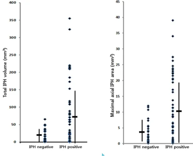
Fig. 2. Intraplaque hemorrhage (IPH) volume and maximal axial wall area of IPH between positive halo sign and negative halo sign on maximum intensity projection (MIP) images of time-of-flight MR angiography. (a) Total IPH volume is significantly larger in the positive halo sign group than in the negative halo sign group on MIP images. (b) The maximal axial wall area of IPH is also significantly larger in the positive halo sign group than in the negative halo sign group on MIP images.
a b
The MR comparison between the positive halo sign and negative halo sign groups is shown in Table 2. The total volume of IPH for patients with a positive halo sign was significantly higher than that of patients without a halo sign (75.0 ± 86.8 vs. 16.3 ± 18.2, P = 0.001) (Fig. 2). Overall carotid stenosis was similar between the two groups. The optimal cut-off value of total IPH volume between the two groups, as determined by ROC curve analysis, was 47.7 mm3 (sensitivity 49.1% and specificity 88.0%). The maximal axial wall area of IPH with a positive halo sign was significantly higher than that of patients without a halo sign (11.3 ± 9.9 vs. 3.7 ± 3.6, P = 0.000). The optimal cut-off value of the maximal axial IPH area between the two groups, as determined by ROC curve analysis, was 5.4 mm2 (sensitivity 62.3% and specificity 76.0%).
The total IPH volume and maximal axial wall area of IPH of the carotid arteries with 16-50% and 51-99% stenosis
were significantly larger than those of the carotid arteries with 0-15% stenosis (Table 3) (Fig. 3). The total IPH volume and maximal axial wall area of the positive halo sign group with 16-50% and 51-99% carotid stenosis were significantly larger than those of the negative halo sign group (P < 0.05).
DISCUSSION
Carotid IPH has been shown to be a significant predictor of future cerebral infarction and stroke in patients with symptomatic carotid artery stenosis (3, 4, 12). A recent study by Hosseini et al. (4) demonstrated that carotid IPH, measured as high signal intensity on MRI, predicted stroke in moderate degree stenosis. In the subgroup of 72 participants with 50% to 59% stenosis, IPH was significantly associated with future ipsilateral infarction and stroke. Saam et al. (3) observed that the presence of IPH was associated with ~6-fold higher risk for cerebrovascular events. According to this study, the annualized event rate in subjects with detectable IPH was 17.71%, compared to 2.43% in patients without IPH. In addition, the development of carotid IPH had immediate and long-term promoting effects on plaque progression and altered the biology and natural history of carotid atherosclerosis (2, 13). Carotid IPH Table 1. Patient Demographics
Patients (n = 65 patients) Patient demographics
Median age, y 73
Age range, y 59-87
Sex (male) 54 (83.1%)
Carotid risk factor
Diabetes 19 (29.2%)
Hypertension 35 (53.8%)
Current smoking 23 (35.4%)
Hyperlipidemia 18 (27.7%)
Previous heart disease 21 (32.3%) Previous stroke history 14 (21.5%) Location of plaque
Right 25 (38.5%)
Left 27 (41.5%)
Bilateral 13 (20.0%)
Table 2. IPH and Carotid Stenosis: Positive Halo Sign and Negative Halo Sign on MIP Images of TOF MRA
Positive halo sign
(n = 53)
Negative halo sign
(n = 25) P Total IPH volume, mm3 75.0 ± 86.8 16.3 ± 18.2 0.001 Maximal axial IPH area, mm2 11.3 ± 9.9 3.7 ± 3.6 0.000 IPH length, mm 10.0 ± 5.4 8.3 ± 7.1 0.246 Stenosis, % 41.0 ± 31.9 44.6 ± 33.8 0.649 IPH = intraplaque hemorrhage; MIP = maximum intensity projection; TOF = time of flight
Table 3. Carotid Stenosis and IPH on MIP Images of TOF MRA Stenosis,
% N* (%) N (%) with positive halo sign on MIP images
Total IPH volume (mm3) Maximal axial IPH area (mm2) Positive halo sign Negative halo sign P Positive halo sign Negative halo sign P
0-15 15 (19.2) 8 (53.3) 31.7 ± 26.9 17.3 ± 17.4 0.248 6.1 ± 3.8 4.1 ± 3.6 0.316
16-49 43 (55.1) 31 (72.1) 79.3 ± 86.81 13.4 ± 15.1 0.013 11.9 ± 9.61 3.2 ± 3.4 0.004
50-99 20 (25.5) 14 (70.0) 90.2 ± 100.31 21.0 ± 23.1 0.117 12.9 ± 11.81 4.0 ± 3.8 0.094
*N = number of carotid plaque
IPH = intraplaque hemorrhage; MIP = maximum intensity projection; MRA = magnetic resonance angiography; TOF = time of flight
is a strong inflammatory stimulant, leading to macrophage accumulation and fibrous cap degradation, which separates the thrombogenic core of the plaque from the lumen (14). As such, early detection of IPH on MRI is crucial for preventing future cerebrovascular events and plaque progression.
A variety of T1-weighted sequences can be used to detect carotid IPH. These include conventional T1-weighted fast spin echo, TOF MRA, or MPRAGE sequences. Several studies have used TOF sequencing to detect carotid IPH, and a routine MRA protocol without additional sequences to evaluate carotid atherosclerosis (1, 5, 9, 15). The appearance of a halo sign around the carotid artery on the MIP of the TOF MRA was useful for the noninvasive detection of a fresh or recent carotid IPH on histologic analysis (6, 9). The sensitivity of the high signal intensity halo for detecting carotid IPH was 91%. Saito et al. (16) reported that carotid IPH signal differences between spine echo, MPRAGE, and TOF by 1.5-T MR show similar accuracies for characterizing plaque components using histopathological information.
However, Ota et al. (8) reported the diagnostic performance of three T1-weighted MR sequences, 2D fast spin-echo,
3D TOF, and 3D MPRAGE, on carotid IPH imaging, using histologic analysis as the standard of reference. Among the three tested T1-weighted MR sequences, the MPRAGE sequence yielded the best agreement with histologic findings for detecting and quantifying IPH, when compared with fast spine echo and TOF sequencing. Therefore, the MPRAGE sequence was included in the routine MR protocol for the early detection of IPH, although this takes approximately three to four minutes (17-19).
Yamada et al. (1) performed a quantitative evaluation of halo signs on MIP images of carotid atherosclerotic plaques from routine TOF MRA. Overall, a halo sign on MIP had high specificity (100%) but relatively low sensitivity (32%) for detecting IPH. The sensitivity had significant positive relationships with the underlying IPH volume and degree of stenosis. The mean IPH volume was 2.7 times larger in those with the halo sign than in those without it. In our study, the sensitivity of high signal intensity on MIP images of TOF MRA for detecting IPH was 67.1%. This sensitivity was lower than that of previous studies that only used TOF MRA to evaluate IPH (6, 9), and higher than a previous comparison study between TOF MRA and carotid plaque MR Fig. 3. Intraplaque hemorrhage (IPH) volume and maximal axial wall area of IPH as carotid stenosis categories. (a) Total volume of IPH of carotid arteries with 16-50% and 51-99% was significantly larger than that of carotid arteries with 0-15%. (b) Maximal axial wall area of IPH of carotid arteries with 16-50% and 51-99% was significantly larger than that of carotid arteries with 0-15%.
a b
imaging (1).
In our study, the total IPH volume and maximal axial area was significantly higher with a positive halo sign on MIP of TOF MRA than with a negative halo sign. This result was consistent with a previous study (1). The sensitivity of the halo sign for detecting IPH did not differ based on the degree of stenosis, but there was a significant increasing trend between stenosis categories and IPH volumes on TOF MRA. These findings suggest that even with low-grade stenosis (0-15%), high IPH volume and axial wall thickness in the carotid plaque were characterized by a halo sign on MIP images of TOF MRA. We used the MPRAGE sequence as a standard of reference for detecting IPH because of its higher sensitivity and compared it to histological findings.
MPRAGE sequence results exhibited the best agreement with histologic findings, and consistently yielded the highest κ value, which showed the highest sensitivity for detecting IPH, and it also presented both the highest correlation and smallest mean difference when compared to histologic findings. The nonselective inversion pulse and spectrally selective water excitation or fat suppression facilitates MPRAGE imaging to suppress signals from background tissues. Such qualities make MPRAGE the optimal tool for the detection and quantification of IPH. In our study on detection using TOF MRA among patients with IPH based on MPRAGE, the sequence was missed in 32.9% of cases. In particular, patients with low grade carotid stenosis should be cautious when using TOF MRA for dynamic IPH changes.
There were some limitations to this study. First, there is not currently a histological gold standard. Histology provides sound confirmation for the presence, absence, and size of IPHs. Our study did not include patients who had received carotid endarterectomy, therefore histologic results were not available. Previous studies have demonstrated the diagnostic usefulness of MPRAGE sequence for IPH detection (8, 16). Second, a relatively small number of patients were examined, and it was a retrospective design.
In conclusion, a high signal intensity halo of IPH on MIP of TOF MRA is associated with increased total volume and maximal axial wall area of IPH. Since samples without the halo sign on TOF sequence exhibited IPH that was apparent on the MPRAGE sequence, we suggest that future diagnostic procedures include MRPAGE to detect IPH rather than the presently-used TOF sequence.
REFERENCES
1. Yamada K, Song Y, Hippe DS, et al. Quantitative evaluation of high intensity signal on MIP images of carotid atherosclerotic plaques from routine TOF-MRA reveals elevated volumes of intraplaque hemorrhage and lipid rich necrotic core. J Cardiovasc Magn Reson 2012;14:81 2. Sun J, Underhill HR, Hippe DS, Xue Y, Yuan C, Hatsukami
TS. Sustained acceleration in carotid atherosclerotic plaque progression with intraplaque hemorrhage: a long-term time course study. JACC Cardiovasc Imaging 2012;5:798- 804
3. Saam T, Hetterich H, Hoffmann V, et al. Meta-analysis and systematic review of the predictive value of carotid plaque hemorrhage on cerebrovascular events by magnetic resonance imaging. J Am Coll Cardiol 2013;62:1081-1091 4. Hosseini AA, Simpson RJ, Altaf N, Bath PM, MacSweeney
ST, Auer DP. Magnetic resonance imaging plaque hemorrhage for risk stratification in carotid artery disease with moderate risk under current medical therapy. Stroke 2017;48:678-685
5. Yoon W, Kim SK, Park MS, Chae HJ, Kang HK. Safety of protected carotid artery stenting in patients with severe carotid artery stenosis and carotid intraplaque hemorrhage.
AJNR Am J Neuroradiol 2012;33:1027-1031
6. Yoshimura S, Yamada K, Kawasaki M, et al. High-intensity signal on time-of-flight magnetic resonance angiography indicates carotid plaques at high risk for cerebral embolism during stenting. Stroke 2011;42:3132-3137
7. van den Bouwhuijsen QJ, Selwaness M, Tang H, et al.
Change in carotid intraplaque hemorrhage in community- dwelling subjects: a follow-up study using serial MR imaging. Radiology 2017;282:526-533
8. Ota H, Yarnykh VL, Ferguson MS, et al. Carotid intraplaque hemorrhage imaging at 3.0-T MR imaging: comparison of the diagnostic performance of three T1-weighted sequences. Radiology 2010;254:551-563
9. Yim YJ, Choe YH, Ko Y, et al. High signal intensity halo around the carotid artery on maximum intensity projection images of time-of-flight MR angiography: a new sign for intraplaque hemorrhage. J Magn Reson Imaging 2008;27:1341-1346
10. Yarnykh VL, Terashima M, Hayes CE, et al. Multicontrast black-blood MRI of carotid arteries: comparison between 1.5 and 3 tesla magnetic field strengths. J Magn Reson Imaging 2006;23:691-698
11. Underhill HR, Yarnykh VL, Hatsukami TS, et al. Carotid plaque morphology and composition: initial comparison between 1.5- and 3.0-T magnetic field strengths. Radiology 2008;248:550-560
12. Singh N, Moody AR, Gladstone DJ, et al. Moderate carotid artery stenosis: MR imaging-depicted intraplaque hemorrhage predicts risk of cerebrovascular ischemic events in asymptomatic men. Radiology 2009;252:502-508 13. Sun J, Balu N, Hippe DS, et al. Subclinical carotid
atherosclerosis: short-term natural history of lipid-rich necrotic core--a multicenter study with MR imaging.
Radiology 2013;268:61-68
14. Mughal MM, Khan MK, DeMarco JK, Majid A, Shamoun F, Abela GS. Symptomatic and asymptomatic carotid artery plaque. Expert Rev Cardiovasc Ther 2011;9:1315-1330 15. Gupta A, Baradaran H, Kamel H, et al. Intraplaque high-
intensity signal on 3D time-of-flight MR angiography is strongly associated with symptomatic carotid artery stenosis. AJNR Am J Neuroradiol 2014;35:557-561
16. Saito A, Sasaki M, Ogasawara K, et al. Carotid plaque signal differences among four kinds of T1-weighted magnetic
resonance imaging techniques: a histopathological correlation study. Neuroradiology 2012;54:1187-1194 17. Kwak HS, Yang HJ, Hwang SB, Chung GH. Carotid wall
imaging with routine brain MRI to facilitate early detection of carotid plaque and intraplaque hemorrhage. J Stroke 2017;19:107-108
18. McNally JS, Kim SE, Yoon HC, et al. Carotid magnetization- prepared rapid acquisition with gradient-echo signal is associated with acute territorial cerebral ischemic events detected by diffusion-weighted MRI. Circ Cardiovasc Imaging 2012;5:376-382
19. Park JS, Kwak HS, Lee JM, Koh EJ, Chung GH, Hwang SB. Association of carotid intraplaque hemorrhage and territorial acute infarction in patients with acute neurological symptoms using carotid magnetization- prepared rapid acquisition with gradient-echo. J Korean Neurosurg Soc 2015;57:94-99