pISSN 1598-9992 eISSN 2233-6869
CASE REPORT
직장암 수술 후 하부 장간막동맥 절단부의 외상신경종:
증례 보고 및 문헌 고찰
전성미, 이재영1, 변선주2
서울대학교병원 교육수련부, 영상의학과1, 서울아산병원 병리과2
Traumatic Neuroma at the Inferior Mesenteric Artery Stump after Rectal Cancer Surgery:
A Case Report and Literature Review
Sung Mi Jeon, Jae Young Lee1, and Sun-Ju Byeon2
Department of Education and Training, Department of Radiology1, Seoul National University Hospital, Department of Pathology, Asan Medical Center2, Seoul, Korea
Traumatic neuroma results from regeneration attempts of the proximal end of an injured or severed nerve, resulting in a non-neoplastic nodular lesion. The lower extremity after amputation is the most common site, followed by the head and neck.
Traumatic neuromas occurring in the abdomen, however, are rare. In the abdominal region, traumatic neuromas occur in the cystic duct stump and the common bile ducts as well as around the celiac trunk. This study reports a case of a 59-year-old man who presented with a traumatic neuroma arising at the stump of the inferior mesenteric artery after rectal cancer surgery.
Traumatic neuromas at the stump of the inferior mesenteric artery have not been previously reported. The lesion exhibited atypical imaging features, including a well-enhanced nodule, a significant interval growth in size and a mild increase in 18F-fluo- rodeoxyglucose uptake, resembling lymph node metastasis. This case report will help physicians understand the sites of occur- rence and imaging features of traumatic neuromas in the abdomen. (Korean J Gastroenterol 2016;68:279-283)
Key Words: Neuroma; Trauma; Injuries; Rectal neoplasms; Inferior mesenteric artery
Received April 14, 2016. Revised September 12, 2016. Accepted September 22, 2016.
CC This is an open access article distributed under the terms of the Creative Commons Attribution Non-Commercial License (http://creativecommons.org/licenses/
by-nc/4.0) which permits unrestricted non-commercial use, distribution, and reproduction in any medium, provided the original work is properly cited.
Copyright © 2016. Korean Society of Gastroenterology.
교신저자: 이재영, 03080, 서울시 종로구 대학로 101, 서울대학교병원 영상의학과
Correspondence to: Jae Young Lee, Department of Radiology, Seoul National University Hospital, 101 Daehak-ro, Jongno-gu, Seoul 03080, Korea. Tel: +82-2-2072- 3073, Fax: +82-2-743-6385, E-mail: leejy4u@snu.ac.kr
Financial support: None. Conflict of interest: None.
INTRODUCTION
Traumatic neuroma occurs by regeneration attempts of the proximal end of an injured or severed nerve, resulting in a non-neoplastic nodular lesion.1,2 The lesion typically arises one to 12 months after transection and exhibits slow or no interval size changes over months to years.3 This condition was first described by Odier in 1811 in war-wounded veterans with amputated limbs presenting with disabling pain.4,5 Traumatic neuromas can occur in any part of the body. The
lower extremity after amputation is the most common site, followed by the head and neck (frequently after tooth ex- traction or radical dissection).6 Traumatic neuromas occur- ring in the abdomen, however, are rare.
In the abdominal region, traumatic neuromas occur in the cystic duct stump and less commonly in the common bile ducts (CBD), or a choledochoenteral anastomotic site after cholecystectomy, bile duct surgery or blunt abdominal trauma.7-12 Other sites reported are around the celiac trunk after gastrectomy or in the rectal wall after polypectomy.13,14
Fig. 1. An ill-defined left para-aortic lesion on CT images obtained two weeks after surgery. (A) Non-contrast CT reveals an ill-defined infil- trative para-aortic soft tissue attenuation lesion (arrow, 30.53±
27.25 HU) at the inferior mesenteric artery stump area. (B) Portal- venous phase CT indicates that the lesion (arrow) is persistently enhancing (53.64±28.65 HU). This lesion was interpreted as a postoperative granulation tissue because the occurrence of a lesion at a stump is considered a typical finding.
Fig. 2. A left para-aortic lesion changing into a discrete nodule on follow-up CT scans. (A) Portal-venous phase CT obtained seven months after operation depicts a 1.2 cm discrete enhancing nodule (arrow) at the inferior mesenteric artery stump area (73.17±10.37 HU). (B) Portal-venous phase of a contrast-enhanced CT obtained 20 months after operation depicts a 1.2 cm discrete enhancing mass (arrow) at the inferior mesenteric artery stump area (96.16±14.04 HU) at the portal-venous phase. No significant interval change in size is noted compared with the previous CT (Fig. 2A). (C) Portal-venous phase of a contrast-enhanced CT obtained 32 months after operation reveals a 1.8 cm discrete enhancing mass (arrows) at the inferior mesenteric artery stump area (110.00±14.30 HU) at the portal-venous phase. An interval increase in size is noted compared with the previous CT (1.2 cm to 1.8 cm). Coronal portal-venous phase image also reveals a well-demarcated slightly elongated mass (arrows) at the left para-aortic area.
We present a case of traumatic neuroma at the stump of the inferior mesenteric artery (IMA) that has rapid interval growth in size and hypermetabolic activity in a patient with prior rectal cancer surgery. To the best of our knowledge, this is the first report of traumatic neuroma occurring after rectal cancer surgery.
CASE REPORT
A 59-year-old Korean male underwent an ultra-low ante- rior resection (U-LAR) with colo-anal anastomosis for rectal cancer (an annular constricting mass located 5 cm above the anal verge, 4 cm segment involvement of the mid-rectum). He did not have any underlying disease or significant medical history at the time of diagnosis of rectal cancer. According to the TNM staging system of the American Joint Committee on Cancer and the International Union Against Cancer, the diag- nosed rectal cancer was clinically determined as stage 2 (cT3N+M0). After neoadjuvant chemoradiotherapy, the le- sion was downgraded to stage 1 (ypT2N0M0), which was sur- gically confirmed. The patient received adjuvant chemo- therapy after U-LAR.
The patient exhibited no apparent symptoms, and physical examinations remained normal during later visits. Laborato-
Fig. 3. A hypermetabolic lesion at the left para-aortic area. A 18F-fluorodeoxyglucose PET-CT scan depicts a hypermetabolic lesion (arrows) at the left para-aortic inferior mesenteric artery stump area (standardized uptake value, 2.87).
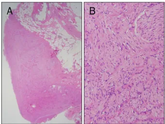
Fig. 4. Microscopic findings (H&E). (A) The lesion is composed of variably sized proliferating nerve fascicles in a background of collagen and adipose tissue (×12). (B) The image reveals haphazard proliferation of nerve fascicles including axons, Schwann cells, and fibroblasts (×200).
ry tests revealed no abnormal findings, and carcinoem- bryonic antigen level ranged from 0.5 ng/mL to 2.3 ng/mL (normal range, <5.0 ng/mL).
In the first postoperative CT obtained two weeks after U-LAR, a soft tissue lesion at the stump of IMA was detected (Fig. 1). The lesion had an ill-defined margin and was slightly contrast-enhanced. The lesion, which was irregular in margin and slightly contrast-enhanced, was diagnosed as a post- operative granulation tissue. When viewed at seven and 20 months after U-LAR, the lesion changed into a nodular, well-circumscribed, homogenously enhancing lesion meas- uring 1.2 cm in diameter (Fig. 2A, B). By 32 months, the lesion size increased from 1.2 cm to 1.8 cm in diameter (Fig. 2C).
The interval change in size raised a strong suspicion of a metastatic lymph node.
A PET-CT scan revealed that the lesion was hypermetabolic (standardized uptake value [SUV], 2.87) (Fig. 3). This finding strengthens our suspicion of a metastatic lymph node. To confirm whether the lesion was a nodal metastasis, a surgi- cal biopsy was performed. Histological examination of the surgical specimen revealed haphazardly arranged pro- liferation of variably sized nerve fascicles, including axons, Schwann cells, and fibroblasts, but no evidence of malignant cells (Fig. 4). The lesion was pathologically confirmed as trau- matic neuroma.
DISCUSSION
Most intra-abdominal traumatic neuromas are asympto- matic.4,13,14 Some traumatic neuromas cause symptoms, such as epigastric pain, vomiting, or obstructive jaundice, with varying degrees and time of onset.8 Although asympto- matic traumatic neuromas do not require treatment, as their natural history is benign, accurate differentiation of traumatic neuromas from recurring tumors or metastatic lymph nodes in the clinical context of prior malignancy is crucial.1,2,4,8,13
Imaging is one of the most valuable examination methods, paired with pathologic examination, to confirm the diagnosis of traumatic neuromas.1,2,4,15 Radiologic descriptions of trau- matic neuromas are primarily based on cases involving am- putated extremities or the head and neck.1-3,6,15 Characteris- tics of traumatic neuromas on ultrasonography include a small short/long diameter ratio, a small short-axis diameter, and the presence of a central hyperechoic area.2 On CT imag- ing, traumatic neuromas are characteristically stable nod- ules with a low attenuation.1,2 MRI features of traumatic neu- romas include a signal intensity that is similar to that of mus- cle on T1-weighted images, contrast enhancement, and slightly heterogeneous, intermediate to high signal intensity frequently combined with a hypointense rim on T2-weighted images.2,3,15 On metabolic imaging using 18F-fluorodeox- yglucose (FDG) PET, traumatic neuromas exhibit no in- creased uptake of FDG.13,16
Although understanding the imaging characteristics of traumatic neuroma is important to make a correct diagnosis and differentials, the imaging features need further descrip- tion in the literature. In a report of traumatic neuromas occur- ring in four patients after neck dissections, the characteristic CT features were described as a nodule with a central hypo- attenuation, a hyperattenuating rim, and intact overlying fat.1 However, another author asserted that a hyperattenuating rim on CT cannot be used as a differentiating radiologic fea- ture because this feature is observed in both traumatic neu- roma and recurrent lymphadenopathy.2 Traumatic neuroma is currently described as a stable nodule with low attenuation on a contrast-enhanced CT scan that occurs one to 12 months after the clinical setting related to its development, trauma or surgery.2,13
The MRI features of traumatic neuromas are described in several studies.2,3,15 Although no significant difference in the signal intensity on T1- and T2-weighted images is apparent between traumatic neuroma and recurrent lymphadenop- athy, a hypointense rim on T2-weighted MRI is seen only in traumatic neuromas.2,3 A recent review of 13 subjects with 20 neuromas concluded that traumatic neuromas exhibit en- hancement and lack a T2 hyperintense rim surrounding a central area of low signal, called a target sign.15
Altered glucose metabolism is characteristic of numerous malignancies, and FDG-PET is increasingly used for staging, restaging, detecting recurrence or metastasis, or differ- entiating between benign and malignant lesions. However, because FDG uptake is not tumor-specific, false positive re- sults are possible in many non-neoplastic and benign dis- eases, such as inflammation or posttraumatic repair.16 Com- pared with other conventional modalities, data on FDG-PET features of traumatic neuromas have been extremely limited.
The report that traumatic neuromas exhibited no increased uptake on FDG-PET scans is consistent with the natural his- tory of traumatic neuroma as a benign entity.13
We present a case of a traumatic neuroma at the stump of the IMA that presented radiologic findings inconsistent with prior reports, making it difficult to differentiate this lesion from a malignancy pre-operatively. First, a contrast-enhanced CT scan exhibited a well-enhanced nodule (73 HU to 110 HU at portal-venous phase CT). This finding is not typical for a traumatic neuroma, usually described as hypoattenuating in the arterial, portal, and equilibrium phases of an enhanced
CT scan.13 Second, a FDG-PET scan revealed a hyper- metabolic nodule in the left para-aortic IMA stump area (SUV 2.87), later found to be a false positive after surgical excision.
Third, the lesion increased from 1.2 cm to 1.8 cm in diameter over one year. The growth rate during the year prior to neuro- ma resection (0.6 cm/year) raised strong suspicion of malignancy.
Only one case of a traumatic neuroma exhibiting imaging findings consistent with our case has been reported. A 70- year-old man presented with late-onset jaundice as a result of a polypoid mass in CBD.8 The patient underwent a chol- ecystectomy 25 years prior to presentation and a chol- edochojejunostomy 12 years prior. A contrast-enhanced ab- dominal CT scan exhibited a polypoid mass in mid-CBD with contrast enhancement. Magnetic resonance cholangiog- raphy exhibited a polypoid filling defect in the mid-CBD, and the PET-CT scan showed a mild hypermetabolic lesion in the CBD (SUV 1.9). An extensive dissection called pylorus-pre- serving pancreatico-duodenectomy was performed under the impression of CBD cancer, but the lesion was finally con- firmed as a CBD neuroma. Therefore, traumatic neuroma can show contrast enhancement on CT and mild hypermeta- bolism on PET-CT, even though it is not typical for traumatic neuroma.
With regard to its anatomic location, a traumatic neuroma at the IMA stump has not been reported in the literature. The lesion may involve the inferior mesenteric plexus (intercon- nected with the celiac plexus) surrounding the root of IMA that is resected during U-LAR performed in the rectal cancer patient of this case. The risk of biliary traumatic neuromas can be increased due to any injury to the surrounding nerves (mainly derived from the celiac plexus) or arteries or the bile ducts themselves during surgical manipulations, such as vas- cular ligation, thermocoagulation, and excessive exploration.8 Thus, the traumatic neuroma of our case likely resulted from injury to the inferior mesenteric plexus during the IMA re- section or lymph node dissection for rectal cancer surgery.
In this patient, an infiltrative soft tissue lesion was seen at the IMA stump area two weeks after rectal cancer surgery (Fig. 1). This lesion showed typical imaging findings of post- operative granulation tissue that is commonly seen after lymph node dissection. Therefore, the lesion is not thought to be the initial finding of traumatic neuroma. A well-defined nodular mass developed seven months after surgery, possi-
bly the beginning of traumatic neuroma.
In conclusion, a traumatic neuroma occurred at the stump of the IMA. Traumatic neuromas are rarely observed at this site. The lesion exhibited atypical imaging features, such as a well-enhanced nodule, rapid interval growth in size and a mild increase of FDG uptake. Understanding this case will help enhance our knowledge of imaging features of trau- matic neuromas.
REFERENCES
1. Huang LF, Weissman JL, Fan C. Traumatic neuroma after neck dissection: CT characteristics in four cases. AJNR Am J Neuroradiol 2000;21:1676-1680.
2. Yabuuchi H, Kuroiwa T, Fukuya T, Tomita K, Hachitanda Y.
Traumatic neuroma and recurrent lymphadenopathy after neck dissection: comparison of radiologic features. Radiology 2004;
233:523-529.
3. Boutin RD, Pathria MN, Resnick D. Disorders in the stumps of amputee patients: MR imaging. AJR Am J Roentgenol 1998;171:
497-501.
4. Brogan DM, Kakar S. Management of neuromas of the upper extremity. Hand Clin 2013;29:409-420.
5. Swanson HH. Traumatic neuromas. A review of the literature.
Oral Surg Oral Med Oral Pathol 1961;14:317-326.
6. Murphey MD, Smith WS, Smith SE, Kransdorf MJ, Temple HT.
From the archives of the AFIP. Imaging of musculoskeletal neuro- genic tumors: radiologic-pathologic correlation. Radiographics 1999;19:1253-1280.
7. Nagata Y, Tomioka T, Chiba K, Kanematsu T. Traumatic neuroma of the common hepatic duct after laparoscopic cholecystec- tomy. Am J Gastroenterol 1995;90:1887-1888.
8. Koh DW, Lee WJ, Kim JH, et al. Amputation neuroma mimicking common bile duct cancer: a case report. Korean J Gastroenterol 2008;52:32-36.
9. Shimura K, Tamada K, Asada M, et al. Intraductal ultra- sonography of traumatic neuroma of the bile duct. Abdom Imaging 2001;26:632-634.
10. van Gulik TM, Brummelkamp WH, Lygidakis NJ. Traumatic neuro- ma giving rise to biliary obstruction after reconstructive surgery for iatrogenic lesions of the biliary tract--a report of three cases.
Hepatogastroenterology 1989;36:255-257.
11. Mentha G, Rubbia-Brandt L, Orci L, et al. Traumatic neuroma with biliary duct obstruction after orthotopic liver transplantation.
Transplantation 1999;67:177-179.
12. Katsinelos P, Dimiropoulos S, Galanis I, et al. Biliary stricture due to neuroma after an innocent blunt abdominal trauma. Surg Endosc 2002;16:1494.
13. Kwon JH, Ryu SW, Kang YN. Traumatic neuroma around the cel- iac trunk after gastrectomy mimicking a nodal metastasis: a case report. Korean J Radiol 2007;8:242-245.
14. Curran T, Poylin V, Kane R, Harris A, Goldsmith JD, Nagle D. Case report of a traumatic rectal neuroma. Gastroenterol Rep (Oxf) 2015. doi: 10.1093/gastro/gov023.
15. Ahlawat S, Belzberg AJ, Montgomery EA, Fayad LM. MRI features of peripheral traumatic neuromas. Eur Radiol 2016;26:1204- 1212.
16. Chang JM, Lee HJ, Goo JM, et al. False positive and false negative FDG-PET scans in various thoracic diseases. Korean J Radiol 2006;7:57-69.