대한소화기학회지 2008;51:45-47
접수: 2007년 3월 16일, 승인: 2007년 6월 26일 연락처: 김용훈, 700-712, 대구시 중구 동산동 194번지
계명대학교 동산의료원 외과
Tel: (053) 250-7388, Fax: (053) 250-7322 E-mail: [email protected]
Correspondence to: Yong Hoon Kim, M.D.
Department of Surgery, Keimyung University College of Medicine, 194, Dongsan-dong, Jung-gu, Daegu 700-712, Korea Tel: +82-53-250-7388, Fax: +82-53-250-7322
E-mail: [email protected]
충수절제술 후 발생한 잔존 충수염 1예
계명대학교 의과대학 외과학교실, 내과학교실*, 진단방사선과학교실†
백성규ㆍ김미선ㆍ김용훈ㆍ정우진*ㆍ권중혁
†A Case of Stump Appendicitis after Appendectomy
Seong Kyu Baek, M.D., Mi Sun Kim, M.D., Yong Hoon Kim, M.D., Woo Jin Chung, M.D.*, and Jung Hyeok Kwon, M.D.†
Departments of Surgery, Internal Medicine*, Diagnostic Radiology†, Keimyung University College of Medicine, Daegu, Korea
Stump appendicitis is an acute inflammation of the residual appendix and a rare complication after an appendec- tomy. Although the signs and symptoms do not differ from acute appendicitis, the diagnosis is often not consid- ered because of the past surgical history. Only a small number of stump appendicitis cases have been reported, but there has been no report of stump appendicitis in Korea. Herein, we report a case of stump appendicitis. A 28-year-old female was admitted to our hospital due to right lower quadrant abdominal pain. Fifteen months ago, the patient had a laparoscopic appendectomy under the diagnosis of an acute appendicitis, but she subsequently suffered from intermittent abdominal pain and fever. Abdominal ultrasonography and CT scan showed an inflamed appendiceal stump. Laparoscopic stump appendectomy was done and the biopsy revealed stump appendicitis.
(Korean J Gastroenterol 2008;51:45-47)
Key Words: Appendectomy; Appendicitis; Stump
서 론
젊은 성인에서 우하복부 통증이 있는 경우 가장 먼저 감 별해야 할 질환은 급성 충수염일 것이다. 그러나 충수절제 술을 받은 기왕력이 있는 환자에서는 통상적으로 다른 질환 의 가능성을 먼저 생각하게 된다.
저자들은 복강경 충수절제술을 시행 받은 후, 1년 이상 간헐적인 우하복부 통증을 호소하는 28세 여자 환자에서 잔 존 충수염(stump appendicitis)을 진단하고 복강경을 이용한 잔존 충수절제를 시행한 예를 경험하였다.
증 례
환자는 28세 여자환자로 약 1년간의 간헐적인 우하복부 통증을 주소로 내원하였다. 과거력에서 약 1년 3개월 전 국 내 모 대학병원에서 급성 충수염에 의한 우하복부 통증으로 복강경 충수절제술을 시행 받은 병력이 있었다. 수술 후 환 자는 특별한 합병증 없이 퇴원하였다. 이후 간헐적인 우하 복부 통증과 함께 미열이 있어 개인 의원을 수 차례 방문하 였으나 정확한 원인을 밝히지 못하고 보존 치료만을 받았 다. 최근 통증이 심해지고 열감도 있어 본원 응급실을 방문 하였다. 당시 37.8도의 열이 있었고 다른 신체 활력 징후는 정상이었다. 신체검사에서 우하복부 압통과 반발통이 있었
46 대한소화기학회지: 제51권 제1호, 2008
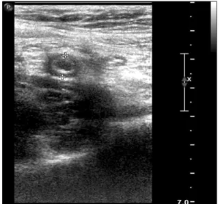
Fig. 1. A transverse ultrasound scan shows a dilated appendiceal stump measuring 8 mm in diameter.
Fig. 2. Abdominal CT findings.
(A) Axial CT scan of abdomen after administration of intravenous contrast material demonstrates a dilated appendiceal stump (black arrow) in the retrocecal region as- sociated with thickening of the peritoneal reflection (white arrow- heads). (B) Coronal reformatted image from CT scan reveals the dilated apendiceal stump (white ar- row) with periappendiceal inflam- mation. A prior staple line (white arrowhead) is also visualized.
Fig. 3. Gross finding of resected appendiceal stump. Total length of the specimen is 4.0 cm. A surgical clip is visible from initial operation (arrow).
다. 복부 초음파 검사에서 충수로 여겨지는 관상 구조가 약 0.77 cm 크기로 관찰되었으며 부종을 동반하고 있어 급성 충수염에 합당하다(Fig. 1).
병변의 전체적인 복강 내 양상을 조사하고 다른 질환의 가능성을 감별하기 위해 복부 전산화단층촬영을 시행하였 다. 맹장 뒤쪽에 위치한 충수가 팽창되어 있으나 주위의 체 액 저류는 없었다(Fig. 2). 수술은 계획수술로 복강경 충수절 제술을 시행하였다.
수술 소견으로 맹장 뒤쪽에 위치한 충수는 길이가 3.5 cm 직경이 약 0.8 cm 로 비후되어 있고 섬유성의 염증삼출막 (fibrinopurulent exudates)으로 덮여 있었다(Fig. 3). 주위에 삼 출액은 많지 않았다. 수술 후 제2일에 환자는 특별한 합병 증 없이 퇴원하였고 현재 외래 추적 관찰 중이며 8개월 동 안 통증의 재발은 없었다. 조직검사에서 중성구의 염증세포 의 침윤을 특징으로 하는 급성화농충수염으로 진단되었다.
고 찰
급성 충수염 치료는 항생제를 이용한 내과 방법이 시도된 적이 있으나 재발률이 높아 특별한 경우를 제외하고 외과 절제를 하는 것이다.1,2 충수절제술 후 남은 돌기에서 발생 하는 잔존 충수염은 Pubmed를 검색결과 약 30여 편의 논문 에 37 증례가 보고되어 있으며3,4 국내에서는 보고된 바 없 다. 잔존 충수염은 수술 당시 잔존부위를 길게 남길 수 밖에 없는 상황 즉, 염증이 심하여 불완전한 절제술이 이루어 졌 거나, 충수-맹장 접합부를 정확하게 확인하지 못하고 수술 을 시행한 후 이차 감염이 된 경우에 발생할 수 있다. 잔존 충수염 증례 보고에서는 잔존부의 길이가 0.5-6.5 cm (중간 값 3.3 cm)으로 다양하게 보고하고 이를 예방하기 위해 충 수 기저부의 확인과 완전한 충수 절제술이 중요하다고 하였 다.4 한편, 다른 보고에서는 복강경 충수절제술 중 충수 기 저부를 확인하지 못 할 경우 개복술로 전환하고 남아있는 충수 기저부가 3 mm를 넘지 않아야 한다고 주장하였다.5 따 라서 잔존 충수염의 발생가능성을 줄이기 위해서는 1차 충
백성규 외 4인. 충수절제술 후 발생한 잔존 충수염 1예 47
수 절제술 시 잔존부위를 가능하면 짧게 남기는 것이 중요 할 것이다.
충수 잔존부위를 처리하는 방식에 따라 잔존충수염 발생 위험이 다를 수 있으나 단순 결찰(simple ligation) 방법과 기 저부 함입술(stump invagination into the cecum) 사이에 잔존 충수염 발생의 차이를 증명할 만한 보고는 없다. 그리고 두 술식 간의 수술 후 합병증 발생도 전향 연구에서 유의한 차 이가 없었다.5,6
양성질환에 대해서 최소 칩습 수술에 대한 관심과 요구가 많아지면서 충수절제술에도 복강경 수술이 많이 이루어지 고 있다. 개복술과 비교할 때 복강경 충수절제술은 충수 기 저부를 박리할 때 맹장의 손상위험 때문에 충분히 기저부를 노출시키지 못하고 절제를 시도 할 가능성이 있으며, 기술 적으로 잔존부위를 함입시키기 어렵기 때문에 잔존충수염 의 발생률이 높을 가능성이 있지만, 아직까지 검증된 바는 없다.4,7 잔존충수염의 임상 양상은 일반적인 충수염과 차이 가 없다. 그러나 충수절제술의 기왕력 때문에 진단 과정에 서 다른 소화기 질환을 먼저 생각하는 경우가 대부분이며, 이로 인한 진단의 지체로 잔존충수염 환자의 약 70%에서 천공 후 발견되는 경우가 많다.4,5 진단은 대부분 우하복부 맹장 주위의 염증, 농양, 체액 저류, 염증성의 종괴 등의 간 접적인 소견을 보이며, 잔존충수의 길이가 충분하지 않은 경우에는 수술 전에 잔존충수염으로 진단하는 예가 드물다.
진단 방법은 초음파보다는 다중검출 복부 전산화단층촬영 (multi-detector computed tomography)을 통한 경우가 더 많았 다.8-12
결론적으로, 급성 충수염의 수술 시 잔존부위를 최소한으 로 남겨 완전히 절제하는 것이 향후 발생할지도 모르는 잔 존충수염의 발생을 줄일 수 있는 방법이라고 생각한다. 또 한 충수절제술을 시행 받은 젊은 성인 환자에서 우하복부 통증을 호소하는 경우 감별진단으로 잔존충수염의 가능성 을 고려하여야 하며, 진단 검사를 의뢰할 때도 영상의학과 의사에게 정보를 충분히 알려주어 정확한 진단 및 수술이 지연되는 일이 없어야 할 것이다.
참고문헌
1. Eriksson S, Granstrom L. Randomized controlled trial of ap- pendicectomy versus antibiotic therapy for acute appendicitis.
Br J Surg 1995;82:166-169.
2. Mazuski JE, Sawyer RG, Nathens AB, et al. The Surgical Infection Society guidelines on antimicrobial therapy for in- tra-abdominal infections: evidence for the recommendations.
Surg infect 2002;3:175-233.
3. Yigit T, Mentes O, Eryilmaz M, Balkan M, Uzar AI, Kozak O. Stump resections resulting from incomplete operations.
Am Surg 2007;73:75-78.
4. Liang MK, Lo HG, Marks JL. Stump appendicitis: a compre- hensive review of literature. Am Surg 2006;72:162-166.
5. Mangi AA, Berger DL. Stump appendicitis. Am Surg 2000;
66:739-741.
6. Lee WB, Lee JK, Lee BC. Simple ligation of stump without embedding suture during appendectomy caused no increased complication rates. J Korean Surg Soc 2004;66:46-49.
7. Walsh DC, Roediger WE. Stump appendicitis-a potential problem after laparoscopic appendicectomy. Surg Laparosc Endosc 1997;7:357-358.
8. Thomas SE, Denning DA, Cummings MH. Delayed pathol- ogy of the appendiceal stump: a case report of stump appen- dicitis and review. Am Surg 1994;60:842-844.
9. Rao PM, Sagarin MJ, McCabe CJ. Stump appendicitis diag- nosed preoperatively by computed tomography. Am J Emerg Med 1998;16:309-311.
10. Baldisserotto M, Cavazzola S, Cavazzola LT, Lopes MH, Mottin CC. Acute edematous stump appendicitis diagnosed preoperatively on sonography. AJR Am J Roentgenol 2000;
175:503-504.
11. Aschkenasy MT, Rybicki FJ. Acute appendicitis of the appen- diceal stump. Am J Emerg Med 2005;28:41-43.
12. Shin LK, Halpern D, Weston SR, Meiner EM, Katz DS.
Prospective CT diagnosis of stump appendicitis. AJR Am J Roentgenol 2005;184:S62-S64.