Copyright © 2015 The Korean Society of Radiology
1253
INTRODUCTION
Infection is one of the leading causes of morbidity and mortality in patients with severe acute pancreatitis (1).
Although a healthy pancreatic gland is relatively resistant to invasion by fungal organisms, an inflamed gland has a higher rate of infection, which is directly proportional to the extent of necrosis. Thus, pancreatic fungal infection is associated with severe acute pancreatitis, in addition to continuous use of prophylactic fluconazole and surgical or endoscopic pancreatic intervention (2). The increase in the population of patients with altered immunity because of organ transplantation (3) or other causes of an acquired immunocompromised condition (4) has resulted in a steady
Pancreatic Candidiasis That Mimics a Malignant Pancreatic Cystic Tumor on Magnetic Resonance Imaging: A Case Report in an Immunocompetent Patient
Minjung Seong, MD
1, Tae Wook Kang, MD
1, Sang Yun Ha, MD
21Department of Radiology and Center for Imaging Science and 2Department of Pathology, Samsung Medical Center, Sungkyunkwan University School of Medicine, Seoul 06351, Korea
Candida is a commensal organism that is frequently found in the human gastrointestinal tract. It is the most common organism that causes pancreatic fungal infections. However, magnetic resonance imaging findings of Candida infection in the pancreas have not been described. We report imaging findings of pancreatic candidiasis in a patient in immunocompetent condition. It presented as a multi-septated cystic mass with a peripheral solid component in the background of pancreatitis and restricted diffusion on diffusion-weighted image that mimicked a malignant pancreatic cystic tumor.
Index terms: Pancreas; Candida; Infection; Magnetic resonance imaging
Received May 23, 2015; accepted after revision July 15, 2015.
Corresponding author: Tae Wook Kang, MD, Department of Radiology and Center for Imaging Science, Samsung Medical Center, Sungkyunkwan University School of Medicine, 81 Irwon-ro, Gangnam-gu, Seoul 06351, Korea.
• Tel: (822) 3410-0518 • Fax: (822) 3410-6368
• E-mail: [email protected]
This is an Open Access article distributed under the terms of the Creative Commons Attribution Non-Commercial License (http://creativecommons.org/licenses/by-nc/3.0) which permits unrestricted non-commercial use, distribution, and reproduction in any medium, provided the original work is properly cited.
Korean J Radiol 2015;16(6):1253-1256
increase in the incidence of opportunistic infections of the pancreas.
Among various sources of pancreatic fungal infections, Candida is the most common causal organism, followed by Torulopsis species (5). The incidence of Candida infection in acute pancreatitis has been reported to range from 12%
to 41%. Diagnosis of pancreatic infection is made when infective organisms are identified from aspirate or blood culture. However, routine blood cultures are positive in only 50% of patients with tissue-proven disseminated candidiasis (6). Thus, imaging could potentially help to differentiate fungal infection of the pancreas from other causes in patients with negative or ambiguous laboratory results. However, to the best of our knowledge, the magnetic resonance (MR) imaging findings of pancreatic candidiasis have not yet been described. We report here the MR imaging findings of a patient with pancreatic candidiasis and provide a review of the relevant medical literature.
CASE REPORT
Institutional Review Board approval was obtained, and the requirement for informed consent was waived for this retrospective case report. This report followed the proposed
http://dx.doi.org/10.3348/kjr.2015.16.6.1253pISSN 1229-6929 · eISSN 2005-8330
Case Report
|Gastrointestinal Imaging
1254
Seong et al.
Korean J Radiol 16(6), Nov/Dec 2015
kjronline.org CARE guidelines (7).
A 56-year old male visited the emergency department because of epigastric pain for the past week; he also
complained of radiating pain to his back. The patient had a history of endoscopic ampullectomy for an ampulla of Vater adenoma four years prior and subsequent transduodenal
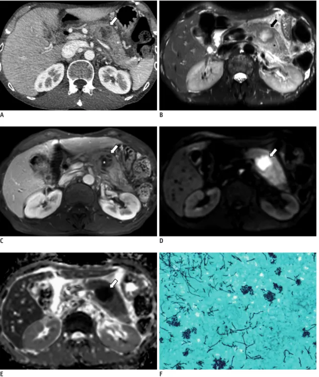
Fig. 1. Pancreatic candidiasis in 56-year-old man.
A. On axial CT scan obtained at portal phase, 3.4 cm multi-septated cystic lesion with lobulating contour (white arrow) is observed between body and tail of pancreas. Pancreas tissue around lesion is mildly swollen (asterisk). B. On T2-weighted axial MR image, cystic mass (black arrow) has relatively low signal intensity component compared with adjacent swollen pancreas (asterisk) at periphery and internal thick septa. C. In dynamic enhancement study, thick peripheral portion and internal septa of tumor (white arrow) are enhanced. Non-enhanced portion (asterisk) matches region of hyperintensity on T2-weighted image, indicating necrotic portion or cystic component of mass. D. On diffusion-weighted image with b-value of 800, lesion (white arrow) shows high signal intensity. E. Apparent diffusion coefficient value of corresponding area is low, indicating presence of diffusion restriction (white arrow). F. Gomori’s methenamine-silver staining highlights multiple fungal hyphae, consistent with Candida species (x 200).
A
C
E
B
D
F
1255 Pancreatic Candidiasis on MRI
Korean J Radiol 16(6), Nov/Dec 2015
kjronline.org
ampullectomy because of a recurrence of tubular adenoma with high-grade dysplasia. On physical examination, tenderness at the epigastrium and upper left quadrant of the abdomen was present. Except for mild elevation in inflammatory markers (C-reactive protein, 8.97 mg/dL;
erythrocyte sedimentation rate, 43 mm/hr), the other lab results at presentation were unremarkable, including serum amylase and lipase levels (47.5/25.6 U/L).
After the patient’s admission, computed tomography (CT) was performed to assess abdominal pain using a 64-slice multidetector CT scanner (LightSpeed VCT; GE Healthcare;
Milwaukee, WI, USA). Contrast-material-enhanced axial CT images showed a 3.4-cm multi-septated low-density lesion with a lobulating contour between the body and tail of the pancreas. Pancreas tissue around the lesion was mildly swollen, and peripancreatic fat infiltration was noted. However, the main pancreatic duct was not dilated.
There was no regional lymph node enlargement around the pancreas (Fig. 1A). Based on these CT findings, we considered a pseudocyst associated with pancreatitis, an unusual form of focal pancreatitis including an abscess, and pancreatic cystic tumor as part of the suite of differential diagnosis.
To further characterize the lesion, an MR study was performed using a 3T whole-body MR system (InteraAchieva 3T, Philips Healthcare, Best, the Netherlands) with a 16-channel phased-array receiver coil. For the enhancement studies, gadoxetic acid as a contrast agent (Gd-EOB-DTPA, Primovist or Eovist, Bayer Healthcare, Berlin, Germany) was administered intravenously. On a T2-weighted image, there was a relatively well-defined pancreatic cystic mass with irregular internal thick septa in the body portion of the pancreas that had a peripheral low signal intensity component compared with the adjacent swollen pancreas (Fig. 1B). During dynamic enhancement, the thick peripheral portion of the tumor and internal septa were enhanced. An internal non-enhancing portion within the mass that matched the high signal intensity on the T2- weighted image was detected, indicating a necrotic portion or cystic component (Fig. 1C). On the diffusion-weighted image with a b-value of 800, the lesion showed high signal intensity (Fig. 1D), and the corresponding area had a low value on the apparent diffusion coefficient map, indicating the presence of restricted diffusion compared with the surrounding inflamed pancreas parenchyma (Fig. 1E).
Based on these imaging findings, unusual pancreatitis with pancreatic abscess formation, solid pseudo-papillary
neoplasm, neuroendocrine tumor with cystic degeneration and malignant pancreas cystic tumor including mucinous cystic neoplasm and intraductal papillary mucinous neoplasm with an associated invasive carcinoma were considered in the differential diagnosis. Under suspicion of a malignant cystic tumor of pancreas based on the imaging findings, distal pancreatectomy with splenectomy was performed. At operation, this cystic lesion was palpated as a solid mass. In addition, severe pancreatitis with adhesion to the adjacent stomach and the liver was seen. An abscess was confirmed by frozen biopsy.
On histopathologic analysis, multifocal abscesses were seen in the pancreas parenchyma and peripancreatic tissue. Dilatation of branch ducts with intraluminal fungal organisms was noted, consistent with Candida infection.
In addition, Gomori’s methenamine-silver and periodic acid Schiff stains revealed fungal organisms that showed hyphae (Fig. 1F).
DISCUSSION
Risk factors for pancreatic fungal infection include prophylactic broad-spectrum antibiotics in patients with severe acute pancreatitis and abdominal surgical manipulation after an episode of acute pancreatitis.
Endoscopic retrograde cholangiopancreatography, use of total parenteral nutrition, and mechanical ventilation are additional risk factors (2). Other conditions that result in an altered host immune system such as steroid therapy, chemotherapy, acquired immunodeficiency syndrome (AIDS), immunosuppression therapy after organ transplantation, advanced age, and comorbid illnesses such as diabetes mellitus also predispose patients to fungal infection (5).
In our case, a history of repeated ampullectomy may have contributed to opportunistic infection of the pancreas by Candida despite the patient’s immunocompetent condition.
The increase in prevalence of solid organ transplantation has increased the number of patients who receive
immunosuppressive therapy. The Transplant-Associated Infection Surveillance Network reported a one-year cumulative incidence of pancreatic fungal infection of 3.4%; the most common invasive fungal infection was invasive candidiasis (53%) (8). In addition, AIDS patients are more susceptible to opportunistic infections including fungal infections but often do not present with symptoms.
Thus, the role of imaging studies is becoming increasingly
important (4). Although three recognizable forms of
1256
Seong et al.
Korean J Radiol 16(6), Nov/Dec 2015
kjronline.org pancreatic infection–infected pancreatic necrosis, infected
pseudocyst, and pancreatic abscesses–are known (5), the radiologic features of pancreatic candidiasis have been described in only one case report based on CT scans (9).
In our study, a cystic mass with an enhanced peripheral portion and necrotic inner portion that showed restricted diffusion on a background of pancreatitis was observed in MR images. Although there were no laboratory findings of pancreatitis, these findings could suggest the possibility of pancreatic abscess first. In addition, malignant pancreatic cystic tumors such as solid pseudo-papillary neoplasms, neuroendocrine tumors with cystic degeneration, and malignant pancreas cystic tumors (i.e., mucinous cystic neoplasms or intraductal papillary mucinous neoplasms with an associated invasive carcinoma) could be considered as differential diagnoses based on imaging findings because of the presence of diffusion-restriction of the lesion and a relatively thick enhanced peripheral portion with irregular septa (10). However, poor enhancement of a peripheral solid portion and male predominance are not common in cases of neuroendocrine tumor with cystic degeneration, solid pseudo-papillary neoplasm, or mucinous cystic neoplasm (10).
Other differentiating features may be the presence of accompanying pancreatitis in the setting of a patient with predisposing factors for pancreatic fungal infections compared with pancreatic cystic tumors. Overlap of diffusion restriction between a fungal abscess and malignant cystic tumor can be explained as follows. The high viscosity of inflamed materials may restrict the freedom of motion of water molecules within the abscess cavity similar to the compact tumor cellularity of a malignant pancreatic tumor (11). Considering the limitation of diffusion-weighted imaging for the diagnosis of pancreatic candidiasis,
diagnosis should be based on a combination of conventional MR imaging and clinical setting.
We suggest that a relatively well-defined cystic mass with internal septation and diffusion restriction in the background of pancreatitis could be MR imaging findings of pancreatic candidiasis manifesting as a pancreatic abscess.
If a proper diagnosis is possible prior to surgical resection of the pancreas, the extent of surgery may differ from that of surgical resection to save more unaffected pancreatic
tissue because most pancreatic fungal infections can be managed by less invasive therapies such as necrosectomy, necrostomy, and/or percutaneous catheter drainage rather than anatomical pancreatic resection (2, 5).
In conclusion, pancreatic candidiasis can manifest as a solitary abscess that should be differentiated from malignant cystic tumor of the pancreas. Therefore, pancreatic candidiasis could be considered in the
differential diagnosis of a pancreatic cystic mass, especially in patients with factors that predispose them to pancreatic fungal infections.
REFERENCES
1. Hoerauf A, Hammer S, Müller-Myhsok B, Rupprecht H. Intra- abdominal Candida infection during acute necrotizing pancreatitis has a high prevalence and is associated with increased mortality. Crit Care Med 1998;26:2010-2015 2. Kochhar R, Noor MT, Wig J. Fungal infections in severe acute
pancreatitis. J Gastroenterol Hepatol 2011;26:952-959 3. Pugliese F, Ruberto F, Cappannoli A, Perrella SM, Bruno K,
Martelli S, et al. Incidence of fungal infections in a solid organ recipients dedicated intensive care unit. Transplant Proc 2007;39:2005-2007
4. Reeders JW, Yee J, Gore RM, Miller FH, Megibow AJ.
Gastrointestinal infection in the immunocompromised (AIDS) patient. Eur Radiol 2004;14 Suppl 3:E84-E102
5. Shanmugam N, Isenmann R, Barkin JS, Beger HG. Pancreatic fungal infection. Pancreas 2003;27:133-138
6. Gregory DW. Saturday Conference: Candida infections. South Med J 1982;75:339-345
7. Gagnier JJ, Kienle G, Altman DG, Moher D, Sox H, Riley D; CARE Group. The CARE guidelines: consensus-based clinical case report guideline development. J Clin Epidemiol 2014;67:46-51
8. Pappas PG, Alexander BD, Andes DR, Hadley S, Kauffman CA, Freifeld A, et al. Invasive fungal infections among organ transplant recipients: results of the Transplant-Associated Infection Surveillance Network (TRANSNET). Clin Infect Dis 2010;50:1101-1111
9. Howard JM, Bieluch VM. Pancreatic abscess secondary to Candida albicans. Pancreas 1989;4:120-122
10. Kalb B, Sarmiento JM, Kooby DA, Adsay NV, Martin DR. MR imaging of cystic lesions of the pancreas. Radiographics 2009;29:1749-1765
11. Wang Y, Miller FH, Chen ZE, Merrick L, Mortele KJ, Hoff FL, et al. Diffusion-weighted MR imaging of solid and cystic lesions of the pancreas. Radiographics 2011;31:E47-E64