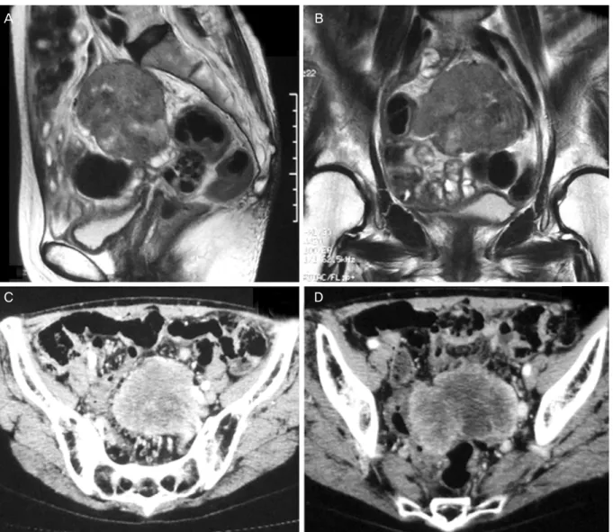
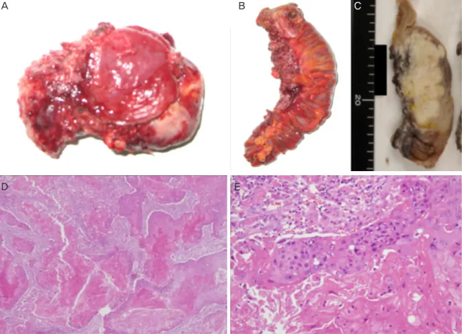
Long-term recurrence-free survival of a patient with advanced pure primary ovarian squamous cell carcinoma treated with dose-dense paclitaxel combined with carboplatin
6
0
0
전체 글
(2)
(3)
(4)
(5)
(6)
수치
관련 문서
임계 에너지는 핵분열 하는 핵종에 따라서 다르므로 중성자를 흡수했다 해서 모든 핵종이 핵분열을 일으키는 것은 아님.. 235 U와 같이 정지상태의 중 성자를
Median overall survival of 28 multiple myeloma patients treated with Cyclophosphamide-Prednisone combination regimens as a primary therapy was 115 weeks.. Median
In addition, the expression of REDD1 was increased when Huh7 cells were treated with PA, and overexpression of REDD1 affected cell survival and lipid
Concentration-Dependent inhibition of cell viability by adenosine in human oral fibroblast and FaDu human head and neck squamous cell carcinoma · · ·
It also suggest that cell proliferation inhibits by a novel signal transduction for adenosine effect in human FaDu hypopharynx squamous cell carcinoma cells....
Differential Expression of Desmoglein1, Desmoglein3, Epithelial Membrane Antigen, Ber-EP4 and CD10 in Basal Cell Carcinoma and Squamous Cell Carcinoma
Objective: The purpose of this study was to analyze recent trend in incidence of basal cell carcinoma and squamous cell carcinoma in patients from the Gwangju City
유도 핵반응은 자발적인 핵분열 (Spontaneous Nuclear Reaction)과는 달리 다른 물질과의 작 용으로 원자가 반응하여 여러 가지 생성물을 만들어
