Original Articles Korean Circulation J 1999;;;;29((((4))))::::382-391
만성 신부전증에서 좌심실 이완기능의 심초음파 지표에 미치는 혈액투석의 효과
세종병원 내과,
1이화여자대학교 의과대학 내과학교실
2전 성 희
1
·박 성 훈2
The Effect of Hemodialysis on the Echocardiographic Indexes of Left Ventricular Diastolic Function in Chronic Renal Failure
Seong Hee Jeon, MD
1and Seong Hoon Park, MD
21
Department of Internal Medicine, Sejong General Hospital, Puchon,
2
Department of Internal Medicine, Ewha Womans University, College of Medicine, Seoul, Korea
ABSTRACT
Background and Objectives:The assessment of left ventricular (LV) diastolic function is important in chronic renal failure because abnormal LV diastolic function has been frequently described in patients on maintenance hemodialysis both during the dialysis and in the dialysis-free interval despite the normal LV systolic function.
But the echocardiographic indexes of LV diastolic function is known to be affected by several factors such as loading condition, LV compliance and heart rate. The purpose of this study is to investigate the effect of hemodialysis on the echocardiographic indexes of left ventricular diastolic function in chronic renal failure.
Materials and Methods:We examined transmitral flow velocity, pulmonary venous flow velocity, and mitral annulus velocity in 20 patients (15 men and 5 women, average 50±14, range 19-69 years) of chronic renal failure with normal LV systolic function by echocardiography before and after hemodialysis. Results:
1) According to the body weight change (from 59.5±8.3 to 57.2±8.1 kg, p=0.0001), after hemodialysis, inferior vena cava dimension (from 18±4 to 13±5 cm, p=0.0001), left ventricular end-diastolic dimension (from 57±6 to 53±7 cm, p=0.0001), and left ventricular outflow tract (LVOT)-time velocity integral (TVI, from 26±5 to 23±5 cm, p=0.004), which reflect intravascular blood volume, decreased significantly. 2) The peak velocity of early transmitral flow (E, from 0.79±0.14 to 0.64±0.11 m/s, p=0.0001), the peak velocity of late transmitral flow (A, from 0.84±0.21 to 0.78±0.21 m/s, p=0.011), and E/A ratio (from 0.99±0.25 to 0.87±0.27, P=0.007) decreased significantly, and deceleration time (DT, from 241±48 to 267±59 ms, p=
0.055) showed tendency of prolongation after hemodialysis. 3) Peak systolic velocity of pulmonary venous flow decreased significantly after hemodialysis (from 0.65±0.11 to 0.59±0.12 m/s, p=0.042). 4) The difference between duration of reversal flow of pulmonary vein and duration of transmitral flow during atrial contraction (ADD) did not change significantly after hemodialysis (from 5±31 to 1±29 ms, p=0.502), and did not correlate with the change of peak velocity of early transmitral flow during hemodialysis (DMVE, r=
0.390, p=0.089). 5) The peak early diastolic velocity (Ean, from 0.07±0.02 to 0.06±0.02 m/s, p=0.002)and Ean/the peak late diastolic velocity (Aan) ratio (from 0.78±0.27 to 0.62±0.19, p=0.003) of medial annulus
논문접수일:1998년 12월 19일 심사완료일:1999년 4월 2일
교신저자:박성훈, 158-050 서울 양천구 목동 911-1 이화여자대학교 의과대학 내과학교실 전화:(02) 650-5018・전송:(02) 650-2076
E-mail:[email protected]
of mitral valve decreased significantly after hemodialysis. Conclusion:Hemodialysis, which reduces LV preload by fluid removal, changes the echocardiographic indexes of left ventricular diastolic function in chronic renal failure. Preload condition need to be accounted for when we evaluate the LV diastolic function with echocardiography. ((((Korean Circulation J 1999;29((((4)))):382-391))))
KEY WORDS:Echocardiography·Hemodialysis·LV diastolic function·Preload.
서 론
만성 신부전증에서는 고혈압, 체액저류, 빈혈, 동맥 경화증, 허혈성 심질환, 동정맥루, 대사불균형, 심근 석 회화, 심낭염 등에 의해 좌심실기능이 저하될 수 있 다.
1)만성 신부전증 환자의 좌심실 수축기능은 혈액투 석에 의해 호전된다고 알려졌으나,
2-4)좌심실 이완기능 에 대한 연구는 드물다. 그러나 혈액투석 환자에서 좌 심실 수축기능이 정상이더라도, 좌심실 이완기능 장애 가 혈액투석 중이나
5)투석과 투석사이의 기간에
1)6)발 견되는 빈도가 높고, 신이식 수술 전후 폐부종의 발생 은 좌심실 수축기능 보다는 좌심실 이완기능의 장애와 관련이 높기 때문에
7)만성 신부전증 환자에서 좌심실 이완기능에 대한 정확한 평가가 필요하다.
좌심실 이완기능의 평가방법은 심도자술, 방사성 동 위원소를 이용한 심혈관 조영술, 도플러 심초음파를 이 용하는 방법 등이 있으며, 이중 도플러 심초음파는 비 관혈적이며 반복, 추적검사가 용이하고, 관혈적 방법인 심도자술로 측정한 지표와 잘 일치하므로
8-10)좌심실 이완기능의 평가에 흔히 이용된다. 특히 도플러 심초음 파를 이용하여 승모판 혈류속도나 폐정맥 혈류속도를 측정하거나 도플러 조직 영상(Doppler tissue imaging) 을 시행하여 승모판륜의 심근속도 등을 측정함으로써 좌심실 이완기능을 평가할 수 있다.
8)9)11-15)한편 좌심 실 이완기능은 부하조건에 의해 영향받는다고 알려져
있는데,
14)16-19)특히 만성 신부전증 환자는 혈액투석으
로 과다했던 유효순환 혈액량, 즉 전부하가 감소되므로 혈액투석 후 좌심실 이완기능의 평가결과가 달라질 수 있음을 예측할 수 있다. 그러나 지금까지 폐정맥 혈류 속도나 승모판륜의 심근속도를 측정하여 혈액투석 즉 전부하 감소로 인한 좌심실 이완기능 지표의 변화에 대 해 관찰한 연구는 없다.
따라서 본 연구는 만성 신부전증 환자를 대상으로 혈
액투석 전후에 도플러 심초음파를 이용하여 승모판 혈 류속도와 폐정맥 혈류속도를 측정하고, 도플러 조직 영 상을 이용하여 승모판륜의 심근속도를 측정하여, 만성 신부전증 환자에서 혈액투석이 좌심실 이완기능 지표 에 미치는 영향에 대해 알아보고자 하였다.
대상 및 방법
대 상
이화여자대학교 부속목동병원 인공신장실에서 유지 혈액투석을 받고 있는, 혈역학적으로 안정되고, 좌심실 구혈률이 50% 이상이며 국소벽운동기능장애(regional wall motion abnormality)가 없는 만성 신부전증 환자 20명을 대상으로 하였다. 대상환자들은 모두 본 임상 연구에 동의하였다. 대상환자는 모두 20명(남자 15명, 여자 5명)이었고, 연령은 평균 50±14세로 19세에서 69세까지 분포하였다. 임상적으로 관상동맥질환, 선천 성 심질환, 판막질환, 중등도 이상의 심낭삼출액, 부정 맥, 폐울혈이나 하지부종이 동반된 환자들은 연구대상 에서 제외하였다.
방 법
혈액투석 전후 각각 30분 이내에 체중, 혈압, 심박수 측정 및 심초음파 검사를 시행하였다. 검사자간의 오차 (interobserver variation)를 줄이기 위하여 심초음파 검사에 충분히 훈련된 심장내과 의사 1명이 지속적으 로 시행하였고, 측정오차를 줄이기 위해 모든 심초음파 도 측정치는 5회 연속된 심주기의 값을 측정하여 평균 값을 사용하였다.
M형 심초음파 검사
심초음파 기기(Acuson 128 XP/10, Acuson Cor-
poration, California)의 2.5 MHz 탐촉자를 이용하여
미국 심초음파 학회가 제시한 방법으로
20)좌심실의 수
축기말 직경(LVESD)과 확장기말 직경(LVEDD)을 측 정하고, 좌심실구혈률(LVEF)을 다음의 공식으로 계산 하였다.
LVEF(%)=(LVEDD
2-LVESD
2)/LVEDD
2×100 하대정맥 직경은 subcostal view에서, 호기말에 심 전도의 P파가 시작되는 때에, 우심방에서 1~2 cm 떨 어진 부위의 하대정맥 직경을 측정하였다(Fig. 1).
이면성 및 간헐파형 도플러 심초음파 검사
좌심실유출로 TVI는 심첨 오방도(apical 5-chamber view)에서 sample volume을 좌심실유출로에 두고 간 헐파형 도플러로 측정하였다. Canadian Consensus Recommendations에서 제시한 방법에 따라,
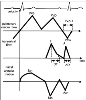
21)환자에 게 긴장을 피하게 하기 위해 정상호흡을 시킨 상태에서 호기말에 기록하였으며, 승모판 혈류 파형은 심첨 사방도 (apical 4-chamber view)에서 sample volume(size 2 mm)을 승모판 첨부(leaflet tip)에 위치시켰고, 폐정 맥 혈류 파형은 같은 단면도에서 색채혈류를 참조하여 벽운동흔적을 최소화하기 위하여 sample volume을 좌 심방에서 1 cm 지난 부위의 우측 상폐정맥(right su- perior pulmonary vein)에 위치시켜서 가능한 한 초음 파의 진행방향이 혈류와 평행하도록 한 후 초당 50 mm 의 속도로 기록하였다(Fig. 2).
승모판 혈류 파형으로부터 초기 급속 충혈기 유입에 해당하는 이완기의 최고 혈류속도(E), 후기 심방 수축 기의 최고 혈류속도(A), E/A 비, E파의 감속시간(de- celeration time, DT)과 A파의 기간(AD)을, 폐정맥
혈류 파형으로부터는 수축기혈류의 최고속도(PVS), 이 완기혈류의 최고속도(PVD), 좌심방 수축에 따른 역류 혈류의 최고속도(PVA)와 PVA파의 기간(PVAD)을 측정하였다.
ADD(A Duration Difference)는 폐정맥 역류혈류의 기간(PVAD)에서 승모판 혈류의 A파의 기간(AD)을 뺀 값으로, DMVE(Difference of transmitral Velocity E)는 투석 전 E에서 투석 후 E를 뺀 값으로 정의하였다.
도플러 조직 영상(Doppler tissue imaging, DTI)
환자를 좌측와위로 하고 호기말 상태에서, 같은 기기 에서 DTI 기능을 활성화시킨 후, 심첨 사방도(apical 4-chamber view)에서 3 mm의 sample volume을 승모판 외륜(lateral mitral annulus) 혹은 승모판 내륜 (medial mitral annulus)의 심근에 위치하게 한 후 초 당 50 mm의 속도로 기록한 승모판륜의 심근속도 파형 에서, 수축기의 승모판륜의 최고 심근속도(San), 초기 급속 충혈기에 해당하는 이완기의 승모판륜의 최고 심 근속도(Ean), 후기 심방수축기의 승모판륜의 최고 심 근속도(Aan)와 Ean/Aan 비를 측정하였다(Fig. 2).
통계처리
각각의 지표는 5회 연속된 심주기의 값을 측정하여 평균과 표준편차로 나타내었다. 혈액투석 전후의 측정 치의 비교는 paired Student’s t-test를, ADD와 DMVE 의 상관관계는 Pearson 상관분석으로 유의성을 검정 하였다. p값이 0.05이하이면 통계적으로 유의한 것으로
Fig. 1. Measurement of the diameter of inferior ve- nacava. A, Two-dimensional echocardiogram;B, M-mode echocardiogram. IVC diameter was me- asured at the beginning of the p wave on the electrocardiogram with the patient holding breath at end-expiration. IVC:inferior vena cava, RA:
right atrium.
A A
A A B B B B
판정하였다.
결 과
체중, 혈압 및 심박수
혈액투석 후 환자들의 체중은 59.5±8.3 kg에서 57.2±8.1 kg으로 평균 2.2±0.4 kg 감소하여 유의한 변화를 보였으며(p=0.0001), 수축기 혈압(투석 전 154±26 mmHg, 투석 후 159±27 mmHg, p=0.480), 이완기 혈압(투석 전 93±12 mmHg, 투석 후 94±
15 mmHg, p=0.761) 및 심박수(투석 전 분당 74±
10회, 투석 후 분당 72±6회, p=0.474)는 유의한 변 화가 없었다(Table 1).
M형 심초음파 검사 결과
혈액투석 후 하대정맥 직경은 18±4 mm에서 13±
5 mm로(p=0.0001), 좌심실이완기말 직경은 57±6 mm 에서 53±7 mm로(p=0.0001) 유의하게 감소하 였으며, 좌심실수축기말 직경은 37±4 mm에서 36±8 mm로 유의한 변화를 보이지 않았다. 좌심실구혈률은 58±12 %에서 54±12%로(p=0.008) 유의하게 감 소하였다(Table 2).
간헐파형 도플러 심초음파 검사 결과
혈액투석 후 좌심실유출로 TVI는 26±5 cm에서 23±5 cm로(p=0.004) 유의하게 감소하였다. 승모판 혈류 파형에서 혈액투석 후 E(투석 전 0.79±0.14 m/s, 투석 후 0.64±0.11 m/s, p=0.0001), A(투석 전 0.84±0.21 m/s, 투석 후 0.78±0.21 m/s, p=
0.011)와 E/A 비(투석 전 0.99±0.25, 투석 후 0.87
±0.27, p=0.007)는 유의하게 감소하였고, DT는 241±48 ms에서 267±59 ms로 증가되는 경향을 보 였으나(p=0.055) 통계적으로 유의하지 않았다(Table 3). 폐정맥 혈류 파형에서 혈액투석 후 PVS는 유의한 감소를 보였으나(투석 전 0.65±0.11 m/s, 투석 후 0.59±0.12 m/s, p=0.042) PVD, PVA, PVAD는 유 의한 변화가 없었다(Table 3).
ADD는 투석 전 5±31 ms에서 투석 후 1±29 ms 로 유의한 변화를 보이지 않았다(p=0.502). 전부하 Table 1. Changes of clinical variables obtained before and after hemodialysis in 20 patients
Predialysis Postdialysis p Value Body Weight (kg) 59.5± 8.3 57.2± 8.1 0.0001 Heart Rate (/min) 74 ±10 72 ± 6 NS Systolic BP 154 ±26 159 ±27 NS Diastolic BP 93 ±12 94 ±15 NS Data are expressed as mean values±standard deviations NS:not significant
Table 2. Changes of M-mode echocardiographic va- riables obtained before and after hemodialysis in 20 patients
Predialysis Postdialysis p Value IVC diameter 18± 4 13± 5 0.0001 LVEDD (mm) 57± 6 53± 7 0.0001 LVESD (mm) 37± 4 36± 8 NS LVEF (%) 58±12 54±12 0.008 Data are expressed as mean values±standard deviations NS:not significant, IVC:inferior vena cava, LVEDD:
left ventricular end-diastolic dimension, LVESD:left ventricular end-systolic dimension, LVEF:left ventricular ejection fraction
Fig. 2. Measurement of pulmonary venous flow velocities (upper panel), transmitral flow velocities (middle panel), and mitral annulus velocities (lower panel).
A:peak velocity of late transmitral flow, Aan:peak
late diastolic velocity of mitral annulus, AD:duration
of late transmitral flow, DT:deceleration time of early
transmitral flow, E:peak velocity of early transmitral
flow, Ean:peak early diastolic velocity of mitral an-
nulus, PVA:peak velocity of pulmonary vein reversal
flow during atrial contraction, PVAD:duration of pul-
monary vein reversal flow during atrial contraction,
PVD: peak velocity of diastolic pulmonary venous
flow, PVS:peak velocity of systolic pulmonary venous
flow, San:peak systolic velocity of mitral annulus.
감소로 인한 E의 감소정도가 좌심실 이완기말압의 변 화와 관계가 있는지 알아보기 위해 DMVE와 ADD의 상관관계를 분석해 보았으나, 이 두 변수간에 유의한 상관관계를 보이지 않았다(r=0.390, p=0.089).
도플러 조직 영상 결과
승모판 내륜의 심근속도 파형에서 Ean(투석 전 0.07
±0.02 m/s, 투석 후 0.06±0.02 m/s, p=0.002)와 Ean/Aan 비(투석 전 0.78±0.27, 투석 후 0.62±
0.19, p=0.003)가 의미있게 감소하였으나(Fig. 3) 승 모판 외륜의 심근속도는 유의한 변화를 보이지 않았다
(Fig. 4, Table 3).
고 찰
좌심실 이완기능은 심도자술에 의해 압력과 용적의 변화를 측정하는 관혈적 방법 외에도 방사성 동위원소 를 이용한 심혈관 조영술(radionuclide angiocardio- graphy),
11)12)22)전산화 단층 촬영술, 자기 공명 영상을 이용하여 이완기 심실용적의 변화를 관찰하거나,
23-25)심초음파를 이용한 비관혈적 방법으로 평가할 수 있다.
이중 심초음파 검사는 반복, 추적검사가 용이하고, 환 자에게 부담이 적고, 관혈적 방법인 심도자술에 의한 지표와 잘 일치하므로
8-10)좌심실 이완기능 평가에 흔 히 이용된다. 특히 도플러 심초음파를 이용하여 승모판 혈류속도와 폐정맥 혈류속도를 측정하거나 도플러 조 직 영상을 시행하여 승모판륜의 심근속도를 측정함으 로써 좌심실 이완기능을 평가할 수 있다.
8)9)11-15)최근 까지 만성 신부전증 환자에서 혈액투석 후 좌심실 이완 기능의 변화에 대한 연구들에서는 승모판 혈류 파형만 이용되었으나, 승모판륜의 심근속도가 전부하의 영향을 받지 않으며, 승모판 혈류 파형만으로는 감별이 불가능 Table 3. Changes of echocardiographic variables ob-
tained before and after hemodialysis in 20 patients Predialysis Postdialysis p Value LVOT-TVI (cm) 26±5 23±5 0.004 Transmitral flow
E (m/s) 0.79±0.14 0.64±0.11 0.0001 A (m/s) 0.84±0.21 0.78±0.21 0.011 E/A 0.99±0.25 0.87±0.27 0.007
DT (ms) 241 ±48 267 ±59 NS
Pulmonary venous flow
PVS (m/s) 0.65±0.11 0.59±0.12 0.042 PVD (m/s) 0.45±0.09 0.41±0.10 NS PVA (m/s) 0.27±0.05 0.25±0.06 NS PVAD (ms) 149 ±21 142 ±20 NS Mitral annulus motion
medial (septal) side
San (m/s) 0.07±0.02 0.07±0.02 NS Ean (m/s) 0.07±0.02 0.06±0.02 0.002 Aan (m/s) 0.10±0.02 0.10±0.02 NS Ean/Aan 0.78±0.27 0.62±0.19 0.003 Lateral side
San (m/s) 0.09±0.03 0.10±0.03 NS Ean (m/s) 0.10±0.04 0.10±0.04 NS Aan (m/s) 0.11±0.03 0.11±0.03 NS Ean/Aan 0.99±0.46 0.93±0.40 NS Data are expressed as mean values±standard deviations.
NS:not significant, A:peak velocity of late trans- mitral flow, Aan:peak late diastolic velocity of mitral annulus, DT:deceleration time of early transmitral flow, E:peak velocity of early transmitral flow, Ean peak early diastolic velocity of mitral annulus, PVA:
peak velocity of pulmonary vein reversal flow during atrial contraction, PVAD:duration of pulmonary vein reversal flow during atrial contraction, PVD:peak velocity of diastolic pulmonary venous flow, PVS:
peak velocity of systolic pulmonary venous flow, San:
peak systolic velocity of mitral annulus
Fig. 3. Scatterplots of comparison of the peak early
diastolic velocity (Ean) of medial mitral annulus before
and after hemodialysis.
한 위정상화(pseudonormalization)와 정상 이완기능 을 감별할 수 있다는 보고가 있어,
26)본 연구에서는 혈 액투석과 같이 전부하의 현저한 변화가 발생하는 경우 에도 승모판륜의 심근속도가 전부하의 영향을 받지 않 는지 알아보기 위하여 폐정맥 혈류 파형과 승모판륜의 심근속도도 동시에 평가하였다.
승모판 혈류 파형을 이용한 좌심실 이완기능 평가의 유용성이 고혈압을 비롯한 여러 질환에서 보고 되었으 나,
27)승모판 혈류 파형이 좌심실 이완기능을 대변하기 에는 좌심실 경직도, 좌심실 이완속도, 심낭압, 좌심방 과 좌심실간의 이완기 압력차이, 부하조건, 연령, 심박 수 등에 영향을 받으므로 이것만으로 이완기능을 평가 하는 것은 정확도가 떨어지며, 심방세동이나 동성빈맥, 방실전도장애 등의 환자에서 정확한 측정이 용이하지 않다는 단점이 있다.
28)승모판 혈류 파형을 이용한 혈 액투석 후 좌심실 이완기능의 변화에 대한 이전 연구결 과들은 다양하게 보고되고 있다. Gupta 등은 IVRT (isovolumic relaxation time)와 DT가 감소되었고,
29)Choi 등은 E, A가 감소하였고 E/A 비는 증가하여 좌 심실 이완기능이 호전된 증거라고 한 반면,
30)Rozich 등과 Sadler 등은 A는 변화없이 E와 E/A비가 감소하 였다고 하였다.
31)32)본 연구 결과는 E, A와 E/A비가 모두 감소하였다. 하대정맥 직경, 좌심실이완기말 직경 및 좌심실유출로 TVI의 감소는 유효 순환 혈액량이 감 소되었음을 의미하고 이로 인해 좌심방과 좌심실사이 압력차(transmitral pressure gradient)가 감소하여 초기 급속 충혈기 혈류가 감소되었음에도 불구하고 보 상적으로 증가되어야 할 후기 심방수축기 혈류가 오히 려 감소된 결과로 해석할 수 있다. 이는 정상인에서 lower body negative pressure를 이용하거나, nitro- glycerin를 연속 정주하여 전부하를 감소시켰을 때 E 와 E/A 비가 감소되었다는 이전 연구 결과들과 유사하
며,
17)33)또한 유지 혈액투석으로 체액이 감소되지 않
았을 때와 비교하여 체액이 감소된 경우에만 E와 E/A 비가 감소되었다는 보고도 있으므로,
32)34)혈액투석의 효과는 전부하를 감소시켜 승모판 혈류 파형의 변화를 가져온다고 볼 수 있다.
폐정맥 혈류 파형은 좌심방의 압력이나 좌심실 이완 기말압을 예측하는데 유용함이 알려지면서
10)35)좌심실 이완기능을 평가하는 데 도움을 주고 있으나 승모판 혈 류 파형과 마찬가지로 연령, 부하조건, 좌심방압, 승모 판기능, 좌심실 유순도(compliance) 등에 의해 영향을 받는다.
10)18)36)본 연구결과 PVS, PVD, PVA, PVAD 모두 혈액투석 후 감소되는 경향을 보였으나 PVS 이 외에는 통계학적으로 유의하지 않았다. 좌심방이 폐정 맥과 좌심실의 open conduit이므로, 승모판 혈류 파형 의 초기 급속 충혈기의 최고 혈류속도(E)와 폐정맥 이 완기 혈류의 최고 속도(PVD)는 밀접한 상관관계가 있 으며, E와 PVD는 좌심방압, 심근의 점탄성도(visco- elastic forces) 등의 같은 요인들에 의해 변화한다고 기술한 Nishimura 등의 연구에서도, nitroglycerine 정주 후 E는 감소하였으나 PVD는 유의한 변화를 보이 지 않았다.
18)또한 lower body positive pressure를 적용하거나, nitroglycerine을 투여하여 전부하를 변화 시킨 후, 승모판 혈류 파형과 폐정맥 혈류 파형의 변화 를 동시에 관찰한 다른 연구들에서도, E와 PVD의 변 화가 일치하지 않았다.
37) 38)그러므로 E는 현저한 변화 를 보였음에도 PVD가 유의한 변화를 보이지 않은 것 은 좌심방의, 폐정맥과 좌심실의 open conduit 기능 이 Fig. 4. Scatterplots of comparison of the peak early
diastolic velocity (Ean) of lateral mitral annulus before
and after hemodialysis.
외의 다른 내적 속성이 이와 같은 결과를 보인 것으로 생각되며, 본 연구만으로는 이에 대한 해명이 불가능하 고 좀 더 많은 연구가 진행되어야 할 것이다.
승모판 혈류 파형과 폐정맥 혈류 파형에서 결정되는 ADD(폐정맥 역류혈류의 기간에서 승모판 혈류 A파의 기간을 뺀 값)는 연령 및 부하상태에 영향을 받지 않는 다고 알려져 있는데,
10)35)본 연구에서 ADD가 투석 후 유의한 변화를 보이지 않은 것은 기존의 연구결과와 동 일하였다. 또한 ADD는 심방수축시 좌심실압의 증가정 도와 좌심실 이완기말압과 유의한 상관관계가 있어서, 좌심실 이완기말압이 높으면 좌심실 유순도(compli- ance) 감소로 폐정맥 역류혈류 기간(PVAD)이 심방수 축기의 승모판 혈류 기간(AD) 보다 길어진다.
10)35)39)이를 근거로 본 연구에서는 전부하 감소로 인한 E의 감소정도(DMVE)가 좌심실 이완기말압의 변화와 관계 가 있는지 알아보기 위해 DMVE와 ADD의 상관관계 를 분석해 보았으나, 이 두 변수간에 유의한 상관관계 가 없었다.
1989년부터 Isaaz에 의해 시작된 도플러 조직 영상 은 기존의 색채 도플러 영상기법과 정반대되는 기법을 통하여 혈류에 대한 신호를 제거하고 저속의 심근벽 속 도만을 측정, 이를 이면성 심초음파에 중첩시키는 방법 으로
40)승모판륜이나 좌심실 후벽, 심첨부 등에서 심근 의 속도를 측정하여 이완기능을 평가한다. 승모판륜은, 심첨부가 비교적 고정되어 있기 때문에 좌심실 장축을 따라 운동하여, 동율동 환자에서 수축기에는 심실을 향 하여 움직여서 양성파를, 초기 급속 충혈기와 후기 심 방 수축기에는 심방을 향해 움직여서 승모판 혈류 파형 과 유사한 두 개의 음성파를 보인다. 그러나 승모판륜 의 심근 속도는 structural mechanic을 반영하고 승모 판 혈류파형은 fluid dynamic을 반영하므로 각각의 이 완기 승모판륜의 최고 심근속도(Ean, Aan)와 승모판 혈류의 최고속도(E, A)가 일치하지는 않으나 Ean/Aan 비와 E/A 비는 잘 일치한다고 알려져 있다. 승모판륜 의 심근속도 파형의 Ean파는 승모판 혈류의 E파와 동 시에 시작되나, 최고속도(Ean)는 승모판 혈류의 최고 속도(E) 보다 평균 20 ms정도 먼저 나타나고, 승모판 혈류의 E파가 끝나기전에 끝난다. 이는 초기 급속 충혈 기동안 potential elastic energy가 방출되면서 좌심실 이 확장되면(“elastic recoil” ), 좌심실 이완기압이 감 소되고 좌심방압이 증가하여 좌심방에서 좌심실로의
혈류가 증진(“suction effect” )되기 때문이다.
14)그러 므로 승모판륜의 초기 급속 충혈기의 운동은 좌심실 이 완기능을 결정하는 중요한 요인인 elastic recoil의 지 표로서, 좌심실 비후나 제한성 심근병증에서와 같이 elastic recoil이 감소하면 좌심실 충만은 주로 승모판 전후의 압력차에 의해 수동적으로 이루어져 E가 Ean 보다 먼저 나타나므로 이완기능 장애의 기전을 밝히는 데 도움을 줄 수 있다고 보고되고 있다.
15)41)승모판륜의 심근속도는 승모판 혈류속도와 같이 연 령에 따라 변화하여 40대 이후가 되면 Ean가 감소하 여 Ean/Aan 비가 1보다 작아진다.
15)26)40)승모판륜의 심근속도가 전부하의 영향을 받는지에 관한 연구로서, Garcia 등은 28명의 교착성 심낭염, 제한성 심근병증 및 정상인에서 승모판 외륜(lateral mitral annulus)의 Ean을 측정하여, 승모판 혈류의 E와 상관관계가 없으 므로 Ean은 전부하에 비교적 영향을 덜 받는다고 하였 고,
41)Nagueh 등은 우심도자술을 시행받았던 60명의 환자에서 폐모세혈관 쐐기압과 승모판 외륜(lateral mitral annulus)의 Ean간의 상관관계가 없었으므로 Ean은 전부하의 영향을 받지 않는다고 하였으나,
42)이 두 연구는 부하조건(loading condition)을 변화시키지 않았다.
21명의 환자에서 500~700 ml의 식염수를 정맥주 사(saline loading)하거나 심박수가 기저치에서 분당 10회 이상 증가할 때까지 nitroglycerine을 평균 99±
48 μg/min의 속도로 연속 정주하는 방법으로 전부하 을 변화시켜 승모판 내륜(medial mitral annulus)의 심 근속도 변화에 대해 연구한 Sohn 등은 승모판 혈류의 E가 유의한 변화를 보였음에도 불구하고 Ean은 변화 가 없었다고 하였다.
26)그러나 본 연구에서는 승모판 외륜과 내륜에서 각각의 심근속도를 측정한 결과, 외륜 에서는 전부하의 영향을 받지 않았으나 내륜에서는 전 부하의 영향을 받는 것으로 나타났다. 승모판륜의 심근 속도의 변화가 내륜과 외륜에서 차이를 보인 것은 본 연구대상에서 폐동맥 고혈압이나 우심실 기능의 장애 가 있는 환자들은 제외하였으므로, 승모판 내륜에서 심 근속도 측정시 우심실에 의한 오차가 있었을 가능성은 적으며, Nagueh 등의 연구
42)에서와 같이 혈액투석 전 은 lateral Ean=0.0006+1.4463×medial Ean(r=
0.792, p=0.0001), 투석 후는 lateral Ean=0.0026
+1.6611×medial Ean(r=0.825, p=0.0001)의 상관
관계를 보였으므로 내륜과 외륜 중 어느 쪽 결과가 더 정확하다고 단정지을 수 없다. 본 연구에서는 환자들의 체중이 평균 2.2±0.4 kg 감소하였으므로 Sohn 등의 연구결과와 일치하지 않는 것은 부하조건의 변화정도 에 따른 차이로 사료되나 향후 부하조건의 변화정도를 다양하게 하여 더 많은 환자를 대상으로 한 연구가 필 요할 것으로 생각한다.
본 연구결과, 심박수와 혈압은 변화가 없었고 좌심실 구혈률은 감소하였는데, 이는 좌심실확장기말 내경의 감소로 인하여 Frank-Starling mechanism이 덜 효과 적으로 작용한 것에 기인한다고 생각된다. 이러한 투석 에 의한 혈역학적 효과에 대해서는 각 연구자마다 결과 가 다양하였다. 즉, Fernado 등은 투석후 심박출량, 일 회박출량, 심박동수에 변화가 없었다고 하였고, Cha- ignon 등은 좌심실구혈률의 변화없이 일회박출량 및 혈압이 감소하였다고 하였으며, O’Regan 등은 심박수 와 좌심실구혈률이 증가하고 심박출량은 감소하였다고
하였다.
43-45)이는 관찰대상의 차이와 간접적인 심기능
측정 지표를 사용함에 의한 것으로 생각된다.
결론적으로, 만성 신부전증 환자에서 심초음파로 측 정한 좌심실 이완기능 지표들은 투석에 의한 전부하의 감소로 투석 후 변화된다는 사실을 확인하였으며, 심초 음파를 이용하여 좌심실 이완기능의 평가시 전부하 상 태를 고려해야 할 것으로 생각한다.
본 연구의 제한점으로는 혈역학적으로 불안정하거나 좌심실구혈율이 50% 이하이거나 국소벽운동기능장애 (regional wall motion abnormality)가 있거나 부정맥 이 있는 환자들은 연구대상에서 제외하였으므로, 이러 한 환자들에서는 본 연구결과를 적용시킬 수 없다. 또 한 심초음파검사는 좌심실 이완기능을 비관혈적으로 측정하는 방법으로 본 연구의 대상환자들은 심도자술 의 적응증이 되지 않아서 심도자를 이용하여 혈역학적 지표들을 측정하지 못하였으나 향후 더 많은 환자들을 대상으로 심도자술을 이용한 혈역학적 지표와의 비교 연구가 필요할 것으로 생각된다.
요 약
서 론:
심초음파로 측정한 좌심실 이완기능의 지표들은 부 하조건에 의해 영향받는다고 알려져 있는데, 특히 만성
신부전증 환자는 혈액투석으로 과다했던 체액량, 즉 전 부하가 감소되므로 혈액투석 후 좌심실 이완기능의 평 가결과가 달라 질 수 있음을 예측할 수 있다. 따라서 본 연구는 만성 신부전증 환자에서 혈액투석이 심초음 파로 측정한 좌심실 이완기능 지표에 미치는 영향에 대 해 알아보고자 하였다.
대상 및 방법:
좌심실 구혈률이 50% 이상이며 국소벽운동기능장애 (regional wall motion abnormality)가 없는 만성 신 부전증 환자 20명을 대상으로 혈액투석 전후에 도플러 심초음파를 이용하여 승모판 혈류속도와 폐정맥 혈류 속도를 측정하고, 도플러 조직 영상을 이용하여 승모판 륜의 심근속도를 측정하였다.
결 과:
1) 대상환자들은 모두 20명(남자 15명, 여자 5명) 이었고, 연령은 평균 50±14세로 19세에서 69세까지 분포하였다.
2) 혈액투석 후 체중감소(투석 전 59.5±8.3 kg, 투 석 후 57.2±8.1 kg, p=0.0001)와 함께 하대정맥 직경 (투석 전 18±4 cm, 투석 후 13±5 cm, p=0.0001), 좌심실이완기말 직경(투석 전 57±6 cm, 투석 후 53
±7 cm, p=0.0001), 좌심실유출로 TVI(투석 전 26
±5 cm, 투석 후 23±5 cm, p=0.004) 등 혈관내 혈 액량을 반영하는 지표들이 유의하게 감소하였다.
3) 혈액투석 후 승모판 혈류의 E(투석 전 0.79±
0.14 m/s, 투석 후 0.64±0.11 m/s, p=0.0001), A(투석 전 0.84±0.21 m/s, 투석 후 0.78±0.21 m/s, p=0.011), E/A 비(투석 전 0.99±0.25, 투석 후 0.87±0.27, p=0.007)가 유의한 감소를 보였으며, DT는 증가하는 경향(투석 전 241±48 ms, 투석 후 267±59 ms, p=0.055)을 보였다.
4) 혈액투석 후 폐정맥의 수축기혈류가 유의하게 감 소하였다(투석 전 0.65±0.11 m/s, 투석 후 0.59±
0.12 m/s, p=0.042).
5) 폐정맥 혈류의 PVA파 기간과 승모판 혈류의 A 파 기간의 차(ADD)는 혈액투석 후 유의한 변화를 보 이지 않았으며(투석 전 5±31 ms, 투석 후 1±29 ms, p=0.502), 투석으로 인한 승모판 혈류 E의 변화 (DMVE)와 유의한 상관관계가 없었다(r=0.390, p=
0.089).
6) 혈액투석 후 승모판 내륜의 Ean(투석 전 0.07±
0.02 m/s, 투석 후 0.06±0.02 m/s, p=0.002)와 Ean/
Aan 비(투석 전 0.78±0.27, 투석 후 0.62±0.19, p
=0.003)가 유의한 감소를 보였다.
결 론:
심초음파로 측정한 좌심실 이완기능 지표들은 투석 에 의한 전부하의 감소로 투석 후 변화된다는 사실을 확인하였으며, 만성 신부전증 환자에서 좌심실 이완기능 의 평가시 전부하 상태를 고려해야 할 것으로 생각한다.
중심 단어 :심초음파・전부하・좌심실 이완기능・혈액 투석.
본 연구의 요지는 1997년 대한 순환기학회 제41차 추계학 술대회에서 구연되었음.
REFERENCES
1)
Kramer W, Wizemann V, Lammlein G, Thormann J, Kindler M, Schlepper M, et al. Cardiac dysfunction inpatients on maintenance hemodialysis Ⅱ. Systolic and diastolic properties of the left ventricle assessed by invasive methods. Contrib Nephrol 1986;52:110-24.
2)
Park KS, Kim SA, Tahk SJ, Yang JY, Shim WH, Lee HY, et al. Echocardiographic evaluation of left ventricularfunction in dialysis patients. Korean J Nephrol 1984;3:
46-54.
3)
Koji T, Sugawa M, Izumi K, Takahashi T, Fujii M, Takezawa H. Left ventricular performance in chronicrenal failure before and after hemodialysis assessed by systolic time intervals. Jpn Circ J 1981;45:397-402.
4)
Henrich WL, Hunt JM, Nixon JV. Increased ionizedcalcium and left ventricular contractility during hemodialysis.
NEJM 1984;310:19-23.
5)
Ritz E, Ruffmann K, Rambausek M, Mall G, Schmidli M. Dialysis hypotentionis it related diastolic left ventricularmalfunction? Nephrol Dial Transplant 1987;2:293-7.
6)
Ruffmann K, Mandelbaum A, Bommer J, Schmidli M, Ritz E. Doppler echocardiographic findings in dialysispatients. Nephrol Dial Transplant 1990;5:426-31.
7)
Himelman RB, Landzberg JS, Simonson JS. Cardiacconsequences of renal transplantation changes in left ventricular morphology and function. J Am Coll Cardiol 1988;12:915-23.
8)
Rokey R, Kuo L, Zoghbi WA, Limacher MC, Quinones MA. Determination of parameters of left ventriculardiastolic filling with pulsed Doppler echocardiography:
Comparison with cineangiography. Circulation 1985;71:
543-50.
9)
Appleton CP, Hatle LA, Popp RL. Relation of transmitralflow velocity patterns to left ventricular diastolic function:
new insights from a combined hemodynamic and Doppler echocardiographic study. J Am Coll Cardiol 1988;12:426-40.
10)
Rossvoll O, Hatle LK. Pulmonary venous flow velocitiesrecorded by transthoracic Doppler ultrasound relation to left ventricular diastolic pressures. J Am Coll Cardiol
1993;21:1687-96.
11)
Friedman BJ, Drinkovic N, Miles H, Shih WJ, Mazzoleni A, DeMaria AN. Assessment of left ventricular diastolicfunction: Comparison of Doppler echocardiography and gated pool scintigraphy. J Am Coll Cardiol 1986;8:1348-54.
12)
Spirito P, Maron BJ, Bonow RO. Noninvasive assessmentof left ventricular diastolic function: Comparative analysis of Doppler echocardiographic and radionuclide angio- graphic techniques. J Am Coll Cardiol 1986;7:518-26.
13)
Nishimura RA, Abel MD, Hatle LK, Tajik AJ. Assess-ment of diastolic function of the heart: Background and current applications of Doppler echocardiography. (Part
Ⅱ: Clinical studies). Mayo Clin Proc 1989;64:181-204.