• 검색 결과가 없습니다.

Early Left Ventricular Dysfunction in Children after Hematopoietic Stem Cell Transplantation for Acute Leukemia: A Case Control Study Using Speckle Tracking Echocardiography

N/A
N/A
Protected

Academic year: 2022

Share "Early Left Ventricular Dysfunction in Children after Hematopoietic Stem Cell Transplantation for Acute Leukemia: A Case Control Study Using Speckle Tracking Echocardiography"

Copied!
8
0
0

로드 중.... (전체 텍스트 보기)

전체 글

(1)

Korean Circulation Journal

Introduction

In recent decades, survival from childhood cancer has improved dramatically due to improved supportive care, more precise risk stratification and personalized therapeutic approach, and extended application of allogeneic hematopoietic stem cell transplantation

Print ISSN 1738-5520 • On-line ISSN 1738-5555

Early Left Ventricular Dysfunction in Children after Hematopoietic Stem Cell Transplantation for Acute Leukemia: A Case Control Study Using Speckle Tracking Echocardiography

Ji-Hong Yoon, MD, Hye Jin Kim, MD, Eun-Jung Lee, MD, Sena Moon, MD, Jae Young Lee, MD, Jae Wook Lee, MD, Nack Gyun Chung, MD, Bin Cho, MD, and Hack Ki Kim, MD

Department of Pediatrics, Seoul St. Mary’s Hospital, College of Medicine, The Catholic University of Korea, Seoul, Korea

Background and Objectives: Cardiovascular complications are the leading cause of morbidity and mortality in childhood cancer survivors.

Hematopoietic stem cell transplantation (HSCT) is a curable therapy for pediatric cancer. However, changes in cardiac function in children after HSCT are not well known. We assessed left ventricular (LV) function in children after HSCT using speckle tracking echocardiography (STE).

Subjects and Methods: Forty consecutive patients with median age of 11.9 years (range, 1.5–16 years) who received HSCT for acute leukemia and had comprehensive echocardiography before and after (median 9.2 month) HSCT were included in this study. The LV function parameters including conventional tissue Doppler imaging (TDI) and STE data were collected from pre- and post-HSCT echocardiography.

These data were compared to those of 39 age-matched normal controls.

Results: Compared to normal controls, post HSCT patients had similar (p=0.06) LV ejection fraction. However, the following three LV func- tion parameters were significantly decreased in post HSCT patients: rate-corrected velocity of circumferential fiber shortening (p=0.04), mitral inflow E velocity (p<0.001), and mitral septal annular E’ velocity (p=0.03). The following four STE parameters were also significantly decreased in post HSCT patients: LV global circumferential systolic strain (p<0.01), strain rate (SR, p=0.01), circumferential diastolic SR (p<0.01), and longitudinal diastolic SR (p<0.001). There was no significant change in TDI or STE parameters after HSCT compared to pre- HSCT. Patients with anthracycline cumulative dose >400 mg/m

2

showed significantly (p<0.05) lower circumferential systolic strain and circumferential diastolic SR.

Conclusion: Subclinical cardiac dysfunction is evident in children after HSCT. It might be associated with pre-HSCT anthracycline exposure with little effect of conditioning regimens. Serial monitoring of cardiac function is mandatory for all children following HSCT. (Korean Circ J 2015;45(1):51-58)

KEY WORDS: Childhood leukemia; Stem cell transplantation; Heart function; Speckle tracking; Echocardiography; Strain rate.

Received: September 26, 2014 / Revision Received: October 16, 2014 / Accepted: October 17, 2014

Correspondence: Jae Young Lee, MD, Department of Pediatrics, Seoul St. Mary’s Hospital, College of Medicine, The Catholic University of Korea, 222 Banpo-daero, Seocho-gu, Seoul 137-701, Korea

Tel: 82-2-2588-6189, Fax: 82-2-537-4544, E-mail: [email protected]

• The authors have no financial conflicts of interest.

This is an Open Access article distributed under the terms of the Creative Commons Attribution Non-Commercial License (http://creativecommons.org/licenses/

by-nc/3.0) which permits unrestricted non-commercial use, distribution, and reproduction in any medium, provided the original work is properly cited.

(HSCT) as life-saving therapy.

1)2)

However, mortality rates in child- hood cancer survivors are continuously increased compared to gen- eral populations. The increase in mortality rates in childhood cancer survivors is associated with long-term organ damage and dysfunc- tion.

3-5)

Except for primary or secondary malignant conditions, late cardiac

(2)

complications have been reported to be the leading cause of mor- bidity and mortality following 5-year of survival.

3)4)6)7)

Cardiac com- plications may occur within one year after treatment. They may de- velop insidiously over years or decades in some patients. They can manifest as subclinical myocardial dysfunction or dilated cardio- myopathy with overt heart failure.

8)9)

Anthracyclines, widely used for childhood malignancies, are well documented cardiotoxic agents. It is also well recognized that the risk of congestive heart failure (CHF) caused by anthracycline exposure is dose-dependent in pa- tients not undergoing HSCT. Left ventricular (LV) dysfunction and end-stage cardiomyopathy may occur even at lower doses in some patients.

5)8)9)

Hematopoietic stem cell transplantation is a life-saving therapy for pediatric cancers including acute leukemia. Indications for HSCT are expanding. Population of childhood cancer survivors who had HSCT is increasing.

1)

Incidence and risk factors of CHF in patients who had HSCT are not well known. Patients who had HSCT might be at risk of further cardiac injury by treatment for pre-HSCT con- ditioning therapies including high-dose cyclophosphamide (CY) and total body irradiation (TBI).

10-12)

However, there have been debates on the effect of pre-HSCT conditioning therapies on cardiovascular system. Recent studies showed that the risk of late cardiovascular complications after HSCT was largely due to pre-HSCT therapeutic exposures with little additional risk from conditioning related expo- sures.

13)14)

A study on the change of LV function before and after HSCT using echocardiography parameters demonstrated that chil- dren at 1-year post HSCT had subclinical declines in systolic and diastolic function.

15)

The authors suggested that HSCT conditioning therapies were likely to be other risk factors for LV dysfunction after HSCT. They recommended that serial assessment of cardiac func- tion should be considered for all children following HSCT.

15)

Echocardiography remains the main imaging modality to moni- tor cardiac function in these patients. LV ejection fraction (EF) or fractional shortening (FS), parameters of systolic function measured by conventional M-mode imaging, have been commonly included in long term follow up protocols. However, the need for geometric assumption and variable reproducibility are the limitations.

16)17)

Recently, studies using tissue Doppler imaging (TDI) and/or two-di- mensional (2D) speckle-tracking echocardiography (STE) parame- ters of myocardial deformation have demonstrated that subclinical LV systolic and diastolic dysfunctions were not uncommon in long- term survivors with normal EF.

18-21)

In this study, we compared the LV systolic and diastolic function in children who had HSCT for acute leukemia to those of age matched normal controls using TDI and 2D STE. In addition, we examined the change of cardiac function from pre-HSCT to post-HSCT echocardiographic findings to determine the impact of HSCT on cardiac function.

Subjects and Methods

Study design

This study was a single center, retrospective, and case control study in 40 children (age <18 years) with acute leukemia who had undergone the first HSCT at Seoul Saint Mary’s Hospital between 1 January 2011 and 31 January 2014. According to our institutional protocol, all patients had echocardiography to assess systolic and diastolic function within 3 months before HSCT, and 3, 6, 9, 12 months and annually thereafter. The inclusion criteria were: 1) pa- tients who had a pre-HSCT echocardiography within 3 months be- fore HSCT and a post-HSCT echocardiography performed at least more than 3 months following HSCT, 2) patients whose echocar- diographic TDI (mitral septal annular velocity) and STE {LV longitudi- nal and mid-LV circumferential strain (S), and strain rate (SR)} data were available. Exclusion criteria were: 1) patients who had congeni- tal heart disease, 2) patients who had risk factors for cardiovascu- lar complications such as hypertension, diabetes, or hyperlipidemia, 3) patients who died within 6 months after HSCT. The echocardio- graphic findings of patients were compared to those of 39 age- matched normal controls. The control subjects were children who visited the cardiac outpatient clinic with heart murmur, nonspecific chest pain, palpitation, or syncope with echocardiographic findings showing definitive normal cardiac structure and functions. Pa- tients’ demographic and HSCT characteristics were collected from the database of HSCT program at our institute. Total anthracycline cumulative dose were recorded as doxorubicin cumulative dose by correcting for equivalent doses of commonly used anthracyclines based on substitution rules and hematologic toxicity.

22)

Transplant procedure

Patients received a TBI or a busulfan-based myeloablative condi- tioning regimen. Pre-HSCT conditioning regimens for this study population included busulfan+fludarabine±antithymocyte glob- ulin (ATG) (n=22, 55%) and TBI+CY±cytarabine or ATG (n=18, 45%). Stem cell sources included bone marrow (n=27), peripheral blood stem cells (n=12), and cord blood (n=1). The regimen for graft- versus-host disease (GVHD) prophylaxis included cyclosporine (3 mg/kg/day, intravenous initially) starting from day 1 and 4 doses of mini-dose methotrexate (5 mg/m

2

) at days 1, 3, 6, and 11. For pa- tients who received a transplant from an unrelated donor, ATG was given at a total dosage of 7.5 mg/kg during a period of three days as part of the conditioning procedure.

Echocardiography

According to the protocol of HSCT program, all patients under-

went comprehensive echocardiographic assessment of systolic and

(3)

diastolic function using Vivid 7 ultrasound machine (GE Medical Systems, Horten, Norway) within 3 months before HSCT, and 3, 6, 9, 12 months and annually thereafter. Echocardigraphic data were collected from two studies, a pre-HSCT study performed closest to HSCT and the latest study performed at least three months follow- ing HSCT. Measurements were performed on ultrasound machines and/or customized EchoPAC software (GE Medical Systems, Horten, Norway). Average value of echocardiographic indices from three cardiac cycles was recorded.

For conventional echocardiographic assessment, LV end-systolic and end-diastolic dimensions, LV EF, and FS were derived from M- mode assessment of mid-LV parasternal long axis view. Rate-cor- rected velocity of circumferential fiber shortening (Vcfc) was also derived. Mitral inflow Doppler velocities at early (E) and late (A) di- astole, E/A ratio, and early E deceleration time (DT) were recorded.

From TDI, mitral septal annular velocities, including peak early dia- stolic (E’), late diastolic (A’), peak systolic myocardial tissue velocity (S’), E’/A’ ratio, and mitral septal myocardial performance index (MPI) were recorded. STE for determinations of LV strain and SR was performed using customized EchoPAC software (GE Medical Sys- tems) to track speckles. The apical four-chamber plane was used to assess LV global longitudinal strain, longitudinal systolic SR, and lon- gitudinal diastolic SR. The parasternal short-axis plane at the level of papillary muscle was used to assess LV global circumferential strain, circumferential systolic SR, and circumferential diastolic SR.

Low intra and interobserver variability for the measurement of glob- al longitudinal strain and SR were previously reported.

23)24)

All post- HSCT echocardiographic parameters in patients were compared to those of normal controls and pre-HSCT parameters.

Statistical analysis

Data were expressed as mean±standard deviation. Comparison of demographic and echocardiographic parameters between pa- tients and controls were made using two-tailed independent-sample t-test for normally distributed data and two-tailed Mann-Whitney U test for data without normal distribution, or Fisher’s exact test when appropriate. Changes in echocardiographic parameters from the pre-HSCT to the post-HSCT were compared using paired-sam- ple t-test. To evaluate the potential effect of age, sex, body mass index (BMI), oncologic diagnosis, TBI, use of CY, acute GVHD after transplant, and cumulative dose of anthracyclin on cardiac func- tions (TDI and STE parameters), we used the independent-samples t-test for dichotomous variables and Pearson correlation analysis for continuous variables. Correlations between LV EF and parameters derived TDI and STE were analyzed by Pearson correlation analysis.

As this was an exploratory analysis, we did not adjust parameters for multiple testing. To explore the effect of anthracycline on cardiac

functions, patients were divided into two groups according to their cumulative dose of anthracycline with cutoff points of 200, 300, or 400 mg/m

2

. Echocardiographic parameters were compared using the independent-samples t-test. Data were analyzed using com- mercially available software PASW Statistics (version 18 for Win- dows; IBM, Armonk, NY, USA). Statistical significance was consid- ered when p was less than 0.05.

Results

Demographic and HSCT characteristics of the study cohort are summarized in Table 1. A total of 40 patients (30 males) who had the first HSCT for acute myeloid leukemia (AML, n=20) or acute lym- phoblastic leukemia (ALL, n=20) at the age of 10.2±4.9 years (range, 1.5–16 years) met our inclusion criteria. Of the 40 patients, 18 (45%) had TBI and CY treatment for pre-transplant conditioning. The me- dian equivalent cumulative dose of anthracycline was 293 mg/m

2

(range, 30–820 mg/m

2

). Acute GVHD developed in 16 (40%) patients with a median of 16 days (range, 5–50 days) after HSCT. All patients were asymptomatic without need for cardiac medication. Echocar- diography was done with a median of 13 days (range, 5–55 days) be- fore and 9.2 months (range, 3–13 months) after HSCT. The 39 con- trols (26 males) with age of 9.3±2.77 years (p=0.07) weighed 35.8±

13.0 kg (vs. 42.7±22.0 kg for patients, p=0.17) at the time of study.

The BMI of patients were lower than those of controls (p=0.01).

Table 1. Demographic and HSCT characteristics of the study cohort

Variable Patients

(n=40)

Controls

(n=39) p

Female, n (%) 10 (25) 13 (33) NS

Age at diagnosis (year) 8.9±4.8

Age at HSCT (year) 10.2±4.9

Age at study (year) 10.9±4.9 9.3±2.8 0.07

Body weight (kg) 42.7±22.0 35.8±13.0 NS

BMI (kg/m

2

) 20.4±4.6 23.7±4.7 0.01

Diagnosis

AML, n (%) 20 (50)

ALL, n (%) 20 (50)

Transplant preparative regimen

TBI based, n (%) 18 (45)

Busulfan based, n (%) 22 (55) Median anthracyclin cumulative

dose (mg/m

2

)

293 (30–820)

Acute GVHD, n (%) 16 (40)

HSCT: hematopoietic stem cell transplantation, BMI: body mass index, AML:

acute myeloid leukemia, ALL: acute lymphoblastic leukemia, TBI: total body

irradiation, GVHD: graft-versus-host disease, NS: not significant

(4)

Conventional echocardiographic parameters

Echocardiographic characteristics of patients and controls are summarized in Table 2. Compared to controls, patients had signifi- cantly higher heart rate (p=0.01) and decreased Vcfc (p=0.04) after HSCT. Patients had slightly lower LV EF (p=0.06) and FS (p=0.06).

Patients also had slightly larger LV end-systolic dimension (p=0.06).

However, all patients had LV EF (>55%) and LV FS (>0.28) within normal limits. LV EF showed significant correlation (r=0.905, p<

0.001) with Vcfc in patients. Patients had significantly lower mitral inflow early diastolic E velocity (p<0.001) and E/A ratio (p<0.001) compared to normal controls. In the patient group, the mitral in- flow early diastolic E velocity decreased significantly (p=0.01) after HSCT as compared to pre-transplant assessment. However, other parameters did not show any significant change from pre-HSCT to post-HSCT.

Tissue Doppler imaging and two-dimensional speckle-tracking echocardiography parameters

Compared to the controls, patients had significantly lower mitral septal annular E’ (p=0.03) and E’/A’ ratio (p<0.001) but higher A’

(p<0.001). Mitral septal S’ and MPI on TDI after HSCT were not sig- nificantly different between the two groups (Table 2). On 2D STE, global longitudinal systolic strain and global longitudinal systolic SR were similar between patients and controls. However, global longitudinal diastolic SR (p<0.001), circumferential systolic strain (p<0.001), circumferential systolic SR (p=0.01), and circumferential diastolic SR (p<0.001) were significantly decreased in patients af- ter HSCT compared to controls. LV EF did now show significant cor- relation with any parameters derived from TDI or STE. For patients, there was no significant change in TDI or STE parameters from pre- HSCT to post-HSCT.

Table 2. Echocardiographic characteristics of patients and controls

Echocardiographic characteristics Patients (n=40) Controls (n=39)

Pre-HSCT Post-HSCT p* p

Heart rate (beats/min) 88.2±16.5 91.1±15.6 NS 82.4±12.9 0.01

LV EF (%) 63.3±4.5 63.0±5.0 NS 65.4±5.8 0.06

LV FS (%) 0.3±0.1 0.3±0.1 NS 0.4±0.0 0.06

LVDD (mm) 43.6±7.3 41.8±6.6 NS 40.3±5.1 NS

LVDS (mm) 28.6±4.8 27.9±4.9 NS 26.0±3.7 0.06

Vcfc 1.04±0.12 1.02±0.14 NS 1.09±0.13 0.04

Mitral inflow Doppler measurements

Early diastolic E velocity (m/sec) 0.9±0.2 0.8±0.2 0.01 1.1±0.2 <0.001

Late diastolic A velocity (m/sec) 0.6±0.2 0.5±0.1 NS 0.5±0.1 NS

E/A ratio 1.7±0.6 1.6±0.5 NS 2.1±0.5 <0.001

Early diastolic E DT (ms) 146±44 156±45 NS 155±57 NS

Tissue Doppler imaging

Mitral septal E’ velocity (m/sec) 0.13±0.02 0.12±0.02 NS 0.14±0.02 0.03

Mitral septal A’ (m/sec) 0.08±0.02 0.08±0.02 NS 0.06±0.02 <0.001

E’/A’ ratio 1.72±0.56 1.63±0.38 NS 2.25±0.54 <0.001

Mitral septal S’ velocity (m/sec) 0.10±0.01 0.08±0.02 NS 0.08±0.01 NS

Mitral septal MPI 0.42±0.07 0.39±0.07 NS 0.42±0.07 NS

2D speckle tracking echocardiography

Global longitudinal systolic strain (%) -20.5±3.3 -20.4±3.2 NS -20.7±3.1 NS

Global longitudinal systolic SR (sec

-1

) -1.30±0.23 -1.30±0.21 NS -1.28±0.22 NS

Global longitudinal diastolic SR (sec

-1

) 1.81±0.53 1.90±0.51 NS 2.44±0.71 <0.001

Global circumferential systolic strain (%) -17.3±3.9 -16.8±3.4 NS -20.0±3.1 <0.001

Global circumferential systolic SR (sec

-1

) -1.14±0.24 -1.18±0.24 NS -1.32±0.24 0.01

Global circumferential diastolic SR (sec

-1

) 1.57±0.53 1.70±0.51 NS 2.31±0.57 <0.001

*Comparison between pre-HSCT and post-HSCT parameters in patients,

Comparison between post-HSCT parameters in patients and those in controls. HSCT:

hematopoietic stem cell transplantation, LV EF: left ventricular ejection fraction, LV FS: left ventricular fractional shortening, LVDD: left ventricular end-dia- stolic dimension, LVDS: left ventricular end-systolic dimension, Vcfc: rate-corrected velocity of circumferential fiber shortening, DT: deceleration time, MPI:

myocardial performance index, SR: strain rate, NS: not significant

(5)

Risk factors for left ventricular dysfunction after hematopoietic stem cell transplantation

The potential effect of age, sex, BMI, oncologic diagnosis, TBI, use of CY, acute GVHD after transplant, and cumulative dose of anthra- cyclin on cardiac function were analyzed. Eighteen patients who had TBI and CY as conditioning regimens showed slightly decreased global longitudinal diastolic SR (1.70±0.46 sec

-1

vs. 1.99±0.55 sec

-1

, p=0.078) without statistical significance from patients without TBI.

LV global longitudinal diastolic SR was significantly decreased in patients who had acute GVHD after HSCT (n=16, 1.67±0.55 sec

-1

vs.

1.98±0.48 sec

-1

, p<0.048). LV global longitudinal diastolic SR sh- owed negative correlations with BMI (r=-0.503, p=0.001) and age at diagnosis (r=-0.612, p<0.001). Sex and diagnosis (AML vs. ALL) were not associated with LV dysfunction. Patients with lifetime cu- mulative dose of anthracycline more than 400 mg/m

2

(n=8) had sig- nificantly lower global circumferential systolic strain (-14.7±3.56%

vs. -17.3±3.13%, p=0.03) and global circumferential diastolic SR (1.31±0.45 sec

-1

vs. 1.80±3.13 sec

-1

, p=0.015) (Fig. 1).

Discussion

This is the first study to evaluate LV deformation using STE after HSCT in children with acute leukemia as a homogenous group of study cohort. Our findings in this study suggest that children who had HSCT for acute leukemia had reduced LV systolic and diastolic function based on TDI and STE, albeit having normal range of LV EF or FS compared to normal controls. Patients had small but signifi- cantly lower Vcfc (an afterload-dependent measure of contractili- ty). In addition, patients had decreased mitral inflow Doppler E

velocity and E/A ratio (an early markers of diastolic dysfunction). Mitral septal peak E’ tissue Doppler velocity was significantly decreased in patients. In addition, patients had significantly reduced LV myo- cardial deformation parameters, circumferential strain, SR, and longitudinal SR. These results might have been significantly influ- enced by pre-transplant cumulative dose of anthracyclin (>400 mg/m

2

). The impact of pre-transplant conditioning treatment on cardiac function was minimal. All patients were asymptomatic with- out needing cardiac medication in our study.

Despite a dramatic increase in cure rate of childhood cancer, high long-term mortality and morbidity in this population compared to general populations have been reported to be related to long-term organ damage and dysfunction.

1-3)5)

Oeffinger et al.

25)

reported that 30 years after treatment, the cumulative incidence of chronic health conditions in long-term cancer survivors attained 73%, with a cu- mulative incidence of 42% for severe, disabling, or life threatening conditions or death. Late cardiac complications, except for patients’

own malignant conditions, have been reported to be the leading cause of morbidity and mortality in childhood cancer survivors after 5-year of survival.

3)6)7)

Mertens

4)

reported that childhood cancer sur- vivors are 15 times more likely to develop clinical heart failure with significantly increased mortality rate due to cardiac causes com- pared to controls. Cardiac complications can manifest as subclini- cal myocardial dysfunction, acute toxicity with overt heart failure early after treatment, or insidious onset dilated cardiomyopathy over years or decades after treatment.

8)9)

In this relatively short-term fol- low up study, although there was no patient with clinical heart fail- ure needing cardiac medication, subclinical cardiac systolic and dia- stolic dysfunction assessed by TDI and STE was evident in patients after HSCT compared to normal controls, albeit having normal range of LV EF or FS. A study on the change of LV function before and after HSCT using echocardiography parameters also demonstrated that children had subclinical declines in systolic and diastolic function at 1-year post HSCT.

15)

Despite the uncertainties of the overall clini- cal significance, subclinical cardiac dysfunction is known to be more prevalent than symptomatic heart failure which might become clini- cally important over time.

15)26)

Serial close follow up evaluation of cardiac function should be considered for all children following HSCT.

Hematopoietic stem cell transplantation has an expanding role as a life-saving therapy for pediatric cancers, including acute leu- kemia. The population of childhood cancer survivors who had HSCT is increasing.

1)

However, the incidence and predictors of CHF in childhood cancer survivors with HSCT has not been well known, although more extensive studies have been done in non-HCT set- tings. Risk factors of developing cardiovascular diseases such as hypertension and diabetes has been reported to be increased in childhood HSCT survivors.

25)27)

A study on 1491 patients (mainly Fig. 1. Global left ventricular strain (S), strain rate (SR), and diastolic strain

rate (DSR) in patients with antracycline cumulative dose <400 mg/m

2

(black bar) compared to those in patients with anthracycline cumulative dose

>400 mg/m

2

(white bar).

5 0 -5 -10 -15 -20 -25

-30

Global longitudinal Global circumferential Values (% or sec

-1

)

S SR DSR S SR DSR p<0.05

p<0.05

(6)

adults) who had survived 2 years or longer after HSCT showed that HSCT recipients experienced increased cardiovascular death {adjust- ed incidence rate difference, 3.6 per 1000 person-years (95% con- fidence interval, 1.7 to 5.5)}. In addition, they had an increased cu- mulative incidence of ischemic heart disease, cardiomyopathy or heart failure, stroke, vascular diseases, and rhythm disorders com- pared to the general population.

3)

Besides pre-HSCT chemothera- peutic exposure, especially anthracyclin derivatives that are well- known cardiotoxic agents, conditioning treatment with high-dose CY and TBI with potentially cardiotoxic exposures unique to HCT re- cipients may affect further cardiac injury.

10-12)25)27)

However, there has been debates on this issue regarding the impact of condition- ing treatment on cardiac function.

13)14)28)

Chow et al.

28)

found that no consistent difference in hazards of cardiovascular complications was observed after TBI. In contrast, Daly et al.

15)

showed that LV sys- tolic performance decreased significantly after HSCT in subgroup of patients without exposure to anthracycline chemotherapy and suggested that the decrease in LV contractility was likely to be sec- ondary to other risk factors, such as HSCT conditioning therapy. In our patients, a small but statistically significant decrease in mitral inflow Doppler peak early diastolic E velocity after HSCT was founds, whereas other echocardiographic parameters including TDI and STE did not change significantly after HSCT compared to pre-HSCT parameters. In addition, post-HSCT echocardiographic functional parameters of 18 patients who had TBI and CY as conditioning regi- mens showed no significant difference from those of patients with- out TBI or CY. These findings suggested that the impact of precondi- tioning treatment on cardiac function was minimal in our patients, although longer follow up evaluation is needed to verify this.

Anthracyclines, widely used in childhood cancer treatment, are well known cardiotoxic agents. The risk of CHF caused by anthracy- cline has been known to be dose-dependent. Further studies have demonstrated evidence of cardiac dysfunction at a low cumulative doses of anthracycline <250 mg/m

2

.

5)8)9)

Daly et al.

15)

reported that children had subclinical LV systolic and diastolic dysfunction at 1-year post HSCT. However, cumulative lifetime anthracycline dose did not correlate with observed changes in function. In our study, patients with cumulative doses of anthracycline >400 mg/m

2

had significantly lower LV deformation parameters such as global cir- cumferential systolic strain (p=0.03) and global circumferential di- astolic SR (p=0.015) measured by STE, although they had LV EF and FS within normal range. This finding suggests that pre-HSCT expo- sure to anthracyclines might have played a major role in the devel- opment of cardiac dysfunction after HSCT in our patients. Our pa- tients received relatively higher cumulative anthracycline doses (median, 293 mg/m

2

; range, 30–820 mg/m

2

) compared to those (me- dian, 150 mg/m

2

; range, 120–195 mg/m

2

) in children reported by

Daly et al.

15)

This heterogeneity of anthracycline cardiotoxicity might be partially explained by the presence of genetic polymorphisms that might have altered the metabolism of anthracyclines, the myo- cardial response to the drug, as well as other factors thought to play a role in susceptibility to de novo disease.

5)

Post-HSCT medications (such as immunosuppressive agents and corticosteroids) and complications (such as acute GVHD or renal injury) may increase the risk of late-occurring CV complications.

5)

In this study, 16 patients had acute GVHD needing corticosteroid treat- ment. They also had significantly decreased LV global longitudinal diastolic SR compared to patients without acute GVHD (p<0.048).

LV global longitudinal diastolic SR, an earlier marker of LV diastolic dysfunction, showed negative correlations with BMI (r=-0.503, p=0.001) in acute GVHD patients. These findings suggest that acute GVHD itself or corticosteroids treatment might have affected myo- cardial diastolic function, although further study is needed to clar- ify this. Sex and diagnosis (AML vs. ALL) was not associated with LV dysfunction in this study.

Given that cardiovascular complications are the leading causes of late mortality and morbidity in childhood cancer survivors after HSCT, it is important to monitor cardiac function closely with an adequate diagnostic modality to detect myocardial dysfunction ear- lier in these population. Echocardiography remains the main diag- nostic modality. LV EF or FS derived by M-mode imaging is frequent- ly incorporated into follow up protocols of pediatric oncology to monitor LV systolic function, although it has limitations such as geometric assumption and variable reproducibility.

16)17)

2D STE is a novel imaging technique to quantify complex cardiac motion with the advantage of angle independency that can measure global and regional ventricular deformation in three dimensions: longitudinal, radial, and circumferential directions.

29)

2D STE has been found clini- cally useful in the assessment of cardiac systolic and diastolic func- tion as well as providing new insights in deciphering cardiac phys- iology and mechanics in cardiomyopathies and identifying early subclinical changes in various pathologies, including early detection of cardiotoxicity in patients receiving chemotherapy for cancer.

18)29)

In a study using STE, Cheung et al.

18)

demonstrated impaired LV myocardial deformation and mechanical dyssynchrony in children after anthracycline therapy despite having normal LV FS. They sug- gested that the new echocardiographic techniques may unmask the evidence of LV systolic dysfunction in patients who may otherwise be regarded as having normal LV systolic function based on isolated M-mode findings.

18)

In our study, there was no significant differ- ence in M-mode derived LV EF and FS between patients and con- trols, with all patients having LV EF and FS within normal limits.

However, patients had significantly reduced LV global longitudinal

and circumferential myocardial strain and SR compared to normal

(7)

controls on STE. Since myocardial systolic strain and SR are strong indices of ventricular contractility, it is worth noting that children in this study might experience cardiac dysfunction early after HSCT, although they had normal LV EF and subclinical condition.

18)29)

The 2D STE is a useful tool to detect early myocardial dysfunction. There- fore, 2D STE may be considered as a routine imaging modality to assess cardiac function in this population. To elucidate the impact of this subclinical early cardiac dysfunction on long term cardiovas- cular consequences and to provide evidence as to whether this ap- proach would alter long-term outcome in children after HSCT, fur- ther longitudinal studies are needed.

There are several limitations in this study. First, since survivors at least more than 6 months after HSCT were included in this study cohort, patients who suffered from acute myocardial dysfunction by high dose of CY and died of it during engraftment period after HSCT might have been excluded from the study. Therefore, imme- diate and acute impact of preconditioning treatment on cardiac function could not be evaluated in this study. Second, this is a rel- atively short-term follow up study after HSCT. Therefore, we do not know whether the cardiac functional parameters will change, im- prove, or deteriorate over time. Longitudinal follow up study is needed to verify prognostic implication of findings in this study.

Third, because of the retrospective nature of this study, some pos- sibly important echocardiographic parameters such as TDI veloci- ties of tricuspid valve annulus or the lateral mitral annulus could not be evaluated. However, we focused on the evaluation of LV longi- tudinal and circumferential myocardial deformation using 2D STE because there has been no study on this issue in children with acute leukemia after HSCT. Nonetheless, we could obtain important in- formation from this study.

In conclusion, early subclinical myocardial systolic and diastolic dysfunction is evident in children after HSCT for acute leukemia.

Novel echocardiograpy imaging techniques such as TDI and STE are sensitive and useful for detecting early change in cardiac func- tion in childhood cancer survivors. Although multiple factor may influence cardiac function, pre-HSCT anthracycline exposure was the main risk factor for cardiac dysfunction with minimal effect from pre-HSCT conditioning regimens or complications after HSCT.

Careful longitudinal clinical assessment and serial quantitative echo- cardiography using new imaging techniques to evaluate systolic and diastolic function should be considered for all patients who had HSCT.

References

1. Ballen KK, King RJ, Chitphakdithai P, et al. The national marrow donor program 20 years of unrelated donor hematopoietic cell transplanta- tion. Biol Blood Marrow Transplant 2008;14(9 Suppl):2-7.

2. Pui CH, Mullighan CG, Evans WE, Relling MV. Pediatric acute lympho- blastic leukemia: where are we going and how do we get there? Blood 2012;120:1165-74.

3. Reulen RC, Winter DL, Frobisher C, et al. Long-term cause-specific mortality among survivors of childhood cancer. JAMA 2010;304:172-9.

4. Mertens AC. Cause of mortality in 5-year survivors of childhood can- cer. Pediatr Blood Cancer 2007;48:723-6.

5. Nieder ML, McDonald GB, Kida A, et al. National Cancer Institute-Na- tional Heart, Lung and Blood Institute/pediatric Blood and Marrow Transplant Consortium First International Consensus Conference on late effects after pediatric hematopoietic cell transplantation: long- term organ damage and dysfunction. Biol Blood Marrow Transplant 2011;17:1573-84.

6. Cardous-Ubbink MC, Heinen RC, Langeveld NE, et al. Long-term cause- specific mortality among five-year survivors of childhood cancer. Pedi- atr Blood Cancer 2004;42:563-73.

7. MacArthur AC, Spinelli JJ, Rogers PC, Goddard KJ, Abanto ZU, McBride ML. Mortality among 5-year survivors of cancer diagnosed during childhood or adolescence in British Columbia, Canada. Pediatr Blood Cancer 2007;48:460-7.

8. Kremer LC, van Dalen EC, Offringa M, Voûte PA. Frequency and risk factors of anthracycline-induced clinical heart failure in children: a systematic review. Ann Oncol 2002;13:503-12.

9. Swain SM, Whaley FS, Ewer MS. Congestive heart failure in patients treated with doxorubicin: a retrospective analysis of three trials. Can- cer 2003;97:2869-79.

10. Lähteenmäki PM, Chakrabarti S, Cornish JM, Oakhill AH. Outcome of single fraction total body irradiation-conditioned stem cell transplan- tation in younger children with malignant disease--comparison with a busulphan-cyclophosphamide regimen. Acta Oncol 2004;43:196-203.

11. Murdych T, Weisdorf DJ. Serious cardiac complications during bone marrow transplantation at the University of Minnesota, 1977-1997.

Bone Marrow Transplant 2001;28:283-7.

12. Braverman AC, Antin JH, Plappert MT, Cook EF, Lee RT. Cyclophospha- mide cardiotoxicity in bone marrow transplantation: a prospective eval- uation of new dosing regimens. J Clin Oncol 1991;9:1215-23.

13. Armenian SH, Sun CL, Kawashima T, et al. Long-term health-related outcomes in survivors of childhood cancer treated with HSCT versus conventional therapy: a report from the Bone Marrow Transplant Sur- vivor Study (BMTSS) and Childhood Cancer Survivor Study (CCSS).

Blood 2011;118:1413-20.

14. Armenian SH, Sun CL, Mills G, et al. Predictors of late cardiovascular complications in survivors of hematopoietic cell transplantation. Biol Blood Marrow Transplant 2010;16:1138-44.

15. Daly KP, Colan SD, Blume ED, et al. Changes in echocardiographic mea- sures of systolic and diastolic function in children 1 year after hema- topoietic SCT. Bone Marrow Transplant 2011;46:1532-9.

16. Children’s Oncology Group. Long-term follow-up guidelines for survi- vors of childhood, adolescent and young adult cancers. Version 4.0.

Arcadia, CA: Children’s Oncology Group; 2013. Available from: www.

survivorshipguidelines.org.

17. Lipshultz SE, Easley KA, Orav EJ, et al. Reliability of multicenter pedi-

atric echocardiographic measurements of left ventricular structure and

(8)

function: the prospective P(2)C(2) HIV study. Circulation 2001;104:

310-6.

18. Cheung YF, Hong WJ, Chan GC, Wong SJ, Ha SY. Left ventricular myo- cardial deformation and mechanical dyssynchrony in children with normal ventricular shortening fraction after anthracycline therapy.

Heart 2010;96:1137-41.

19. Alehan D, Sahin M, Varan A, Yıldırım I, Küpeli S, Büyükpamukçu M. Tis- sue Doppler evaluation of systolic and diastolic cardiac functions in long-term survivors of Hodgkin lymphoma. Pediatr Blood Cancer 2012;

58:250-5.

20. Baysal T, Koksal Y, Oran B, Sen M, Unal E, Cimen D. Cardiac functions evaluated with tissue Doppler imaging in childhood cancers treated with anthracyclines. Pediatr Hematol Oncol 2010;27:13-23.

21. Rathe M, Carlsen NL, Oxhøj H. Late cardiac effects of anthracycline containing therapy for childhood acute lymphoblastic leukemia. Pedi- atr Blood Cancer 2007;48:663-7.

22. Shankar SM, Marina N, Hudson MM, et al. Monitoring for cardiovascu- lar disease in survivors of childhood cancer: report from the Cardio- vascular Disease Task Force of the Children’s Oncology Group. Pediat- rics 2008;121:e387-96.

23. Chow PC, Liang XC, Cheung EW, Lam WW, Cheung YF. New two-di-

mensional global longitudinal strain and strain rate imaging for as- sessment of systemic right ventricular function. Heart 2008;94:855-9.

24. Mavinkurve-Groothuis AM, Weijers G, Groot-Loonen J, et al. Interob- server, intraobserver and intrapatient reliability scores of myocardial strain imaging with 2-d echocardiography in patients treated with an- thracyclines. Ultrasound Med Biol 2009;35:697-704.

25. Oeffinger KC, Mertens AC, Sklar CA, et al. Chronic health conditions in adult survivors of childhood cancer. N Engl J Med 2006;355:1572-82.

26. Bu’Lock FA, Mott MG, Oakhill A, Martin RP. Left ventricular diastolic function after anthracycline chemotherapy in childhood: relation with systolic function, symptoms, and pathophysiology. Br Heart J 1995;

73:340-50.

27. van Dalen EC, van der Pal HJ, Kok WE, Caron HN, Kremer LC. Clinical heart failure in a cohort of children treated with anthracyclines: a long-term follow-up study. Eur J Cancer 2006;42:3191-8.

28. Chow EJ, Mueller BA, Baker KS, et al. Cardiovascular hospitalizations and mortality among recipients of hematopoietic stem cell trans- plantation. Ann Intern Med 2011;155:21-32.

29. Biswas M, Sudhakar S, Nanda NC, et al. Two- and three-dimensional

speckle tracking echocardiography: clinical applications and future di-

rections. Echocardiography 2013;30:88-105.

수치

Table 1. Demographic and HSCT characteristics of the study cohort
Table 2. Echocardiographic characteristics of patients and controls

참조

관련 문서

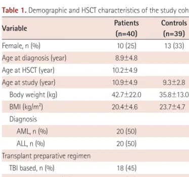
ALL, acute lymphoblastic leukemia; ABL, acute biphenotypic leukemia; AML, acute myeloblastic leukemia; Allo-SCT, allo- geneic stem cell transplantation; BM, bone marrow; PB,

We describe a 37-yr-old man who developed central pontine myelinolysis (CPM) after allogeneic hematopoietic stem cell transplantation (HSCT) for acute lymphoblastic leukemia..

Hematopoietic cell transplantation for children with acute lymphoblas- tic leukemia in second complete remission: similar outcomes in recipi- ents of unrelated marrow and umbilical

Here, we report the iden- tification of a constitutional t(2;11)(q32;q23) chromosomal ab- normality in a pediatric patient with acute megakaryoblastic leu- kemia (AMKL) and a

We report here a case of AML with a solitary t(6;7)(p21.3;p22) passenger translocation that developed at relapse after allo- geneic hematopoietic stem cell transplantation (HSCT) in

The effect of first-line imatinib interim therapy on the outcome of allogeneic stem cell transplantation in adults with newly diagnosed Philadelphia chromosome-positive

Knowledge of the roles of tacrolimus and minidose methotrexate (MTX) in the prevention of acute graft-versus-host disease (aGVHD) in pediatric allogeneic hematopoietic stem

Chronic graft versus host disease (GVHD) is a serious long-term complication of allogeneic hematopoietic stem cell transplantation (HSCT).. Chronic GVHD can occur any time