INTRODUCTION
Central pontine myelinolysis (CPM) is an acute, non-inflam- matory demyelinating disease of the pons of the central ner- vous system (CNS) (1). The etiology and the pathogenesis of CPM remain unclear, but too-rapid correction of hyponatrem- ia is often associated with CPM, which is the result of the selec- tive loss of myelin. Other risk factors identified include alco- holism, malnutrition, liver disease, and interestingly, liver transplant (2, 3). Only one prior case report of subjects asso- ciated with CPM and hepatic veno-occlusive disease after allo- geneic hematopoietic stem cell transplantation (HSCT) has been published (4).
We describe a rare case who developed CPM after allogene- ic HSCT for acute lymphoblastic leukemia (ALL).
CASE REPORT
A 37-yr-old man was admitted to our hospital for HSCT.
He was diagnosed with ALL, presenting with mild fever, fa- tigue, and headache. A bone marrow examination showed acute
precursor B cell leukemia with morphology consistent with FAB L2 subtype. Cytogenetic study demonstrated a clonal abnormality with translocation between chromosomes 9 and 22. BCR/ABL rearrangement was positive by fluorescence in situ hybridization (78.5%). The patient received initial induc- tion chemotherapy, which was composed of vincristine 2 mg on days 1 and 8, daunorubicin 90 mg/m2/day on days 1-3, prednisolone 60 mg/m2/day on days 1-14, and imatinib 600 mg once daily from day 8 with intrathecal methotrexate. He achieved morphological remission. Since an HLA-matched sibling donor was available, allogeneic HSCT was performed with the matched sibling donor.
Following a conditioning regimen of busulfan 0.8 mg/kg/
dose four times daily on days -8, -7, -6, -5 and cytoxan 60 mg/
kg/day on days -3, -2, the patient received 9.23×10⁸nucle- ated cells/kg (10.4×10⁶CD34 cells/kg) from his brother. For prophylaxis of graft-versus-host disease (GVHD), cyclosporine and prednisolone were used. He tolerated this conditioning therapy well, and the post-transplant course was unremark- able until the 12th day. The patient showed altered mentality on the 13th day, and his serum cyclosporine level was increased higher than 1,300 ng/mL (normal range, 150-300 ng/mL).
324
Kyu-Hyoung Lim, Sunyoung Kim, Yoon-Su Lee, Ki Hwan Kim, Jinwon Kim, Ji young Rhee, Hye Jin Kim, Hyeon Gyu Yi, So Yeon Oh, Joo Han Lim, Sae-Won Han, Sangyoon Lee, Inho Kim, Sung-Soo Yoon,
Seonyang Park, and Byoung Kook Kim
Department of Internal Medicine, Seoul National University Hospital, Seoul National University College of Medicine, Seoul, Korea
Address for correspondence Inho Kim, M.D.
Department of Internal Medicine, Seoul National University College of Medicine, 28 Yeongeon-dong, Jongno-gu, Seoul 110-744, Korea
Tel : +82.2-2072-0834, Fax : +82.2-744-6075 E-mail : [email protected] J Korean Med Sci 2008; 23: 324-7
ISSN 1011-8934
DOI: 10.3346/jkms.2008.23.2.324
Copyright � The Korean Academy of Medical Sciences
Central Pontine Myelinolysis in a Patient with Acute Lymphoblastic Leukemia after Hematopoietic Stem Cell Transplantation:
A Case Report
We describe a 37-yr-old man who developed central pontine myelinolysis (CPM) after allogeneic hematopoietic stem cell transplantation (HSCT) for acute lymphoblastic leukemia. After HSCT, desquamation developed on the whole body accompanied by hyperbilirubinemia. The liver biopsy of the patient indicated graft-versus-host dis- ease-related liver disease, and the dose of methylprednisolone was increased. Then, the patient developed altered mentality with eye ball deviation to the left, for which electroencephalogram and magnetic resonance imaging (MRI) scans were done.
Brain MRI scan demonstrated the imaging findings consistent with central pontine myelinolysis and extrapontine myelinolysis. He did not have any hyponatremia episode during hospitalization prior to the MRI scan. To the best of our knowledge, presen- tation of CPM after allogeneic HSCT is extremely rare in cases where patients have not exhibited any episodes of significant hyponatremia. We report a rare case in which hepatic dysfunction due to graft-versus-host disease has a strong association with CPM after HSCT.
Key Words : Myelinolysis, Central Pontine; Hematopoietic Stem Cell Transplantation; Graft versus Host Disease
Received : 20 April 2007 Accepted : 17 July 2007
Central Pontine Myelinolysis After Hematopoietic Stem Cell Transplantation 325
As his altered mentality was attributed to cyclosporine-induc- ed agitation and psychosis, we stopped administering cyclos- porine and replaced cyclosporine with methylprednisolone.
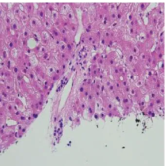
Thereafter, the patient showed much improvement in con- sciousness. On the 25th day, the patient developed desqua- mation at whole body and hyperbilirubinemia with a peak bilirubin level of 29 mg/dL. To rule out the possibility of veno-occlussive disease (VOD), doppler ultrasonography was conducted, which revealed no evidence of VOD. Then, trans- jugular liver biopsy was done. Liver biopsy revealed vacuo- lated cytoplasm and hyperchromasia of the bile duct epithe- lium, which was compatible with GVHD (Fig. 1). Therefore, we conducted high-dose steroid pulse therapy for 6 days and maintained methylprednisolone 2 mg/kg/day since then.
On the 48th day, the patient demonstrated drowsy con- sciousness with Glasglow Coma Scale E4M1V1 and left eye- ball deviation. We conducted cerebrospinal fluid (CSF) tap-
ping and a brain MRI scan. CSF cytology was negative for malignant cells, and the brain MRI scan showed multiple white matter low densities (Fig. 2), which meant a nonspe- cific result with respect to his neurologic problem.
At that time, the patient had a decreased urine output and increased serum blood urea nitrogen (BUN)/Creatinine (Cr) level. The ranges of serum BUN (normal range, 10-26 mg/
dL) and Cr level (normal range, 0.7-1.4 mg/dL) were 125- 165 mg/dL, 2.3-4.3 mg/dL respectively. To rule out the pos- sibility of uremic encephalopathy, he received hemodialysis three times per week from the 53th day. Thereafter, the BUN and Cr level were maintained around 96-120 mg/dL, 1.5- 3.3 mg/dL respectively (Fig. 3).
During these treatments, the patient’s consciousness was unchanged, so we followed up with CSF tapping, electroen- cephalogram, and a brain MRI scan on the 70th day. CSF cytology was negative, and the electroencephalogram find- ing showed low amplitude theta slowings at all leads, which meant diffuse cerebral dysfunction (Fig. 4). The follow-up brain MRI revealed diffuse marked progression of confluent
Fig. 1. Needle biopsy of the liver. The GVHD here is affecting the liver and marked by bile duct damage (hematoxylin and eosin stain, ×400).
Fig. 2. Brain MRI reveals nonspecific findings including multiple white matter low densities.
Fig. 4. Low amplitude theta slowings at all leads on electroen- cephalogram indicate diffuse cerebral dysfunction.
Fig. 3. Serial changes of BUN and Cr.
BUN (mg/dL) Cr (mg/dL)
200 160 120 80 40 0
5 4 3 2 1
45 48 52 54 56 63 70 76 0
BUN Cr
Transplant day
326 K.-H. Lim, S. Kim, Y.-S. Lee, et al.
white matter T2 high-signal lesions in periventricular white matter, basal ganglia, pons, and cerebellum, which was con- sistent with CPM and extrapontine demyelinolysis (Fig. 5).
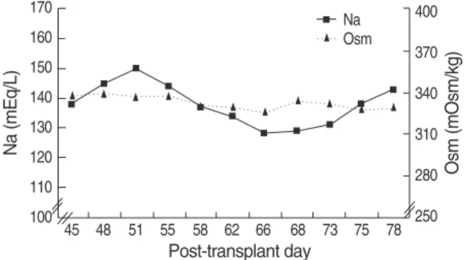
A review of serial serum sodium levels revealed that though serum sodium measurements (normal range, 135-145 mEq/L) had fluctuated between 128-150 mEq/L during 20 days of hospitalization prior to follow-up MRI scan, there was no episode of correcting hyponatremia in our patient.
On the 48th day, the serum sodium level was 145 mEq/L and daily variations of serum sodium level were within the range of 3-7 mEq/L/day since then. Also, the range of serum osmolarity (normal range, 289-302 mOsm/kg) was 328-339 mOsm/L (Fig. 6). During the same period, cyclosporine as another risk factor was not used any longer since the 14th day.
The patient was given plasmapheresis three times per week and steroid therapy as the experimental treatment of CPM, but its effectiveness was insignificant. He died of bilateral intra- ventricular hemorrhage on the 78th day.
DISCUSSION
CPM is a clinical syndrome including disturbances of con- sciousness, disturbed function of caudal cranial nerves with signs of pseudobulbar palsy, dysarthria as well as spastic para- paresis and tetraparesis of various degrees of severity, epilep- tic seizures, and hypotension (5-7).
A clinical diagnosis of CPM can be made, if patients suf- fering from chronic alcoholism, electrolyte disturbance, or other chronic diseases as well as liver transplant patients presenting with massive mental changes show specific neuroradiological findings on MRI scan (2). There is no treatment of choice in CPM.
CPM has been known to result from the rapid correction
of hyponatremia and merge as a well-recognized neurologi- cal complication following hepatic dysfunction such as chron- ic liver disease and liver transplant (6, 8). In one series of post- mortem examinations of liver transplant patients, a 29% inci- dence of CPM was reported (9). It has been revealed that the use of cyclosporine following liver transplant increases the risk of development of CPM (10).
Neurological deterioration was a prominent feature of our patient’s post-transplant course. In such a critically ill patient, it is likely that several factors contributed to his encephalopa- thy, including liver dysfunction, uremia, cyclosporine, and CPM.
Other causes of encephalopathy in the setting of HSCT in- clude electrolyte disturbances, cerebrovascular events or infec- tion, and sedative drugs; but laboratory parameters and imag- ing findings did not support a role for these possibilities in this case.
Despite the likely multifactorial etiology of the encephalopa- thy, the clinical signs of left eyeball deviation and the loss of consciousness in conjunction with suggestive neuroimaging are best explained by the diagnosis of CPM.
To the best of our knowledge, presentation of CPM after allogeneic HSCT is extremely rare in cases where patients have not exhibited any episodes of significant hyponatremia. Only one case was reported that severe hepatic VOD and cyclosporine therapy after HSCT induced the development of CPM with- out any episodes of significant hyponatremia (4).
Given the known association with liver disease as the cause of CPM, we concluded that our patient’s severe hepatic dys- function due to GVHD might have predisposed him to the development of CPM.
REFERENCES
1. Messert B, Orrison WW, Hawkins MJ, Quaglieri CE. Central pon- tine myelinolysis. Considerations on etiology, diagnosis, and treat- ment. Neurology 1979; 29: 147-60.
2. Lampl C, Yazdi K. Central pontine myelinolysis. Eur Neurol 2002;
47: 3-10.
3. Ashrafian H, Davey P. A review of the causes of central pontine myeli- Fig. 5. Follow-up Brain MRI showed diffuse marked progression of
confluent white matter T2 high-signal lesion in both periventricular white matter, basal ganglia, pons, and cerebellum, consistent with CPM.
Fig. 6. Serial changes of sodium (Na) and osmolarity (Osm).
Na (mEq/L) Osm (mOsm/kg)
170 160 150 140 130 120 110 100
400 370 340 310 280 45 48 51 55 58 62 66 68 73 75 78 250
Na Osm
Post-transplant day
Central Pontine Myelinolysis After Hematopoietic Stem Cell Transplantation 327
nolysis: yet another apoptotic illness? Eur J Neurol 2001; 8: 103-9.
4. Fraser C, Charnas L, Orchard P. Central pontine myelinolysis follow- ing bone marrow transplantation complicated by severe hepatic veno- occlusive disease. Bone Marrow Transplant 2005; 36: 733-4.
5. McCormick WF, Danneel CM. Central pontine myelinolysis. Arch Intern Med 1967; 119: 444-78.
6. Karp BI, Laureno R. Pontine and extrapontine myelinolysis: a neu- rologic disorder following rapid correction of hyponatremia. Medicine (Baltimore) 1993; 72: 359-73.
7. Sterns RH, Cappuccio JD, Silver SM, Cohen EP. Neurologic sequelae after treatment of severe hyponatremia: a multicenter perspective. J Am Soc Nephrol 1994; 4: 1522-30.
8. Bronster DJ, Emre S, Boccagni P, Sheiner PA, Schwartz ME, Miller CM. Central nervous system complications in liver transplant recip- ients-incidence, timing, and long-term follow-up. Clin Transplant 2000;
14: 1-7.
9. Singh N, Yu VL, Gayowski T. Central nervous system lesions in adult liver transplant recipients: clinical review with implications for man- agement. Medicine (Baltimore) 1994; 73: 110-8.
10. Fryer JP, Fortier MV, Metrakos P, Verran DJ, Asfar SK, Pelz DM, Wall WJ, Grant DR, Ghent CN. Central pontine myelinolysis and cyclosporine neurotoxicity following liver transplantation. Transplan- tation 1996; 61: 658-61.