• 검색 결과가 없습니다.

Diagnosis of Incomplete Kawasaki Disease in Infants Based on an Inflammation at the Bacille Calmette-Guérin Inoculation Site

N/A
N/A
Protected

Academic year: 2022

Share "Diagnosis of Incomplete Kawasaki Disease in Infants Based on an Inflammation at the Bacille Calmette-Guérin Inoculation Site"

Copied!
7
0
0

로드 중.... (전체 텍스트 보기)

전체 글

(1)

Korean Circulation Journal

Introduction

The etiology of Kawasaki disease (KD) is still unknown and no sin- gle pathognomonic clinical or laboratory finding for its definitive diagnosis has been identified. To establish a diagnosis, we inevita- bly depend on the diagnostic criteria that include the typical con- stellation of symptoms and signs noted by the Japanese Kawasaki

Print ISSN 1738-5520 • On-line ISSN 1738-5555

Diagnosis of Incomplete Kawasaki Disease in Infants Based

on an Inflammation at the Bacille Calmette-Guérin Inoculation Site

Ji Hye Seo, MD 1 , Jeong Jin Yu, MD 1 , Hong Ki Ko, MD 1 , Hyung Soon Choi, MD 2 , Young-Hwue Kim, MD 1 , and Jae-Kon Ko, MD 1

1

Department of Pediatrics, College of Medicine, University of Ulsan, Seoul,

2
Department of Pediatrics, Kosin University Gospel Hospital, Busan, Korea

Background and Objectives: This study was intended to test how the inflammation at the Bacille Calmette-Guérin (BCG) inoculation site (BCGitis) can be a useful a diagnostic feature of Kawasaki disease (KD).

Subjects and Methods: All subjects were infants at the time of admission, and had received BCG vaccination during their neonatal peri- od. There were 54 patients with complete KD (group 1) and 29 patients with incomplete KD (group 2). All 83 patients had BCGitis during the acute phase of illness. Data regarding the coronary artery diameters in 31 age-matched controls were used for comparison.

Results: The 2 patient groups did not differ in clinical and laboratory variables. During the acute phase, the median z scores of the left anterior descending coronary artery (LAD) diameter were 0.20, 0.42, and -0.48 in groups 1, 2, and control respectively, and that of right co- ronary artery (RCA) diameters were -0.15, -0.16, and -1.17 respectively. The z scores in both patient groups were greater than those in con- trols (p=0.0014 in LAD and p<0.0001 in RCA between group 1 and controls; p=0.0023 in LAD and p<0.0001 in RCA between group 2 and controls). A similar pattern was observed during the subacute and convalescent phases.

Conclusion: BCGitis is a useful feature in the diagnosis of incomplete KD in infants who received BCG vaccine during neonatal period.

(Korean Circ J 2012;42:823-829)

KEY WORDS: Mucocutaneous Lymph Node Syndrome; Coronary vesseles.

Received: May 24, 2012 Accepted: July 31, 2012

Correspondence: Jeong Jin Yu, MD, Department of Pediatrics, College of Me- dicine, University of Ulsan, Asan Medical Center, 88 Oylimpic-ro 43-gil, Song- pa-gu, Seoul 138-736, Korea

Tel: 82-2-3010-3924, Fax: 82-2-473-3725 E-mail: pediatrist@medimail.co.kr

• The authors have no financial conflicts of interest.

This is an Open Access article distributed under the terms of the Creative Commons Attribution Non-Commercial License (http://creativecommons.

org/licenses/by-nc/3.0) which permits unrestricted non-commercial use, distribution, and reproduction in any medium, provided the original work is properly cited.

Disease Research Committee

1)

or by the American Heart Association (AHA).

2)

However, pediatricians occasionally encounter febrile chil- dren who do not fulfill the diagnostic criteria but who have several findings compatible with those of KD. In this type of situation, the diagnosis of incomplete KD is a clinical challenge that cannot be avoided by delaying the diagnosis because the risk of coronary complications is seen even in incomplete presentation of the dis- ease.

3-5)

Since 1966, the Korean Pediatric Society has recommended that

a routine universal Bacille Calmette-Guérin (BCG) vaccination be gi-

ven to neonates.

6)

In Korea, nearly all neonates receive the BCG vac-

cine by one month of age. Erythema, induration, or crust at the BCG

inoculation site (BCGitis) is listed as a significant finding among the

diagnostic guidelines for KD by the Kawasaki Disease Research Com-

mittee in Japan

1)

and by the AHA.

2)

Recently, BCGitis was reported

to be a useful diagnostic tool for identifying incomplete KD in sev-

eral cases.

7)8)

We have considered BCGitis as a pathognomonic find-

ing of KD, and have applied it to the diagnosis of KD in infants, as

BCGitis was reported to have been observed in more than 50% of

(2)

KD patients 1 to 12 months following BCG inoculation.

9)

Our study was designed to test how the BCGitis can be a diag- nostic feature of KD by comparing patients with complete disease presentation and those in whom incomplete KD was diagnosed based on a BCGitis.

Subjects and Methods

Study population

From January 2006 to February 2012, 490 patients with KD were admitted to Asan Medical Center in Seoul, Korea. Of these patients, 101 were less than 12 months of age at the time of admission. Six- ty-seven patients met the AHA diagnostic criteria of KD.

2)

Fifty-four patients showed BCGitis during the acute phase of illness, and were assigned to the complete presentation group. Of the remaining 34, 29 patients showed BCGitis during the acute phase of illness, and were assorted to the incomplete presentation group. All 83 enroll- ed patients had been inoculated with BCG during the neonatal pe- riod. If a patient had been examined initially by a clinically experi- enced pediatrician, a fever of 3 or 4 days’ duration was permitted to establish a diagnosis. As controls for evaluating the diameters of co- ronary arteries, we enrolled 31 healthy infants, who had been referr- ed to Asan Medical Center for evaluation of a cardiac murmur dur- ing the past year and for whom the clinical and echocardiographic ex- aminations showed no evidence of cardiac disease.

The Institutional Review Board of the Medical Center approved this retrospective study (2012-0011) and waived the need for patient consent. Demographic, laboratory, and clinical data were obtained from patients’ medical records.

Echocardiographic data

Echocardiographic examinations were performed 3 times using a Vivid 7 Vantage machine (GE Vingmed, Horten, Norway), i.e., during the acute phase, the subacute phase (a subsiding of fever 4 weeks after the onset of disease), and the convalescent phase (4-12 weeks after the onset of disease). Stored echocardiographic images were reviewed by one author who was blinded to patient status. The fol- lowing diameters were measured by EchoPAC PC, Version 7 (GE Vingmed, Horten, Norway) using the method of de Zorzi et al.

10)

: of the left main coronary artery (LMCA), the left anterior descending artery (LAD), and the right coronary artery (RCA).

We randomly selected 19 images of coronary arteries to deter- mine the intra- and inter-observer variability in the measurement of the coronary artery diameters. The intra-observer percent precision in diameter measurement of LMCA, LAD, and RCA were 5.4%, 8.7%, and 5.1%, respectively, and the inter-observers percent precisions were 5.8%, 10.4%, and 7.0%, respectively.

The diameters of the coronary arteries were converted to a z score using the regression equations from the study of Olivieri et al.

11)

Application of the American Heart Association algorithm for diagnosing incomplete Kawasaki disease

The AHA recommended an algorithm that included laboratory and echocardiographic criteria for the diagnosis of incomplete KD.

2)

La- boratory criteria include albumin ≤3 g/dL, anemia for age, elevation of alanine aminotransferase, platelets after 7 days ≥450000/mm

3

, white blood cell count ≥15000/mm

3

, and urine ≥10 white blood cells/

high-power field. An echocardiogram is considered positive for the purposes of this algorithm if any of 3 conditions had been met, i.e., a z-score of LAD or RCA ≥2.5, coronary arteries meet the Japanese Ministry of Health criteria for aneurysms,

12)

or ≥3 other suggestive features exist, including perivascular brightness, lack of tapering, de- creased left ventricular function, mitral regurgitation, pericardial ef- fusion, and a z score of LAD or RCA of 2-2.5. We applied this algo- rithm to 29 patients that had an incomplete presentation of BCGitis.

However, in our clinical practice, we do not use 2 variables of the echocardiographic criteria, i.e., perivascular brightness and lack of tapering, as we consider that the former is inappropriate

13)

and the latter is too subjective. Therefore, we did not perform an investiga- tion of these 2 variables.

Statistical analysis

All the data are presented as medians (range). All statistical analy- ses were performed using SAS Version 9.1 (SAS Institute, Cary, NC, USA), and statistical significance was defined as a p<0.05. Categori- cal variables were compared using Fisher’s exact test. Comparisons of numerical variables between groups were performed using the Wilcoxon rank sum test.

Results

The clinical and laboratory findings of patients with KD are pre-

sented in Table 1. The frequencies of the classic, principal diagnostic

features in the incomplete presentation group were appropriately

lower than those in the complete presentation group. The less fre-

quently appearing features included cervical lymphadenopathy

(3.4%) and palmar erythema (20.7%) in the incomplete presenta-

tion group. No other clinical or laboratory findings differed between

the 2 groups. In the incomplete presentation group, there were 17

(58.6%) patients with 3 principal features, 8 (27.6%) with 2 principal

features, and 4 (14.0%) with 1 principal feature. In the control sub-

jects, the median age was 7.2 months (p=0.8663 compared to the

complete presentation group, and p=0.1524 compared to the incom-

plete presentation group), and there were 14 (45.2%) females (p=

(3)

0.1533 and p=0.7997, respectively).

Echocardiographic features were compared between the patient gr- oups and the controls (Table 2) (Fig. 1). The systolic function vari- ables and the frequencies of mitral regurgitation and pericardial ef- fusion did not differ between the patient groups. The z scores of the LMCA diameters (p=0.0192 in the complete presentation group and p=0.0022 in the incomplete presentation group during the acute phase, p=0.0073 and p=0.0234, respectively, during the subacute phase and p=0.0053 and p=0.0290, respectively, during the conva- lescent phase) and the RCA (p<0.0001 and p<0.0001, respectively, during the acute phase, p=0.0003 and p<0.0001, respectively, during the subacute phase, and p=0.0030 and p=0.0104, respectively, dur- ing the convalescent phase) of the patients were consistently great- er than those of the controls throughout the 3 phases of illness. The LAD (p=0.0014 and p=0.0023, respectively, during the acute phase, and p=0.0085 and p=0.0317, respectively, during the subacute ph- ase) of the patients were also greater than those of the controls, al- though they (p=0.2209 and p=0.0617, respectively) were not signi- ficantly greater during the convalescent phase. The z scores of the diameters of coronary arteries did not differ between the 2 patient groups. Only the z score of RCA in the incomplete group (p=0.0447)

was greater than that of the complete group during the subacute phase. Comparisons of the diameters of coronary artery between groups showed a similar pattern.

We divided the incomplete presentation group into subgroups with 3 and with <3 classic diagnostic principal features. We com- pared all of the variables presented in Table 1 and 2 between the subgroups; however, we could not find any variables showing a sig- nificant difference between the subgroups. Additionally we com- pared all of the variables between the complete presentation group and 13 excluded patients with complete presentation who did not have BCGitis, we could not find any variables showing a significant difference. These results are not presented in this paper.

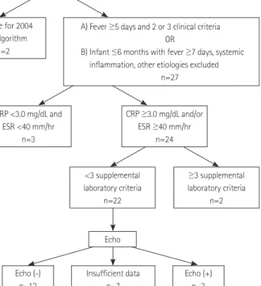
We applied the AHA algorithm for diagnosis of incomplete KD to 29 patients in the incomplete presentation group. The results are summarized in Fig. 2. Two patients were not eligible for use of the AHA algorithm.

2)

Incomplete KD could be diagnosed in 5 (18.5%) of the 27 patients, i.e., 2 (7.4%) based on laboratory criteria and 3 based on echocardiographic criteria. In 15 patients (55.6%), a diag- nosis of incomplete KD was not possible; 12 patients did not fulfill the laboratory and echocardiographic criteria, and in 3 patients, the values of acute-phase reactants were less than the threshold Table 1. Clinical and laboratory findings of Kawasaki disease patients with BCGitis distinguished by presentation

Complete KD (n=54) Incomplete KD (n=29) p

Age (month) 7.4 (2.0-12.1) 6.3 (2.5-11.4) 0.0984

Female (%) 15 (27.8) 12 (41.4) 0.2279

Body weight (kg) 8.3 (5.2-11.5) 7.5 (5.5-10.5) 0.0639

Height (cm) 72 (59-80) 69 (58-90) 0.0817

Fever duration (day) 5 (3-9) 5 (4-10) 0.2773

IVIG refractory cases (%) 4 (7.4) 3 (10.3) 1.0

Classic principal features (%)

Conjunctival injection 54 (100) 22 (75.9) 0.0004

Red lips/Oral mucosa 53 (98.1) 23 (79.3) 0.0066

Palmar erythema 50 (92.6) 6 (20.7) <0.0001

Polymorphous rash 52 (96.3) 19 (65.5) 0.0003

Cervical lymphadenopathy 16 (29.6) 1 (3.4) 0.0042

Laboratory features

WBC (×10

3

/uL) 12.4 (5.8-24.6) 12.7 (8.2-22.3) 0.2731

Anemia (%) 6 (11.1) 4 (13.8) 0.7339

Platelet (×10

3

/uL) 355 (164-864) 343 (172-696) 0.3899

ESR (mm/hr) 59 (6-105) 58 (11-120) 0.9126

CRP (mg/dL) 5.59 (0.74-24.40) 6.25 (0.54-16.44) 0.9734

Albumin (g/dL) 3.6 (2.5-4.2) 3.4 (2.7-4.1) 0.1217

Sodium (mmol/L) 136 (132-139) 136 (131-139) 0.8198

GPT (IU/L) 28 (8-362) 26 (10-192) 0.4001

Pyuria (>5/HPF) (%) 15 (27.8) 4 (13.8) 0.1794

Data are medians (range) or numbers of frequency (%). KD: Kawasaki disease, IVIG: intravenous immunoglobulin, WBC: white blood cells counts, ESR:

erythrocyte sedimentation rate, CRP: C-reactive protein, GPT: glutamate-pyruvate transaminase, BCGitis: Bacille Calmette-Guérin inoculation site

(4)

value. In 7 patients, the diagnosis was inconclusive because of the absence of data regarding 2 echocardiographic variables.

Discussion

When the clinical, laboratory, and echocardiographic variables of patients with incomplete KD were compared with those of patients with complete disease presentation, there were no significant differ- ences between the 2 groups. In addition, the z scores of coronary arteries in patients with incomplete presentation were significantly greater than those of the controls throughout the three phases of illness. The z score of LAD during the convalescent phase was an ex- ception. In this study, the 2 patient groups differed only in the fre- quencies of the principal features of disease.

Sudo et al.

14)

collected data from a total of 23263 patients with KD in a nationwide survey of Japan from 2007 through 2008. In the study, incomplete KD was defined as the presence of ≤4 principal

symptoms defined according to the Japanese diagnostic criteria

1)

(corresponding to ≤3 principal features of the AHA diagnostic cri- teria

2)

) regardless of whether the patient had coronary artery ab- normalities; their incomplete presentation group included 271 ch- ildren (1.2%) with only one or 2 principal symptoms.

14)

Currently, a diagnosis of incomplete KD might be performed in cases with fewer classical diagnostic criteria and several compatible clinical, labora- tory, or echocardiographic findings, based on the exclusion of an- other febrile illness.

15)

The above-mentioned results of the survey by Sudo et al.

14)

seem to reflect the current diagnosing practice for in- complete KD. To date, there have been 2 guidelines for the diagnosis of incomplete KD. According to the Japanese diagnostic criteria,

1)

a diagnosis of incomplete KD is possible in a case with 4 principal sym- ptoms and a coronary artery aneurysm. However, many authors be- lieve that this definition is too restrictive and specific.

16)

Pediatric cli- nicians frequently encounter KD in suspicious cases with <4 prin- cipal symptoms. In addition, a coronary artery abnormality included Table 2. Echocardiographic features of Kawasaki disease patients with BCGitis by the type of presentation

Complete KD (n=54) Incomplete KD (n=29) Controls (n=31)

LVEDD (mm) 24.8 (16.6 - 31.8) 24.3 (18.2 - 29.3) 24.1 (21.6 - 29.4)

FS (%) 38.2 (24.1 - 74.0) 36.9 (30.0 - 64.7) 36.2 (27.0 - 51.8)

Mitral regurgitation (%) 16 (29.6) 11 (37.9)

Pericardial effusion (%) 4 (7.4) 4 (13.8)

Acute phase

LMCA Diameter (mm)

†‡

1.96 (1.20 - 2.90) 2.00 (1.20 - 3.00) 1.49 (1.00 - 2.22)

Z-score

†‡

0.16 (-2.76 - 2.68) 0.41 (-3.14 - 3.08) -0.66 (-2.11 - 0.71)

LAD Diameter (mm) 1.60 (1.00 - 2.30) 1.60 (1.00 - 3.50) 1.47 (1.08 - 2.77)

Z-score

†‡

0.20 (-2.77 - 2.33) 0.42 (-2.61 - 5.23) -0.48 (-2.79 - 2.72)

RCA Diameter (mm)

†‡

1.58 (0.80 - 3.20) 1.60 (0.90 - 2.63) 1.26 (0.82 - 1.65)

Z-score

†‡

-0.15 (-3.61 - 3.03) -0.16 (-2.47 - 2.63) -1.17 (-3.84 - 0.0)

Subacute phase

LMCA Diameter (mm)

†‡

2.00 (1.14 - 3.39) 1.84 (1.40 - 3.20) 1.49 (1.00 - 2.22)

Z-score

†‡

0.11 (-3.10 - 3.31) -0.02 (-1.56 - 3.35) -0.66 (-2.11 - 0.71)

LAD Diameter (mm) 1.60 (0.80 - 2.64) 1.51 (0.90 - 3.19) 1.47 (1.08 - 2.77)

Z-score

†‡

0.03 (-3.42 - 3.17) 0.09 (-2.76 - 3.86) -0.48 (-2.79 - 2.72)

RCA Diameter (mm)

†‡

1.50 (0.97 - 3.11) 1.57 (1.20 - 3.02) 1.26 (0.82 - 1.65)

Z-score*

†‡

-0.43 (-2.52 - 3.30) -0.25 (-1.28 - 2.85) -1.17 (-3.84 - 0.0)

Convalescent phase

LMCA Diameter (mm)

†‡

1.99 (1.35 - 2.69) 2.00 (1.40 - 2.65) 1.49 (1.00 - 2.22)

Z-score

†‡

0.11 (-2.23 - 1.83) 0.10 (-1.61 - 2.11) -0.66 (-2.11 - 0.71)

LAD Diameter (mm) 1.50 (0.70 - 2.35) 1.59 (0.80 - 2.40) 1.47 (1.08 - 2.77)

Z-score -0.15 (-3.17 - 2.30) 0.13 (-3.48 - 2.22) -0.48 (-2.79 - 2.72)

RCA Diameter (mm)

†‡

1.49 (1.01 - 2.73) 1.47 (0.83 - 2.34) 1.26 (0.82 - 1.65)

Z-score

†‡

-0.75 (-2.59 - 2.55) -0.63 (-3.25 - 1.52) -1.17 (-3.84 - 0.0)

Data are medians (range) or numbers of frequency (%). *p<0.05, group 1 vs. group 2,

p<0.05, group 1 vs. controls,

p<0.05, group 2 vs. controls. KD: Kawa-

saki disease, LVEDD: left ventricle end-diastolic dimension, FS: fraction shortening, LMCA: left main coronary artery, LAD: left anterior descending coronary

artery, RCA: right coronary artery, BCGitis: Bacille Calmette-Guérin inoculation site

(5)

in the definition of incomplete presentation is itself, a bad outcome of the disease. AHA proposed a diagnostic algorithm of incomplete KD in 2004,

2)

details of which were presented in the Methods sec- tion. Newburger et al.

2)

acknowledged this algorithm as only an in- formed opinion of a Committee of Experts (evidence level C). Al- though Yellen et al.

17)

tested the performance of the AHA diagnostic criteria and reported its 97% applicability, their study subjects were restricted to cases with coronary artery aneurysm. This study was the first attempt to test the performance of this algorithm in the diagnosis of incomplete KD in infants. It is well known that the in- cidence of incomplete presentation is relatively higher in younger patients

18)19)

and that there is a risk of delayed diagnosis and man- agement in infants with incomplete presentation.

20)21)

In this study, 5 (18.5%) of the 27 patients with incomplete presentation that was eligible for AHA algorithm application could be diagnosed, and the laboratory criteria was only fulfilled in 3 (7.4%) patients. This disap- pointing result is inconsistent with the result reported by Yellen et

al.

17)

In their study, the laboratory criteria were fulfilled in all patients with incomplete KD. This difference may have been caused by the differences in subjects. In the report by Yellen et al.

17)

patients who had coronary artery aneurysms were selected as subjects and the median duration of fever in the incomplete presentation group was 9 days, which seems to be longer than the 5 days encountered in our study. A high prevalence of BCGitis in KD patients who visited a hospital between 1 and 4 days after the onset of illness was re- ported.

22)

BCGitis has been considered to be an early manifestation of KD.

22)23)

To avoid coronary complications, we can use BCGitis as a specific finding in early diagnosis of incomplete KD.

The less frequently appearing classical principal features of incom- plete KD have been reported to be cervical lymphadenopathy (19- 38.6%) and changes in extremity (21-44.3%). These patterns and frequencies were also identified in our study and are similar to those seen in patients with complete presentation.

3)17)24)25)

Uehara et al.

26)

reported that redness or crust formation at the BCGitis was ob- Fig. 1. Z-scores of the diameters of coronary arteries (left main coronary artery in A, left anterior descending artery in B, and right coronary artery in C) dur- ing 3 phases of illness. *p<0.05, complete presentation group vs. incomplete presentation group,

p<0.05, complete presentation group vs. controls,

p<0.05, incomplete presentation group vs. controls.

0.5

0

-0.5

-1.0

-1.5

0.5

0

-0.5

-1.0

-1.5

1.0 0.5 0 -0.5 -1.0 Complete Incomplete Controls -1.5

Complete Incomplete Controls

Complete Incomplete Controls

*†

‡ ‡

Acute Subacute Convalescent

Acute Subacute Convalescent

Acute Subacute Convalescent

A  

C  

B  

(6)

served in ≥70% of complete KD patients aged 3 to 20 months. Al- though the detection rate of BCGitis in patients with incomplete presentation has not been reported, we assume that it may not dif- fer significantly from that seen in patients with complete presenta- tion. In our study, the results showed 85.3% (29/34), which is com- patible with the results of Manlhiot et al.

24)

who suggested that complete and incomplete KD are 2 sides of the same disease. BCGitis cannot be a global standard criterion for the diagnosis of KD be- cause BCG vaccination is not performed globally; in patients who re- ceived BCG vaccine, the occurrence of BCGitis decreases beginning 12 months after inoculation.

9)

Uehara et al.

26)

selected children with KD aged 3 to 20 months as subjects of their study, because it is rec- ommended for children to undergo BCG vaccination by 6 months of age in Japan. However, as in Korea, nearly all neonates receive the BCG vaccine by one month of age, BCGitis could be considered to be a useful feature for the diagnosis of incomplete KD in infants.

This study has several limitations. First, the number of study sub- jects was relatively small. In the future, additional studies with a greater number of study subjects will be required. Second, it is pos- sible that we underestimated the z score of the diameters of coro- nary arteries because the z scores in our controls were lower than zero. In calculating the z scores of the coronary artery diameters, we used the regression equations seen in a report

11)

in which ≥90 infants were included as study subjects. This underestimation of

the z scores for the coronary artery diameters in controls may be the result of a racial difference in this study and it could be causatively associated with the low performance of the application of AHA echocardiographic criteria in the diagnosis of incomplete KD.

In summary, clinical, laboratory, and echocardiographic variables of patients with incomplete KD and BCGitis, did not differ from th- ose of patients with complete presentation. The z scores of their coronary artery diameters were significantly greater than those of the controls. BCGitis is a robust and useful feature in the diagnosis of incomplete KD in infants who have received BCG vaccine during their neonatal period.

References

1. Ayusawa M, Sonobe T, Uemura S, et al. Revision of diagnostic guide- lines for Kawasaki disease (the 5th revised edition). Pediatr Int 2005;

47:232-4.

2. Newburger JW, Takahashi M, Gerber MA, et al. Diagnosis, treatment, and long-term management of Kawasaki disease: a statement for health professionals from the Committee on Rheumatic Fever, Endo- carditis and Kawasaki Disease, Council on Cardiovascular Disease in the Young, American Heart Association. Circulation 2004;110:2747-71.

3. Sonobe T, Kiyosawa N, Tsuchiya K, et al. Prevalence of coronary artery ab- normality in incomplete Kawasaki disease. Pediatr Int 2007;49:421-6.

4. Witt MT, Minich LL, Bohnsack JF, Young PC. Kawasaki disease: more patients are being diagnosed who do not meet American Heart Asso- ciation criteria. Pediatrics 1999;104:e10.

5. Cho MA, Choi YJ, Jung JW. Affects of “age at diagnosis” on coronary artery lesions in patients with incomplete kawasaki disease. Korean Circ J 2010;40:283-7.

6. Lee HJ. Classification of vaccination and immunization schedule rec- ommended by the Korean Pediatric Society, 2008. J Korean Med Assoc 2008;51:104-9.

7. Chalmers D, Corban JG, Moore PP. BCG site inflammation: a useful diagnostic sign in incomplete Kawasaki disease. J Paediatr Child Health 2008;44:525-6.

8. Sinha R, Balakumar T. BCG reactivation: a useful diagnostic tool even for incomplete Kawasaki disease. Arch Dis Child 2005;90:891.

9. Hsu YH, Wang YH, Hsu WY, Lee YP. Kawasaki disease characterized by erythema and induration at the Bacillus Calmette-Guérin and purified protein derivative inoculation sites. Pediatr Infect Dis J 1987;6:576-8.

10. de Zorzi A, Colan SD, Gauvreau K, Baker AL, Sundel RP, Newburger JW.

Coronary artery dimensions may be misclassified as normal in Kawa- saki disease. J Pediatr 1998;133:254-8.

11. Olivieri L, Arling B, Friberg M, Sable C. Coronary artery Z score regres- sion equations and calculators derived from a large heterogeneous population of children undergoing echocardiography. J Am Soc Echo- cardiogr 2009;22:159-64.

12. Research Committee on Kawasaki Disease. Report of Subcommittee on Standardization of Diagnostic Criteria and Reporting of Coronary Ar- tery Lesions in Kawasaki Disease. Tokyo: Ministry of Health and Wel- fare;1984.

Incomplete KD n=29

Echo (-) n=12

Insufficient data n=7

Echo (+) n=3 Echo

Ineligible for 2004 AHA algorithm

n=2

CRP <3.0 mg/dL and ESR <40 mm/hr

n=3

CRP ≥3.0 mg/dL and/or ESR ≥40 mm/hr

n=24

≥3 supplemental laboratory criteria

n=2

<3 supplemental laboratory criteria

n=22

A) Fever ≥5 days and 2 or 3 clinical criteria OR

B) Infant ≤6 months with fever ≥7 days, systemic inflammation, other etiologies excluded

n=27

Fig. 2. Flow diagram of the application of the American Heart Asscociation

(AHA) algorithm in patients with incomplete Kawasaki disease (KD). CRP: C-

reactive protein, ESR: erythrocyte sedimentation rate, Echo: echocardiography,

OR: odds ratio.

(7)

13. Yu JJ, Jang WS, Ko HK, et al. Perivascular brightness of coronary arter- ies in Kawasaki disease. J Pediatr 2011;159:454-7.

14. Sudo D, Monobe Y, Yashiro M, et al. Coronary artery lesions of incom- plete Kawasaki disease: a nationwide survey in Japan. Eur J Pediatr 2012;171:651-6.

15. Yun SH, Yang NR, Park SA. Associated symptoms of Kawasaki disease.

Korean Circ J 2011;41:394-8.

16. Barone SR, Pontrelli LR, Krilov LR. The differentiation of classic Kawa- saki disease, atypical Kawasaki disease, and acute adenoviral infection:

use of clinical features and a rapid direct fluorescent antigen test. Arch Pediatr Adolesc Med 2000;154:453-6.

17. Yellen ES, Gauvreau K, Takahashi M, et al. Performance of 2004 Ameri- can Heart Association recommendations for treatment of Kawasaki dis- ease. Pediatrics 2010;125:e234-41.

18. Chang FY, Hwang B, Chen SJ, Lee PC, Meng CC, Lu JH. Characteristics of Kawasaki disease in infants younger than six months of age. Pediatr Infect Dis J 2006;25:241-4.

19. Chuang CH, Hsiao MH, Chiu CH, Huang YC, Lin TY. Kawasaki disease in infants three months of age or younger. J Microbiol Immunol Infect 2006;39:387-91.

20. Burns JC, Wiggins JW Jr, Toews WH, et al. Clinical spectrum of Kawasa- ki disease in infants younger than 6 months of age. J Pediatr 1986;

109:759-63.

21. Rowley AH, Gonzalez-Crussi F, Gidding SS, Duffy CE, Shulman ST. In- complete Kawasaki disease with coronary artery involvement. J Pedi- atr 1987;110:409-13.

22. Takayama J, Yanase Y, Kawasaki T. A study on erythematous change at the site of the BCG inoculation. Acta Pediatr Jpn 1982;86:567-72.

23. Yanagawa H, Nakamura Y, Kawasaki T, Shigematsu I. Nationwide epi- demic of Kawasaki disease in Japan during winter of 1985-86. Lancet 1986;2:1138-9.

24. Manlhiot C, Christie E, McCrindle BW, Rosenberg H, Chahal N, Yeung RS. Complete and incomplete Kawasaki disease: two sides of the same coin. Eur J Pediatr 2012;171:657-62.

25. Perrin L, Letierce A, Guitton C, Tran TA, Lambert V, Koné-Paut I. Com- parative study of complete versus incomplete Kawasaki disease in 59 pediatric patients. Joint Bone Spine 2009;76:481-5.

26. Uehara R, Igarashi H, Yashiro M, Nakamura Y, Yanagawa H. Kawasaki

disease patients with redness or crust formation at the Bacille Cal-

mette-Guérin inoculation site. Pediatr Infect Dis J 2010;29:430-3.

수치

Fig. 2. Flow diagram of the application of the American Heart Asscociation  (AHA) algorithm in patients with incomplete Kawasaki disease (KD)

참조

관련 문서

After choosing the type of bike, the next step is the right bike size. the right size for you from

44 글의 첫 번째 문장인 The most important thing in the Boat Race is harmony and teamwork.을 통해 Boat Race에서 가장 중요한 것은 조 화와 팀워크임을

Now that you have the right bike for you, it is important to learn the right riding position.. A poor riding position can lead to injuries

Modern Physics for Scientists and Engineers International Edition,

웹 표준을 지원하는 플랫폼에서 큰 수정없이 실행 가능함 패키징을 통해 다양한 기기를 위한 앱을 작성할 수 있음 네이티브 앱과

_____ culture appears to be attractive (도시의) to the

• The avoidance of unwanted heat loss or gain, and the positive use of sun energy for heating and cooling … become compelling principles of site

- quadriceps tendon 이 슬개골 하연에서 tibial tuberocity에 부착.