The Effect of Anteromedial Tibial Cortex Angle on Change of Posterior Tibial Slope Angle in PTO(Proximal Tibial Osteotomy) using
Computer Assisted Surgery(CAS)
Ho-Sang Lee
*, Cheol-Woong Kim , Ji-Hoon Bae
**, Joon-Ho Wang
**, Jong-Woong Park
**and Dong-Joon Oh
***Key Words: High Tibial Osteotomy, Anteromedial Cortex Oblique, Posterior Tibial Slope
Abstract
An upper tibial opening wedge osteotomy is an operation to cure a malalignment and a degenerate arthritis. To prevent the postoperative malalignment caused by the upper tibial opening wedge osteotomy, the research to define the relationship between a Hinge Axis Angle and a Posterior Slope Angle is needed. The effect of the relationship between the hinge axis angle and the gap angle on the posterior slope angle is studied. After 3-D Compute Tomography (CT) scanning image is reconstructed, the virtual surgery is performed by the reconstructed 3-D tibia model. It was proved that the relationship between the hinge axis angle and the gap angle were constant and the simple mathematical model could be derived. To verify the suggested mathematical model, it compared with the measured data from the virtual surgery. In conclusion, while the deviation between the data from the virtual surgery and ones of the mathematical model under the gap angle<10° was less than 1%.
† Member, Korea Univ. & Triple-C Medical Corp.
E-mail : [email protected]
TEL : +82-2-929-9844 FAX : +82-2-929-9842
* Triple-C Medical Corp. R&D Center in Korea Univ.
** Orthopedic Surgery, Korea Univ. Medical Center
*** Dept. of Mechanical Edu., Andong National Univ.
1. Introduction
The radiographs of all pelvic limb to evaluate the alignment of the pelvic limb in 1970 were so difficult and their accuracy also became a problem.
At that time, the tibiofemoral angle just by the anterior/posterior radiographs of the knee was tried to correct and the concept of 7°+ was set up. After the effective pressures on the medial/lateral and anterior/posterior of the knee were measured accurately during the weight-loading to consider the alignment of all pelvic limb, one of the orthopedist's wishes was to get the information of the reasonable osteotomy to consider the dynamic
pressure change during walking, that was to decrease the pressure at the clinical change part when real-walking and to prevent the transfer the excessive pressure to the opposite part. In the case of High Tibial Osteotomy (HTO), after the merit and the weakness of both Opening Wedge Osteotomy (OWO) and Closed Wedge Osteotomy
(CWO) were co-existed, OWO becomes the current trend. Moreover, the treatment of the medial collateral ligament during HTO was not solved yet.
The patients number of HTO, which was the real operation of the translated knee ailment, is being decreased by the artificial transplant of the knee such as Total Knee Arthroplasty (TKA). During TKA, Anterior Cruciate Ligament (ACL) was removed. When it was working for a long time, many problems such as the loosening of the artificial bone, the insensibility of the distal femur and proximal tibia, the stiffness of the hyper knee flex, the friction noise from the metal to metal, were existed. Since the failure of the artificial
1467 대한기계학회 2008년도 추계학술대회 논문집
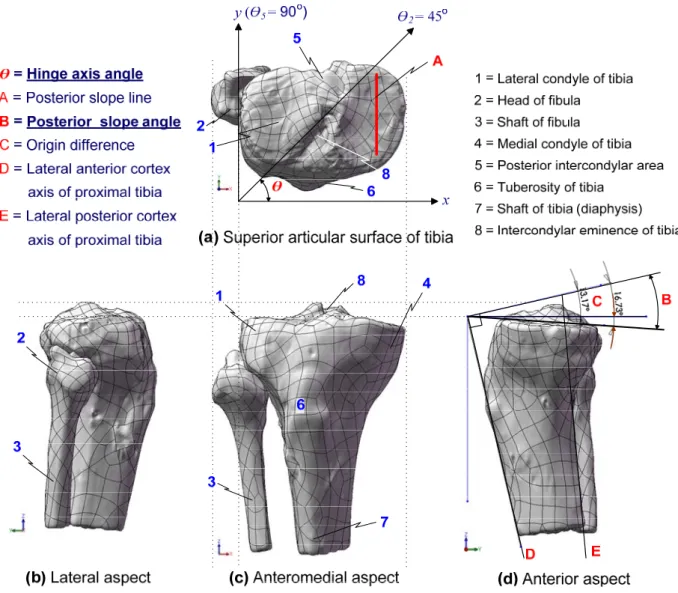
Fig. 1 Terminologies of proximal tibia using CT scan reconstruction (superior, anteromedial and lateral aspects)
transplant was inevitable for the high active patient or the special patient to have the axial dislocation from the bone, it was not believed that it was permanent. HTO is the operation to correct the malalignment by the anatomic axis dislocation from the femur and the tibia (See Fig.1(d)). This operation is to prevent the degenerate arthritis by the distribution of the abnormal excessive loading from the medial tibial plateau to the lateral tibial plateau.(1,2) Therefore, our research group performed the computation aided virtual osteotomy using the reconstruction of 3-D CT images. Shown in Fig.
1(d), Upper Tibial Open Wedge Osteotomy (UTOWO) increases the anterior oblique angle during the re-alignment of the anatomical axis and
it also increases the posterior slope. Since it results in the increase of the ACL tension, the rupture possibility of ACL is greatly increased during the sports activity.(3) Frank et. als.(4) studied the effect of the oblique angle and the wedge angle on the posterior slope angle by the clinical approach but it was restricted in the 90° of the hinge axis angle.
Nowadays, the posterolateral hinge axis osteotomy, where the hinge axis angle was 45°, becomes the standard so Frank's study has the limitation to apply UTOWO. Moreover, the results were verified by the computation aided virtual osteotomy. The parameters to affect the increase of the posterior slope angle, were not only the hinge axis angle, the gap angle but also the oblique angle and the
1468
wedge angle. For this reason, it the co-relationship of the above 4 angles were defined, it is believed that the bio-mechanical approach to the osteotomy is even clearer. Genu varum is one of the main causes of degenerative arthritis by stress concentration on the medial compartment of knee joint.(1~3) A genu varum increases the weight from a medial femoral condyle to a meniscus as a posterior slope angle, which is defined as the slope of a medial tibial plateau, is increased freakishly. It increases the rupture possibility of the meniscus and it becomes a degenerate arthritis when it is left alone. The correction of the varus is the most important knee operation and recently the upper tibial opening wedge osteotomy is performed. It was proved from the recent performed virtual osteotomy of the upper tibial opening wedge osteotomy that the relationship between the hinge axis and the gap angle effected on the posterior slope angle as the hinge axis such as 45°, 60°, 75°, 90° is increased and as the gap angle is increased from 5° to 20°. They are greatly effected each other. If the aperture widths of the upper tibia and the lower tibia were defined as Ymax and
Ymin,, it was found that the open wedge angle
caused by the difference between two parameters effected greatly on the posterior slope angle. For this study, all effective parameters on the posterior slope angle were called co-correction angles and the relationships of these co-correction angles such as the gap angle, the open wedge angle, the hinge axis angle and the sagittal plane medial proximal tibial Oblique angle, (hereafter, it is called the oblique angle) were defined. Proximal tibial osteotomy would be the good alternative option for the treatment of degenerative arthritis. Because of certain known advantages, open wedge osteotomy is generally preferred over closing wedge osteotomy in proximal tibia. Unfortunately, open wedge osteotomy has the drawback of increasing the posterior slope of proximal tibia by opening of transverse anteromedial cortex with posterolateral
cortical hinge. The purpose of this study was to compare the change of posterior slope of the osteotomy with posterolateral hinge axis and the osteotomy with lateral hinge axis. Ten paired legs of five fresh frozen cadavers were used for this study. The osteotomy with posterolateral hinge axis was performed on five legs(group I). The osteotomy with lateral hinge axis was performed on the other five legs(group II). Anteroposterior and lateral radiographs were obtained to evaluate medial
(a) (b)
Fig. 2 (a) The conventional anterior-tilting osteotomy method was performed according to the transverse line on anteromedial surface(red dotted line) with posterolateral hinge(red line) (b) The alternative non-anterior-tilting osteotomy method which makes the saw blade completely transverse on sagital plane was performed according to the oblique line(red notted line) with true lateral hinge(red line)
Fig. 3 Measurement of medial proximal angle and posterior slope using tibial anatomincal axis
1469
(a) (b)
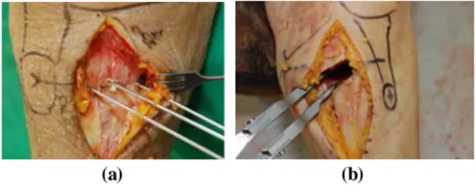
Fig. 4 (a) Three K-wires are inserted through the transverse line according to the anteromedial surface of tibia (b) Osteotomy gap was spread with speader. Posterior gap is wider than anterior gap without any intended bigger force on posterior gap
(A) (B)
Fig. 5 (A) Vertical gaps of medial open wedge osteotomy were measured. Distance from the hinge axis of lateral cortex to the anterior (X2) and posterior (X2) portion of anteromedial cortex at the level of osteotomy was measured and used for the calculation of the vertical height of the anterior (Y2) portion of anteromedial cortex at the level of osteotomy could be calculated with variables such as X1, X2 & Y1 (the vertical height of posterior portion of anteromedial cortex) (B) The hinge axis was positioned on the posterolateral corner of the tibia. The hinge axis and the anteromedial cortex of the tibia at the level of osteotomy site was parallel(θ=0).
Therefore, distance from the hinge axis of lateral cortex to the anterior (X2) and posterior (X2) portion of anteromedial cortex at the level of osteotomy would be equal. The vertical height of the anterior (Y2) portion of anteromedial cortex would be same with the vertical height of the posterior (Y1) portion of anteromedial cortex
proximal tibial angle and posterior slope before and after the surgery. The changes of medial proximal tibial angle and posterior slope were measured and compared. The mean medial proximal tibial angle of group I was changed from 84.2±2.5 to 91.7±3.8 after surgery(p<0.05). The mean medial proximal tibial angle of group II was changed from 82.8±2.3 to 90.8±2.3 after surgery(p<0.05). The mean change of
posterior slope of group I was statistically significant after surgery(2.7±1.0 p<0.05). But the mean change of posterior slope of group II was not statistically significant after surgery(0.3 ±0.5, p>0.05). In conclusion, the medial open wedge osteotomy with the posterolateral hinge increased the posterior slope of the tibia. But the osteotomy with lateral hinge did not increase the posterior slope. So, the hinge of open wedge osteotomy should be positioned not on the posterolateral side of the tibia, but on the lateral side to avoid the increase of the posterior slope of tibia.
References
(1) Chul-Hong Chun, Jung-Woo Kim, Jin-Young Park, and Chae-Gun Kim, 2006, "The Results of High Tibial Open Wedge Osteotomy with Arthroscopic Surgery for Varus Gonarthrosis," Journal of Korean Orthopedic Association, Vol.41, pp.636-642
(2) Torgerson W.R. Jr, Kettelkamp D.B., Logou R.A. Jr, Leach R.E., 1974, "Tibial Osteotomy for the Treatment of Degenerative Arthritis of the Knee,"
Clinical Orthopedic Relative Research, Vol. 101, pp.46-52.
(3) Dejour H., Bonnin M., 1994, "Tibial Translation after Anterior Cruciate Ligament Rupture: Two Radiological Test Compared," British Journal of Bone Joint Surgery, Vol. 76, pp. 745-749.
(4) Frank R. Noyesm, Steven X. Geobel and John West, 2005, "Opening Wedge Tibial Osteotomy: The 3-triangle Method to Correct Axial Alignment and Tibial Slope", The American Journal of Sports Medicine, Vol. 33, No.3, pp. 378-387.
(5) Asik, M., Sen, C., Kilic, B., Goksan, S. B., Ciftci, F., and Taser, O. F., 2006, "High tibial osteotomy with Puddu plate for the treatment of varus gonarthrosis,"
Knee Surg Sports Traumatol Arthrosc, Vol.14, No.10, pp.948-954.
(6) Briem, K., Ramsey, D. K., Newcomb, W., Rudolph, K.
S., and Snyder-Mackler, L., 2007, "Effects of the amount of valgus correction for medial compartment knee osteoarthritis on clinical outcome, knee kinetics and muscle co-contraction after opening wedge high tibial osteotomy," J Orthop Res, Vol.25, No.3, pp.311-318.
(7) Esenkaya, I. and Elmali, N., 2006 "Proximal tibia medial open-wedge osteotomy using plates with wedges: early results in 58 cases," Knee Surg Sports Traumatol Arthrosc,Vol.14, No.10, pp.955-961.
1470