The Effect of Anatomical Axis and Mechanical Axis on Change of Posterior Tibial Slope Angle in PTO(Proximal Tibial Osteotomy)
Eun-Ji Shin
*, Cheol-Woong Kim , Ho-Sang Lee
*, Ji-Hoon Bae
**, Joon-Ho Wang
**, Jong-Woong Park
**and Dong-Joon Oh
***Key Words: High Tibial Osteotomy, Anatomical Axis, Mechanical Axis, Posterior Slope Angle
Abstract
The purpose of this study was to investigate factors affecting the change of tibial posterior slope and introduce a mathematical model which calculate, through 3-dimensional analysis of the proximal tibia, how the angle of the opening wedge along the anteromedial tibial cortex influences the tibial posterior slope and valgus correction when performing a medial open wedge osteotomy. This mathematical model with navigation system can be guidelines which provide surgeons on preoperative and intraoperative measurements to maintain or correct the tibial slope and to obtain the desired valgus correction of the lower limb during an opening wedge osteotomy.
† Member, Korea Univ. & Triple-C Medical Corp.
E-mail : [email protected]
TEL : +82-2-929-9844 FAX : +82-2-929-9842
* Triple-C Medical Corp. R&D Center in Korea Univ.
** Orthopedic Surgery, Korea Univ. Medical Center
*** Mechanical Edu., Andong National University
1. Introduction
The arthritis can be cured by the uniformal distribution of the eccentric pressure at the medial or lateral side of the knee, which resulted from the varus and valgus, to the medial or lateral meniscus.
To improve its basic cause and to cure the main reason of the degenerate arthritis basically, High Tibial Osteotomy (HTO) has gradually been developed. Since the transferred loading from the medial femoral condyle by the anatomical axis dislocation of the tibia is eccentric to the medial tibial plateau of the knee and since it caused the rupture of the meniscus, the varus is the special anatomic malformation to require the osteotomy.
The surgery techique of HTO is comparably simple and it has few of postoperative complication. In addition to these reasons, since its cost is low, HTO is used to cure the varus. After the Upper Tibial Close Wedge Osteotomy, (UTCWO) was operated by Jackson(1) in the beginning, its technique has been developed gradually. Nowadays, Hernigou et. als.(2) establish the most adequate Upper Tibial Open Wedge Osteotomy (UTOWO) for the patient with the varus. The cut area of UTOWO is smaller than that of UTCWO and UTOWO minimizes the deformation difference between the cortical bone and the spongy bone of the proximal tibia. In addition to these advantages, since it prevents to shorten the pelvic limb, HTO becomes the modern trend.(3) The recent studies showed that the development of the artificial transplant to keep the anatomical axis and angle stable resulted in the effect of the axis at the pelvic limb on the knee pain and its function strongly. The metal fixing component developed too keep the angle of the knee stable, was the first 대한기계학회 2008년도 추계학술대회 논문집
1543
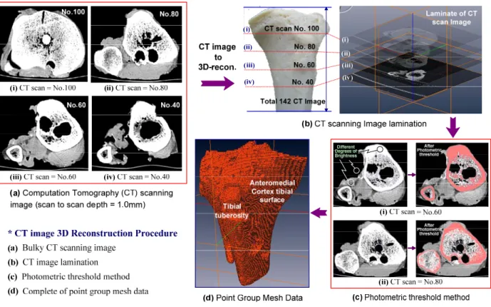
Fig. 1 Computer aided virtual surgery of opening wedge high tibia osteotomy using CT scan images reconstruction reliable method to fix the planned correction
without the second correction until it was cured completely without the damage of the soft tissue during the operation. In reference to the opening osteotomy, the complex correction performed at the close to the knee, comprehensively was able to be made at the many fields and the orthopedists want to use the new and effective medical devices.
Therefore, our research group performed the computation aided virtual osteotomy using the reconstruction of 3-D CT images. As the hinge axis angles shown in Fig. 2 were increased from 45° to 90° by the interval of 15°, the relationship between the posterior slope angle and the gap angle, which were increased from 5° to 20°, were defined and the general-purpose mathematical model was suggested. Moreover, the results were verified by the computation aided virtual osteotomy. The parameters to affect the increase of the posterior slope angle, were not only the hinge axis angle, the gap angle but also the oblique angle and the wedge angle. For this reason, it the co-relationship of the above 4 angles were defined, it is believed
Fig. 2 Schematic view of cross-section A'-A and the definition of hinge axis angle (θ) in the case of θ1=0, θ2=45, θ3=60, θ4=75, θ5=90that the bio-mechanical approach to the osteotomy is even clearer. Therefore, the hinge axis angle, the
gap angle, the oblique angle and the wedge angle were defined as the co-correction angle for our study and their relationship was verified by the computation aided virtual osteotomy. The relationship between the hinge axis angle and the gap angle was again defined and consequently the mathematical model to derive the more accurate results than Frank's were suggested.
1544
Hinge Axis
2mm
Hinge Axis
Saw -cut direc
tion
Fig. 3 Relationship between hinge axis angle = 90° versus hinge axis angle = 90° in high tibial osteotomy
Fig. 4 Stress distributions at the point "A", "B", "C", and
"D", respectively in X type fixation plate
The operation of Upper Tibial Open Wedge Osteotomy (UTOWO) to cure the malalignment and the degenerate arthritis, is easier than that of Upper Tibial Closed Wedge Osteotomy (UTCWO). In addition to this advantage, since the UTOWO did not cause the intraoperating damage of the proximal tibiofibula. It is the effective surgery technique to keep the penoneal nerve. To minimize the deformation of the upper tibia, the shortness of the lower limb can also be prevented. UTOWO is one of the
Check Point A Check Point BC Check Point D Max Stress 500
750 1000 1250 1500 1750 2000 2250 2500 2750 3000
Stress
Check Point & Max Stress Point 70deg
75deg 80deg 85deg 90deg
Fig. 5 Stress value of five different plate angles at the check point A, B, C, D and max. stress, respectively
(c) X-ray image of Ti-alloy fixation plate after osteotomy
in the tibiofemoral site (a) Fixation of Puddue plate
after osteotomy Graft Bone
B A D
C
Graft
(b) Insertion of autogenous cancellous bone graft
Graft Bone
Cortica l Bone Synostosi
s Part Load
Femur
Tibia
Fig. 6 Insertion of Puddue fixation plate and autogenous cancellous bone graft
surgery techniques to transfer the overload of the medial meniscus by the genu varum to the lateral meniscus by the translation of the anatomical axis and uniformly distributes the stress, which is transferred from the trunk to the tibia along the axial direction of the fermur, to the mediolateral menuscus. Considering the mechanical view, the supporting axis of the lower limb is the line to connect the center of the hip joint with the center of the tibiotalar joint. The connecting line is located inside the center of the knee joint and is deviated from about 4±2mm. If the mechanical axis is deviated medially or laterally, the genu varum deviation or the genu valgum deviation can be happened. If the supporting axis proceeds over 15mm from the centre of the knee joint
1545
medially or over 10mm laterally, the genu varum deviation or the genu valgum deviation is severely appeared.(8-9) To view from the lateral surface, tibia plateau is a little bit moved to the back rather than the axis of the femur and about 10° of articular surface in the tibia is angled down. This angle is called the Posterior Slope Angle (PSA) and it is the most important to keep PSA normal during UTOWO to correct the genu varum. However, the angle of the open wedge aperture by the cut of the proximal tibia after the osteotomy, can be changed by the walking/acting of the postoperated patient. After it results in the change of the PSA and the tibial spine deformation of the anatomical axis, the second malalignment and the unstable ligaments can be caused (See Fig.3). Comparing with the exact angle of UTOWO, the maintenance of the open wedge aperture in the proximal upper tibia, which is caused by the deformation of the anatomical axis after the osteotomy, is also very important. At this time, the metal plate is used to keep the open wedge aperture (See Fig.4). If the bone condition of the tibia is normal, the open wedge aperture can be maintained without the special operation to fix it after the osteotomy of 13mm using the metal plate.
References
(1) Chul-Hong Chun, Jung-Woo Kim, Jin-Young Park, and Chae-Gun Kim, 2006, "The Results of High Tibial Open Wedge Osteotomy with Arthroscopic Surgery for Varus Gonarthrosis," Journal of Korean Orthopedic Association, Vol.41, pp.636-642
(2) Torgerson W.R. Jr, Kettelkamp D.B., Logou R.A. Jr, Leach R.E., 1974, "Tibial Osteotomy for the Treatment of Degenerative Arthritis of the Knee,"
Clinical Orthopedic Relative Research, Vol. 101, pp.46-52.
(3) Dejour H., Bonnin M., 1994, "Tibial Translation after Anterior Cruciate Ligament Rupture: Two Radiological Test Compared," British Journal of Bone Joint Surgery, Vol. 76, pp. 745-749.
(4) Frank R. Noyesm, Steven X. Geobel and John West, 2005, "Opening Wedge Tibial Osteotomy: The 3-triangle Method to Correct Axial Alignment and Tibial Slope", The American Journal of Sports Medicine, Vol. 33, No.3, pp. 378-387.
(5) Asik, M., Sen, C., Kilic, B., Goksan, S. B., Ciftci, F., and Taser, O. F., 2006, "High tibial osteotomy with Puddu plate for the treatment of varus gonarthrosis,"
Knee Surg Sports Traumatol Arthrosc, Vol.14, No.10,
pp.948-954.
(6) Briem, K., Ramsey, D. K., Newcomb, W., Rudolph, K.
S., and Snyder-Mackler, L., 2007, "Effects of the amount of valgus correction for medial compartment knee osteoarthritis on clinical outcome, knee kinetics and muscle co-contraction after opening wedge high tibial osteotomy," J Orthop Res, Vol.25, No.3, pp.311-318.
(7) Esenkaya, I. and Elmali, N., 2006 "Proximal tibia medial open-wedge osteotomy using plates with wedges: early results in 58 cases," Knee Surg Sports Traumatol Arthrosc,Vol.14, No.10, pp.955-961.
(8) Cerejo R., D. Dunlop, S. Cahue, D. Channin, J. Song, L.
Sharma, 2002, " The Influence of Alignment on Risk of Knee Osteoarthritis Progression According to Baseline Stage of Disease," Arthritis Rheum, Vol.46, pp.2632-2636.
(9) Cicuttini, F., A. Wluka, J. Hankin, Y. Wang, 2004,
"Longitudinal Study of the Relationship Between Knee Angle and Tibiofemoral Cartilage Volume in Subjects with Knee Osteoarthritis," Rheumatology, Vol.43, pp.321-324.
1546