Introduction
High tibial osteotomy (HTO) can alter the mechanical axis (MA) of a limb so that the weight bearing load is transferred to healthy cartilage during the treatment of osteoarthritis with varus deformity13). In several studies, a proper postoperative MA was achieved using a cable, grid lines, a jig system, and a navigation
system46). Navigation systems provide reliable realtime intraop
erative information and may increase the accuracy of HTO3,4,7). The majority of previous studies reported a positive correla
tion between navigation and radiographic data4,811). However, some studies provided equivocal data12,13). Discrepancies of >2°
between navigation and weight bearing radiographic measure
ments of a corrected alignment have been reported12,13). This may be due to the difficulty of predicting a weight bearing alignment in a supine position under anesthesia, even with the use of a navi
gation system. Another possible reason might be postoperative changes in the tension of soft tissue surrounding the knee joint after HTO9,14).
During computerassisted closedwedge HTO, we observed postosteotomy changes in MA in response to external varus or valgus forces. This change in postosteotomy MA in response to external forces was named “dynamic range”. One question that arises is whether postoperative MA on weight bearing radio
Comparison of Mechanical Axis and Dynamic Range Assessed with Weight Bearing Radiographs and Navigation System in Closed Wedge High Tibial Osteotomy
Dae Kyung Bae, MD, Jong Whan Lee, MD, Seong Jin Cho, MD, and Sang Jun Song, MD
Department of Orthopaedic Surgery, Kyung Hee University College of Medicine, Seoul, Korea
Purpose: To compare navigation and weight bearing radiographic measurements of mechanical axis (MA) before and after closed wedge high tibial osteotomy (HTO) and to evaluate postosteotomy changes in MA assessed during application of external varus or valgus force.
Materials and Methods: Data from 30 consecutive patients (30 knees) who underwent computerassisted closedwedge HTO were prospectively analyzed. Pre and postoperative weight bearing radiographic evaluation of MA was performed. Under navigation guidance, pre and postosteotomy MA values were measured in an unloaded position. Any change in the postosteotomy MA in response to external varus or valgus force, which was named as dynamic range, was evaluated with the navigation system. The navigation and weight bearing radiographic measurements were compared.
Results: Although there was a positive correlation between navigation and radiographic measurements, the reliability of navigation measurements of coronal alignment was reduced after osteotomy and wedge closing. The mean postosteotomy MA value measured with the navigation was 3.5°±0.8°
valgus in an unloaded position. It was 1.3°±0.8° valgus under varus force and 5.8°±1.1° valgus under valgus force. The average dynamic range was >±2°.
Conclusions: Poten tial differences between the postoperative MAs assessed by weight bearing radiographs and the navigation system in unloaded position should be con sidered during computerassisted closed wedge HTO. Care should be taken to keep the dynamic range within the permissible range of alignment goal in HTO.
Keywords: Knee, Osteotomy, Mechanical axis, Navigation, External force, Reliability pISSN 2234-0726 · eISSN 2234-2451
Knee Surgery & Related Research
Received May 12, 2016; Revised July 24, 2016;
Accepted September 9, 2016
Correspondence to: Sang Jun Song, MD
Department of Orthopaedic Surgery, Kyung Hee University College of Medicine, 23 Kyungheedaero, Dongdaemungu, Seoul 02447, Korea Tel: +8229589489, Fax: +8229643865
Email: [email protected]
225
This is an Open Access article distributed under the terms of the Creative Commons Attribution NonCommercial License (http://creativecommons.org/licenses/bync/4.0/) which permits unrestricted noncommercial use, distribution, and reproduction in any medium, provided the original work is properly cited.
Copyright © 2017 KOREAN KNEE SOCIETY www.jksrr.org
graphs is still within the dynamic range of MA assessed by the navigation system.
The primary aim of this study was to analyze the reliability of navigation and weight bearing radiographic measurements of preoperative and postoperative MAs in patients with computer
assisted closed wedge HTO. A secondary aim was to evaluate changes in MA in response to external varus or valgus forces by the navigation system and to compare with values measured on weight bearing radiographs. We hypothesized that there would be changes in reliability of the navigation and weight bearing radio
graphic measurements of the alignment and that postosteotomy MA would exhibit a wide dynamic range under external forces.
Materials and Methods
1. Patients
Data were obtained from patients who underwent computer
assisted closed wedge HTO between August 2012 and Novem
ber 2013. The Vector Vision CTfree navigation system ver. 1.1 (BrainLAB, Heimstetten, Germany) was used for medial com
partment osteoarthritis with a varus deformity >10°. A miniplate staple (U&I Co., Uijeongbu, Korea) was used as a fixative15). Informed consent was obtained from all patients, none of whom declined to participate. Thirty patients (30 knees; 17 left and 13 right) were included; their mean age was 57.7±5.5 years (range, 46 to 68 years) and mean body mass index was 25.2±2.3 kg/m2
(range, 20.5 to 30.3 kg/m2). There were 26 females and 4 males.
2. Radiographic Evaluation
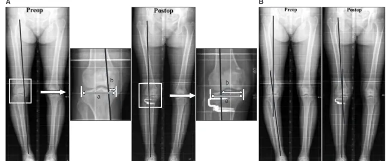
Radiographic measurements of the coronal alignment were performed using a fulllength weight bearing anteroposterior radiograph of the leg, including the hip, knee, and ankle (i.e., an orthoroentgenogram). Lateral radiographs of the knee were obtained and reviewed to assess the tibial posterior slope angle (PSA). Highquality pre and postoperative Xrays were obtained for all patients, and all measurements were performed using the standardized pictureacquiring and communication system (PACS)16). Coronal alignment (expressed as MA and MA% [the proportion of the MA that bisects the total width of the tibial width]) (Fig. 1A) values were evaluated radiographically. The femoral MA was defined as the line adjoining the centers of the hip and distal femoral notch. The tibial MA was defined as the line adjoining the centers of the tibial intercondylar eminences and the talus. The MA was defined as the angle between the femoral and tibial MAs (Fig. 1B)4,6). Postoperative radiographic evaluation was performed using the identical method 3 months after surgery when the patient could stand with complete weight bearing. The PSA was measured with a reference line connecting the center of the medullary canal 10 cm and 20 cm distal to the tibial plateau. Preoperative PSA was defined as the angle between the reference line and a line connecting the anterior and posterior borders of the medial tibial plateau. To reduce observation bias,
Fig. 1. Radiographic measurement of preoperative (preop) and postoperative (postop) mechanical axis (MA) and the percentage of mechanical axis (MA%). (A) The MA% was evaluated on the orthoroentgenogram and expressed in percentage [(b/a)×100]. (B) The MA was defined as the angle between femoral and tibial mechanical axes on the orthoroentgenogram. a: the width of tibia plateau, b: the distance from the medial border of the medial tibial condyle to the point at which the mechanical axis intersects the knee joint line.
a b
A
b a
B
two independent investigators repeatedly performed all radio
graphic measurements. The intra and interobserver reliabilities of all measurements were assessed using intraclass correlation coefficients (ICC)17), which were >0.8 for all measures.
3. Measurement by the Navigation System
Under navigation guidance, the MA was measured before os
teotomy. Postosteotomy MA and MA% values were measured following wedge closing and fixation. After assessing postosteot
omy MA in the unloaded position, we evaluated changes in post
osteotomy MA under conditions of external varus or valgus force (Fig. 2).
4. Statistical Analysis
Measurement data obtained under navigation guidance were compared with radiographic data using paired ttests. The reli
ability of navigation and radiographic measurements was as
sessed using Pearson correlation analysis. ICC values >0.8 were considered very strong; values between 0.6 and 0.8 were mod
erately strong, values between 0.3 and 0.6 were fair, and values
<0.3 were weak18). In addition to data obtained in the unloaded position, mean postosteotomy MA and MA% values, assessed using the navigation system during application of external varus or valgus force, were also compared to the radiographically mea
sured values. They were directly compared in each knee to assess whether postoperative MA values on weight bearing radiographs are within the dynamic range measured using the navigation system during application of external forces. Patients were cat
egorized according to the difference between their postoperative MA values on radiographs and postosteotomy MA values in the unloaded position obtained using the navigation system. When
the difference was between –2° and 2°, patients were assigned to group N (no difference). When the difference was <–2° or >2°, patients were assigned to group D (reduced valgus alignment on weight bearing radiographs) or I (increased valgus alignment on weight bearing radiographs). Preoperative and postoperative MA and PSA values on radiographs, and pre and postosteotomy MA values in the navigation system, were compared between groups N and D (Student ttest) to assess whether correction angle or PSA influences changes in MA on weight bearing radio
graphs.
Statistical analysis was performed using the SPSS ver. 18.0 (SPSS Inc., Chicago, IL, USA). A value of p<0.05 was taken to indicate statistical significance.
5. Surgical Method
A transverse skin incision extending from the tibial tubercle to the fibular neck is made, and the anterior tibialis muscle is subperiosteally dissected4). Partial resection of the fibular head is made to avoid the tethering effect of the fibula during wedge closing in every knee. Two separate reference bases are fixed to the distal femur and to the middle tibia with a pair of halfpins.
Eight kinematic and anatomic landmarks are registered. These include the kinematic hip center, medial and lateral malleoli, me
dial and lateral tibial plateau points, medial and lateral femoral epicondyles, and AP tibial rotation. The surgeon should take care to make the start and end points to be 15 mm distal to the tibial plateau. The far cortical hinge is tailored to be 3 mm in order to help the surgeon to close the osteotomy surface easily. The tibial posterior slope can be finetuned in the planned osteotomy ac
cording to the the PSA measured on the preoperative radiograph.
The navigation system provides information about the deformity,
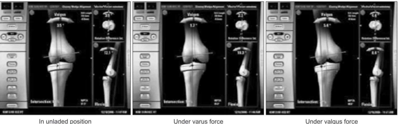
Fig. 2. The alteration of postosteotomy mechanical axis (MA) and percentage of mechanical axis (MA%) on navigation under external force. In this knee, the postosteotomy MA and MA% were 3.5° and 62%, respectively, in the unloaded position. Those values were decreased to 1.7° and 52%, re
spectively, under varus force, and increased to 5.4° and 72%, respectively, under valgus force.
In unladed position Under varus force Under valgus force
level of osteotomy, correction angle, and wedge size. For the op
timum degree of correction, the postoperative MA%, which can be obtained via the navigation system, should be 62%6,19). A pre
calibrated navigated drill guide is used to place two Kwires in the proximal plane of the osteotomy. Another two Kwires are inserted in the distal plane of the osteotomy in the same manner.
This 4 pin technique helps to make the anterior and posterior lengths of the wedge symmetric and to minimize the change in PSA4). The proximal and distal osteotomies are carried out using a sharp electric saw over two Kwires. The wedge is removed and the medial far cortex is then carefully decorticated using a sharp electric saw or an osteotome. A valgus force is applied slowly to the extremity until the proximal and distal osteotomy surfaces are firmly attached. Then, the osteotomy site is rigidly fixed using a miniplate staple of appropriate size15).
The final alignment or postHTO MA and MA% on the com
puter screen were confirmed in the unloaded position. The change of postosteotomy MA and MA% under external varus or valgus force was also evaluated using the navigation system (Fig.
2). A systematic rehabilitation protocol was used: straight leg rais
ing exercises were started 3 days postoperatively; partial weight bearing was started 1 week postoperatively; and full weight bear
ing without crutches was started at 6–12 weeks based on the pa
tient’s condition.
Results
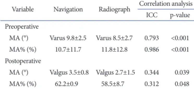
After computerassisted closed wedge HTO, varus deformity was corrected according to both navigation and radiographic data (Table 1). There was a positive correlation between naviga
tion and radiographic measurements of pre and postoperative MA and MA%. According to ICC values, the preoperative cor
relation between measures was strong, and the postoperative correlation was fair (Table 1). The average postosteotomy MA and MA% assessed using the navigation system were 3.5°±0.8°
valgus and 62.2±0.9%, respectively, in the unloaded position. The postosteotomy MA and MA% were changed by external varus and valgus forces. The average change of MA was more than ±2°
(Table 2, Fig. 3). Although the mean postosteotomy MA and MA% obtained using the navigation system in the unloaded po
sition were correlated to those obtained on radiography (Table 2), differences of >2° were observed in several knees (Fig. 3). When patients were categorized according to the difference of postop
erative MA values measured on weight bearing radiographs and using the navigation system in the unloaded position, there were 22 knees in group N with the difference between –2° and 2°, 7 knees in group D, and one knee in group I. The radiographyde
rived values were typically lower than navigationderived values in the unloaded position in group D (Fig. 3). Preoperative MA and PSA values on the radiograph did not differ between groups N and D (Table 3). Pre and postosteotomy MA values by the navigation system were also similar between the two groups, including postosteotomy MA values obtained under varus and valgus forces. The average postoperative MA on weight bearing Table 1. Correlation between Navigation System and Radio graphic
Measurements of Coronal Alignment
Variable Navigation Radiograph Correlation analysis ICC pvalue Preoperative
MA (°) Varus 9.8±2.5 Varus 8.5±2.7 0.793 <0.001 MA% (%) 10.7±11.7 11.8±12.8 0.986 <0.001 Postoperative
MA (°) Valgus 3.5±0.8 Valgus 2.7±1.5 0.344 0.039
MA% (%) 62.2±0.9 58.5±8.7 0.312 0.048
ICC: intraclass correlation coefficient, MA: the angle between the femoral and tibial mechanical axes, MA%: the percentage at which the mechanical axis bisects the total width of the tibia.
Table 2. Comparison of Postosteotomy MA and MA% in Unloaded Po si tion and Under Varus or Valgus External Force on Navigation and in Weight Bearing Radiograph
Post operative Unloaded position Varus force Valgus force Radiograph
MA (°) Valgus 3.5±0.8 Valgus 1.3±0.8 Valgus 5.8±1.1 Valgus 2.7±1.5
ICC 0.344 0.270 0.254
pvalue 0.039 0.149 0.175
MA% 62.2±0.9 50.7±4.5 74.8±0.9 58.5±8.7
ICC 0.312 0.027 0.218
pvalue 0.048 0.886 0.247
MA: the angle between the femoral and tibial mechanical axes, MA%: the percentage at which the mechanical axis bisects the total width of the tibia, ICC: intraclass correlation coefficient.
radiographs was valgus 3.1° in group N and valgus 0.9° in group D, respectively, although navigated values were similar in both groups for postoperative MA (Table 3).
Discussion
The most important finding of this study was that the reliability of MA and MA% values measured using the navigation system decreased after osteotomy and wedge closing, although there was a positive correlation between navigation and radiographic measurements. Based on the assumption that accurate correction involves a weight bearing axis that intersects the tibial plateau at 50%‒70% of the cross sectional tibial width6), navigation mea
surements obtained in a neutral position or during application of external force may be decreased compared to measurements ob
tained radiographically during standing. According to ICC val
ues, the strong preoperative correlation between navigation and radiographic measurements decreased to “fair” postoperatively.
The discrepancy of MA measurement between weight bearing and nonweight bearing condition may be a critical problem in HTO irrespective of the use of a navigation system. The weight bearing postoperative MA is difficult to predict accurately dur
ing osteotomy in the supine position, and this can result in the correction error of alignment20). The present study showed that the reliability of nonweight bearing navigation measurements of MA decreased after osteotomy. Therefore, we think that careful assessment of MA by the navigation system will be necessary.
Fibular management and wedge closing during closed wedge HTO might affect alterations in soft tissue tension and postop
erative alignment during standing assessed by radiographs. Lee et al.13) also reported that the reliability of navigation measurements of alignment after surgery was decreased following computer
assisted open wedge HTO, possibly because superficial medial collateral ligament (MCL) injury sustained during the procedure could cause valgus instability on weight bearing radiographs.
Kendoff et al.9) reported that application of a simulated weight bearing force altered MA values by up to 2.5° during computer
assisted open wedge HTO, due to changes in soft tissue tension, including functional lengthening of the lateral collateral ligament (LCL) exterior to the hinge axis, and MCL release. We hypoth
esized that alterations in soft tissue tension, and its effects on weight bearing alignment, would be detected during computer
assisted closed wedge HTO. Fibular management, including proximal tibiofibular dissociation and partial fibular head resec
tion, could cause LCL laxity after wedge closing, decreasing the valgus correction angle measured by the navigation system under varus external force.
A second important finding was that postosteotomy MA and MA% values measured by the navigation system changed under external varus and valgus forces and were associated with a wide dynamic range compared to the unloaded position (Fig. 3). The change in MA under external force may be explained by func
tional MCL laxity external to the hinge axis or LCL laxity after Fig. 3. Postosteotomy mechanical axis (MA) values on navigation and
weight bearing radiographs. Triangles indicate postosteotomy MA values on navigation in the unloaded position; lines indicate dynamic range during application of varus and valgus force. Squares indicate postoperative MA values during weightbearing on radiographs. In to
tal, eight knees exhibited a difference of >2° between radiography and navigationderived values in the unloaded position (radiographic values are denoted by open squares).
1 6 11 16 21 26
0 9 8 7 6 5 4 3 2 1
30 Patient (no.)
Valgus()
Radiograph
Navigation Table 3. Comparison of Navigation System and Radiographic Measurements of Coronal Alignment between Group N and Group D
Measurement Group N (n=22) Group D (n=7) pvalue Preoperative (°)
Radiograph
MA Varus 8.2±3.0 Varus 9.6±1.6 0.266
PSA 11.3±3.0 11.7±2.8 0.780
Navigation
MA Varus 9.8±2.7 Varus 9.9±1.8 0.888
Postoperative (°) Navigation
MA in unloaded position Valgus 3.4±0.7 Valgus 3.9±0.9 0.119 MA under varus force Valgus 1.3±0.9 Valgus 1.3±0.6 0.855 MA under valgus force Valgus 5.7±1.1 Valgus 5.9±1.1 0.712 Radiograph
MA Valgus 3.1±1.0 Valgus 0.9±0.8 <0.001
PSA 8.6±3.5 9.2±1.7 0.515
MA: the angle between the femoral and tibial mechanical axes, PSA:
posterior tibial slope angle.
closed osteotomy. The average dynamic range of postosteotomy MA was >±2° on navigation, and varied between patients (Fig.
3). Postoperative MA values on weight bearing radiographs were within the dynamic range of postoperative MA values obtained under external force by the navigation system. Although weight bearing radiographic measurements were similar to naviga
tion measurements in the unloaded position in the majority of patients, they were decreased in seven knees, and were similar to navigation measurements obtained during the application of external varus force (Table 3, Fig. 3). This finding accords with previous studies4,14,21) describing a “springback” effect from conditions of LCL laxity and undercorrection of postoperative MA after closed wedge HTO. The ideal postoperative MA angle is between 2°–6°2,22), therefore, surgeons will be able to aim for a corrected angle within this range during the computerassisted closed wedge HTO. Under navigation guidance, postosteotomy MA values in both the unloaded position, and under external varus force, may be corrected to resolve the unpredictability as
sociated with LCL laxity and undercorrection.
Finally, we observed neither group difference (N vs. D) in any confounding variable nor any factor predicting reductions in MA correction. Kendoff et al.9) demonstrated significant shifts in MA during external loading and weight bearing, and further reported that degree of correction, applied magnitude, and ligament integ
rity all affected the alteration of postoperative MA values under external loading conditions. In the present study there was no group difference (N vs. D) in the severity of preoperative varus deformity, correction angle, or alterations in PSA. However, post
operative MA and MA% values on weight bearing radiographs differed between the two groups.
This study has several limitations. First, only closed wedge HTO was included due to our surgical preference. Recognition of changes in postoperative MA values, during weight bearing or under external force conditions during open wedge HTO, is also required to understand changes in correction angle during postoperative weight bearing and in the context of changes in softtissue tension. Second, the characteristics of our prospective cohort differed from those of Western HTO candidates, in whom the gender distribution of osteoarthritis and obesity prevalence may be important factors. Such differences must be considered before generalizing our data to other populations. Third, we did not perform any clinical evaluations. Assuming that proper post
operative alignment results in clinical satisfaction, we focused on the accuracy and reliability of navigated and radiographic measurements of MA. Fourth, the accuracy of percutaneous reg
istration during computerassisted HTO is limited. We marked
landmarks before registration, after careful consideration of sur
face anatomy, to ensure accurate registration. Fifth, preoperative MA was not assessed under external varus and valgus force by the navigation system. It was difficult to explain the reason for reduced reliability of the measurement of MA by the navigation system. However, the preoperative MA under external force was negligible in full extended position of the knees in most patients.
Sixth, the methodological categorization was based on cutoff value of ±2° according to the difference of postoperative MA values between weight bearing radiograph and navigation in un
loaded position. It was thought to have clinical relevance and be within the radiographic measurement accuracy to be detected.
Finally, the accuracy of radiographic measurements was limited.
Slight changes in projection angle and rotation or flexion of the knee may have affected radiographic measurements. Although computed tomography can accurately measure the alignment, its application is limited due to radiation exposure and inability to obtain weight bearing images. We aimed to acquire consistently high quality images in the neutral position and confirmed the intra and interobserver reliability of all measurements.
Conclusions
The reliability of navigation measurements of coronal alignment was reduced after osteotomy and wedge closing. Postosteotomy MA values assessed by the navigation system were altered under the application of external forces and exhibited a dynamic range.
Potential differences of postoperative MA between weight bear
ing radiograph and navigation in unloaded position should be considered during computerassisted closed wedge HTO, and the dynamic range on navigation should be kept within the permis
sible range of alignment goal in HTO.
Conflict of Interest
No potential conflict of interest relevant to this article was re
ported.
References
1. Coventry MB, Ilstrup DM, Wallrichs SL. Proximal tibial osteotomy: a critical longterm study of eightyseven cases. J Bone Joint Surg Am. 1993;75:196201.
2. Lee DC, Byun SJ. High tibial osteotomy. Knee Surg Relat Res. 2012;24:619.
3. Maurer F, Wassmer G. High tibial osteotomy: does naviga
tion improve results? Orthopedics. 2006;29(10 Suppl):S1302.
4. Bae DK, Song SJ, Yoon KH. Closedwedge high tibial os
teotomy using computerassisted surgery compared to the conventional technique. J Bone Joint Surg Br. 2009;91:1164
71.
5. Kim SJ, Koh YG, Chun YM, Kim YC, Park YS, Sung CH.
Medial opening wedge hightibial osteotomy using a kine
matic navigation system versus a conventional method: a 1year retrospective, comparative study. Knee Surg Sports Traumatol Arthrosc. 2009;17:12834.
6. Reising K, Strohm PC, Hauschild O, Schmal H, Khattab M, Sudkamp NP, Niemeyer P. Computerassisted navigation for the intraoperative assessment of lower limb alignment in high tibial osteotomy can avoid outliers compared with the conventional technique. Knee Surg Sports Traumatol Ar
throsc. 2013;21:1818.
7. Hofmann AA, Wyatt RW, Beck SW. High tibial osteotomy:
use of an osteotomy jig, rigid fixation, and early motion versus conventional surgical technique and cast immobiliza
tion. Clin Orthop Relat Res. 1991;(271):2127.
8. Heijens E, Kornherr P, Meister C. The role of navigation in high tibial osteotomy: a study of 50 patients. Orthopedics.
2009;32(10 Suppl):403.
9. Kendoff D, Board TN, Citak M, Gardner MJ, Hankemeier S, Ostermeier S, Krettek C, Hüfner T. Navigated lower limb axis measurements: influence of mechanical weightbearing simulation. J Orthop Res. 2008;26:55361.
10. Lutzner J, Gross AF, Günther KP, Kirschner S. Reliability of limb alignment measurement for high tibial osteotomy with a navigation system. Eur J Med Res. 2009;14:44750.
11. Yamamoto Y, Ishibashi Y, Tsuda E, Tsukada H, Kimura Y, Toh S. Validation of computerassisted openwedge high tibial osteotomy using threedimensional navigation. Ortho
pedics. 2008;31(10 Suppl 1).
12. Kyung BS, Kim JG, Jang KM, Chang M, Moon YW, Ahn JH, Wang JH. Are navigation systems accurate enough to predict
the correction angle during high tibial osteotomy? Com
parison of navigation systems with 3dimensional computed tomography and standing radiographs. Am J Sports Med.
2013;41:236874.
13. Lee DH, Nha KW, Park SJ, Han SB. Preoperative and post
operative comparisons of navigation and radiologic limb alignment measurements after high tibial osteotomy. Ar
throscopy. 2012;28:184250.
14. Shaw JA, Dungy DS, Arsht SS. Recurrent varus angulation after high tibial osteotomy: an anatomic analysis. Clin Or
thop Relat Res. 2004;(420):20512.
15. Bae DK, Mun MS, Kwon OS. A newly designed miniplate staple for high tibial osteotomy. Bull Hosp Jt Dis. 1997;56:
16770.
16. Fowler JR, Ilyas AM. The accuracy of digital radiography in orthopaedic applications. Clin Orthop Relat Res. 2011;469:
17814.
17. Lachin JM. The role of measurement reliability in clinical trials. Clin Trials. 2004;1:55366.
18. Chan YH. Biostatistics 104: correlational analysis. Singapore Med J. 2003;44:6149.
19. Fujisawa Y, Masuhara K, Shiomi S. The effect of high tibial osteotomy on osteoarthritis of the knee: an arthroscopic study of 54 knee joints. Orthop Clin North Am. 1979;10:585
608.
20. Ogata K, Yoshii I, Kawamura H, Miura H, Arizono T, Sugio
ka Y. Standing radiographs cannot determine the correction in high tibial osteotomy. J Bone Joint Surg Br. 1991;73:927
31.
21. Gaasbeek RD, Nicolaas L, Rijnberg WJ, van Loon CJ, van Kampen A. Correction accuracy and collateral laxity in open versus closed wedge high tibial osteotomy: a oneyear ran
domised controlled study. Int Orthop. 2010;34:2017.
22. Jakob RP, Jacobi M. Closing wedge osteotomy of the tibial head in treatment of single compartment arthrosis. Ortho
pade. 2004;33:14352.