Original Article
RECEIVED July 19, 2011, REVISED October 7, 2011, ACCEPTED October 25, 2011 Correspondence to Young-Ju Park
Department of Oral and Maxillofacial Surgery, Kangnam Sacred Heart Hospital 948-1, Daerim 1-dong, Yeongdeungpo-gu, Seoul 150-950, Korea
Tel: 82-2-829-5233, Fax: 82-2-846-9789, E-mail: [email protected]
CC This is an open access article distributed under the terms of the Creative Commons Attribution Non-Commercial License (http://creativecommons.org/licenses/
by-nc/3.0) which permits unrestricted non-commercial use, distribution, and reproduction in any medium, provided the original work is properly cited.
3-Dimensional Micro-Computed Tomography Study on Bone Regeneration with Silk Fibroin,
rh-Bone Morphogenetic Protein Loaded-Silk Fibroin and Tricalcium Phosphate Coated-Silk Fibroin
in Rat Calvaria Defect
Eun-O Pang, Young-Ju Park, Su-Hyun Park, Eung-Sun Kang, Haeyong Kweon
1, Soeng-Gon Kim
2, Chang-Yong Ko
3, Han-Sung Kim
3, Jeong-Hun Nam, Jang-Hun Ahn, Ji-Hyun Chun, Byeong-Min Lee Department of Oral and Maxillofacial Surgery, Kangnam Sacred Heart Hospital, Collage of Medicine, Hallym University,
1
National Academy of Agricultural Science, Rural Department Administration,
2Department of Oral and Maxillofacial Surgery, College of Dentistry, Gangneung-Wonju National University,
3Department of Biomedical Engineering & Institute
of Medical Engeering, Yonsei University
Abstract
Purpose: The purpose of this study was to evaluate the bone regeneration capacity of silk fibroin (SF) when combined with beta tricalcium phosphate (β-tricalcium phosphate [TCP]) and rh-bone morphogenetic protein (BMP) in vivo by micro-com- puted tomography (CT), soft x-ray, and histological analysis.
Methods: A total of 56 critical size defects formed by a trephine bur made on 28 adult female Spague-Dawley rats were used for this study and the defect size was 5.0 mm in diameter. The defects were transplanted with (1) no graft material (raw defect), (2) autogenous bone, (3) SF (10 μg), (4) SF-BMP (10 μg, 0.8 μg each), and (5) SF+β-TCP (10 μg). At 4 and 8 weeks after operation, the experimental animals were sacrificed. Samples were evaluated with soft x-ray, histological examinations and 3-dimensional micro-CT analysis.
Results: In the 3-dimensional micro-CT evaluation, bone volume and bone surface data were higher in the SF-BMP (12.8±1.5, 138.6±45.0 each) ( P <0.05) and SF-TCP (12.3±1.5, 144.9±30.9 each) group than in the SF group (6.1±3.3, 77.2±37.3 each) ( P <0.05), except for the autogenous group (15.0±3.0, 190.7±41.4 each) at 4 weeks. At 8 weeks, SF-BMP (16.8±3.5, 173.9±34.2 each) still revealed higher ( P <0.05) bone volum and surface, but SF-TCP (11.3±1.5, 1132.9±52.1 each) ( P =0.5, P =0.2) revealed the same or lower amount compared with the SF group (13.8±2.7, 127.5±44.8 each). The % of bone area determined by radiodensity was higher in the SF-TCP (31.4±9.1%) and SF-BMP (36.2±16.2%) groups than in the SF (19.0±10.4) group at the period of 4 weeks. Also, in the histological evaluation, the SF-BMP group revealed lower inflammation reaction, lower foreign body reaction and higher bone healing than the SF group at postoperative 4 weeks and 8 weeks.
The SF-TCP group revealed lower inflammation at 4 weeks, but accordingly, as the TCP membrane was absorbed, inflammatory and foreign body reaction are increased at 8 weeks.
Conclusion: The current study provides evidence that the silk fibrin can be used as an effective grafted material for tissue engineering bone generation through a combination of growth factor or surface treatment.
Key words: Silk fibroin, Rat calvarial defect, Bone regeneration, Micro-CT
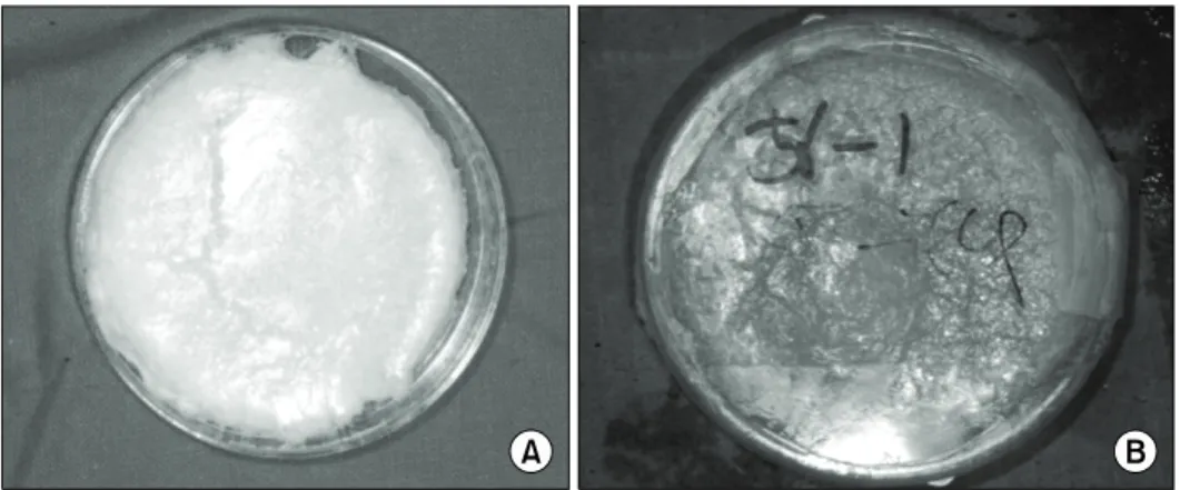
Fig. 1. Sponge-typed graft materials.
(A) Tricalcium phosphate coated silk fibroin. (B) Silk fibroin.
Introduction
Grafts for bone regeneration in pathological or traumatic bone defects have become a common approach used for dental implantations. Autogenous bone, xenobone and syn- thetic polymers such as polylactic-co-glycolic acid, poly- lactic acid and poly glycolic acid are commonly grafted onto bony defect[1-3]. These grafting materials can cause immunological and inflammatory reactions[4]. Sometimes the grafted materials are resorbed before new bone form.
Autogenous bone is a preferred material for bone grafting but it has some problems such as limited available quantity, potential donor site morbidity and surgical inconveniences like prolonged operation time and the necessity of a secon- dary operation site[4].
By contrast, silk is a high molecular weight natural pro- tein polymer that has been approved as a non-absorbable biomaterial by the U.S. Food and Drug Administration[5].
Silk protein spun by the silkworm Bombyx mori mainly consists of fibroin and sericin[5]. Whereas sericin may trig- ger immunological and allergic reactions[6]. In vivo , silk fibroin tends to induce only minimal inflammatory re- actions[7]. It also has other advantageous characteristics, including being biodegradable and biocompatible[8] and having good water vapor and oxygen permeability[9,10].
Previous work has shown, fragmented low molecular weight silk fibroin (SF) effectiveness in bony defect repair both in vivo and in vitro [11-13]. In addition to functioning like slow degrading scaffold with excellent mechanical properties[14], it has worked well in engineering tissues that repair ligaments, tendons, bones, and cartilage[15,16].
Given these beneficial characteristics, this study eval- uated usefulness of rh-bone morphogenetic protein (BMP)-
loaded SF (SF-BMP) and beta tricalcium phosphate-coated SF (SF-tricalcium phosphate [TCP]) as bone graft substitutes for autogenous bone. SF, SF-TCP and rh-BMP loaded SF-BMP were grafted onto bone defects of rat calvaria. To investigate the efficacy of these grafted materials, three-di- mensional microfocus computerized tomography (micro-com- puted tomography [CT] analysis) was performed and the results of the analysis were compared with the result from soft x-ray analysis and histological examination.
Two-dimensional radiographic and histological analyses are commonly used simultaneously for bone regeneration evaluation. These methods are invasive though and only provide indirect two-dimensional images[17-20]. Thus, as a new method, micro-CT was introduced. Not only is mi- cro-CT analysis non-invasive but it also reconstructs three-dimensional images in desired planes with a reso- lution that is one hundred times higher than medical-CT.
These characteristics have caused a significant upswing in micro-CT use in medical and dental studies in recent years[21-24].
Materials and Methods
1. Preparation of SF and SF-TCP
Pure SF and SF-TCP with a molecular weight of-about 350 kDa-were prepared and generously donated by the Rural Development Administration (Suwon, Korea) for this experiment. Briefly, raw silk was degummed twice using Marseilles soap (0.5% fiber by weight) and sodium carbo- nate (0.3% fiber by weight) solution at 100
oC for 1 hour.
The resulting sericin-free silk was then washed with dis-
tilled water. Subsequently, the SF was precipitated into
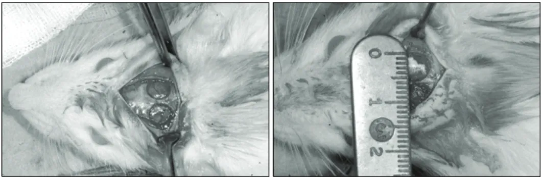
Fig. 2. Photographs of animal surgery.
(5 mm diameter bone defect formed by trephine bur).
Table 1. Animal grouping Group Post-op
4 wk Post-op
8 wk Total Graft quantity AutoBG
RAW SF SF-BMP SF-TCP Sum
3 (6) 3 (6) 3 (6) 3 (6) 3 (6) 15 (30)
3 (6) 2 (4) 3 (6) 3 (6) 2 (4) 13 (26)
6 (12) 6 (12) 6 (12) 6 (12) 6 (12) 28 (56)
Autogenous bone graft None
10 μg 10 μg+0.8 μg
10 μg
AutoBG, autogenous bone graft; RAW, raw defect; SF, silk fibroin; SF-BMP, rh-BMP loaded silk fibroin; SF-TCP, tricalcium phosphate coated silk fibroin.
powder and molded into sponge-typed graft materials (Fig.
1). After separate packings of the SF and SF-TCP onto Petri dishes, they were sterilized using low temperature (36.5
oC) ethylene oxide-gas prior to surgery.
2. Preparation of rh-BMP-2
We used rh-BMP as a constructor’s subscription. The recombinant human bone morphogenetic protein-2 (rh-BMP- 2; R&D, Minneapolis, MN, USA) we used was generated in a Chinese hamster. 10 μg rh-BMP was dissolved in 0.18 mL saline, and injected 0.015 mL each defects.
3. Rat calvarial surgery and animal grouping
Thirty Sprague-Dawley rats weighing 250∼300 g each, (Oriental Bio Co, Charles River Korea, Seoul, Korea) were equally assigned into 5 groups (n=12) as shown in Table 1: 1) the autogenous bone graft (Auto), 2) the raw defect control (Raw), 3) SF graft (10 μg) (SF), 4) SF-BMP (10.8 μg), and 5) SF-TCP (10 μg).
A calvarial critical size defect with an 5.0 mm diameter was created by using trephine bur, and the defect was filled with the prepared graft materials (Fig. 2).
All animals recovered well after surgery, except for two rats treated with raw defect and the SF-TCP respectively that died 2 days later. Thus, the final animal numbers per
group were n=6 for the Auto, SF and SF-BMP groups and n=4 for the Raw and SF-TCP groups (Table 1). The animals were sacrificed by CO2 asphyxiation at 4 weeks and 8 weeks post-surgery and the skulls were harvested for analyses. This study was conducted under an approved protocol for animal care in accordance with guidelines es- tablished by the Institutional Animal Care and Use Committee of Hallym University (No. Hallym 2009-66, July 24, 2009).
4. 3-dimesional micro-CT analysis
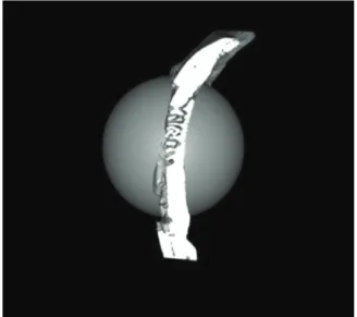
Micro-CT images were obtained using an X-ray micro- tomography system (Skyscan 1072/1172, version. 1.5, Skyscan, Belgium) with source 40 kV/200 uA and, an ex- posure time of 1.5 seconds, a rotational step angle of 0.3 degrees, and no filtering. Samples were fixed to the turn- table inside the CT scanner and then, adjusted to the right place by moving horizontally and vertically. After scanning, images were reconstructed on a personal computer. These raw data images (TIFF files) were transferred into re- constructed digital images (bmp files) using the NRecon software program (Version: 1.6.1, Skyscan, Belgium). The region of interest was the initial bone defect size and shape (Fig. 3).
The new bone formation capacity was analyzed using the morphometric parameters of regenerated new bone volume (bone volume [BV], mm
3) and surface area (bone surface [BS], mm
2). Three-dimensional rendered model were obtained using the CT-AN. 1.10 program (Skyscan NV., Belgium).
5. Soft x-ray analysis
After harvesting the superior aspect of the calvaria in-
cluding the defect, a radiogram was taken with a cab-
inet-style soft x-ray unit (CMB-2, Softex Co., Tokyo, Japan)
Fig. 3. Region of interest. 5 mm diameter round type.
at 20 kVp and 2 mA, with an exposure time of 120 seconds.
The images were transferred into digital image files by digitally scanning film positives using 8 bit and 300 dpi resolution settings. The percentage of the regenerated area was calculated from captured images as the percentage ratio of the new bone area including grafted materials to the experimental area.
After taking the soft x-rays, samples in each group were fixed in a 10% (v/v) neutral buffered formalin solution for histological processing and micro-CT analysis.
6. Histological examination
For the histological examination, fixed samples were de- calcified with 10% formic acid. After bisecting the samples tangential to the sagittal suture, they were embedded with paraffin with the cut surface facing outward 4 μm thick sections, and stained with hematoxylin-eosin and Masson's Trichrome.
7. Statistics
Data obtained were tested by non-parametric statistical analysis (the Kruskal Wallis Test) using Statview statistical software (SAS Institute Inc., Cray, NC, USA). All P value≤
0.05 were considered statistically significant.
Results
1. 3-Dimensional micro-CT analysis
The numerical figure of mean bone volume revealed that both the SF-BMP (12.8 mm
3) and SF-TCP (12.3 mm
3) groups had significantly statistic higher values than the raw defact (RAW) group (9.8 mm
3) at postoperative 4 weeks.
The SF group (6.1 mm
3) group showed lower bone volume than the RAW group did. At postoperative 8 weeks, the SF-BMP group (16.8 mm
3) had the next largest value after the Autogenous group (20.0 mm
3). The value of SF-BMP in bone volume at postoperative 8 weeks, when compared with the RAW group (7.0 mm
3) and the SF-TCP group (11.3 mm
3), was statistically significant (Fig. 4).
The mean bone surface values revealed that the Raw group (117.0 mm
2) had a higher value than the SF group (77.2 mm
2) did at postoperative 4 weeks. The SF-BMP group (138.6 mm
2) and the SF-TCP group (144.9 mm
2), which were compared with the SF group independently, had a significantly lager value in bone surface at postoperative 4 weeks. At postoperative 8 weeks, the SF-BMP (173.9 mm
2) group had the largest value in bone surface excepting the Autogenous group (217.5 mm
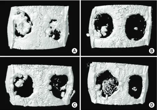
2) (Fig. 5). For a breakdown of the study results, please refer to the attached micro-CT 3-dimensional reconstruction images in Fig. 6 (Table 2).
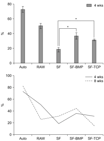
2. Soft x-ray analysis
The mean percent of bone area revealed that the SF-BMP group (36.20%) had a significant higher value than the SF group (19.0%) did at postoperative 4 weeks. The SF-TCP group (31.5%) also had a higher bone percentage area than the SF group. The SF-BMP and SF-TCP groups initially re- vealed similar bone area values: however, the SF-BMP group (44.7%) had a significantly higher bone area than the SF-TCP group (15.7%) at postoperative 8 weeks (Table 3). Interestingly, the SF-TCP group had a lower bone area than the SF group (31.4%) did at postoperative 8 weeks (Fig. 7).
3. Histological examination
From the histological point of view, the SF-TCP group
revealed lower inflammation than the SF group at post-
operative 4 weeks. However, there was higher in-
flammatory change in the SF-TCP group at postoperative
Fig. 4. Three-dimensional analysis: bone volume (post op 4 weeks and 8 weeks). *Statistically significant difference P <0.05. Auto, autogenous; RAW, raw defect; SF, silk fibroin; SF-BMP, rh-BMP loaded silk fibroin; SF-TCP, tricalcium phosphate coated silk fibroin.
Fig. 5. Three-dimensional analysis: bone surface (post op 4 weeks and 8 weeks). *Statistically significant difference P <0.05. Auto, autogenous; RAW, raw defect; SF, silk fibroin; SF-BMP, rh-BMP loaded silk fibroin; SF-TCP, tricalcium phosphate coated silk fibroin.
Fig. 6. Micro-computed tomography rendered images. (A) SF-BMP. (B) RAW.
(C) SF. (D) SF-TCP. SF-BMP, rh-BMP-2 loaded silk fibroin; RAW, raw defect;
SF, silk fibroin; SF-TCP, tricalcium phosphate coated silk fibroin.
Table 2. Micro-computed tomography analysis
Groups Bone volume (mm3) Bone surface (mm2)
4 wk 8 wk 4 wk 8 wk
Auto RAW SF SF-BMP SF-TCP
15.0±3.0 9.8±2.0 6.1±3.3 12.8±3.6 12.3±1.5
20.0±3.1 7.0±1.2 13.8±2.7 16.8±3.5 11.3±1.5
190.7±41.4 117.5±29.5 77.2±37.3 138.6±45.0 144.9±30.9
217.5±35.0 74.1±12.3 127.5±44.8 173.9±34.2 132.9±52.1 Auto, autogenous; RAW, raw defect; SF, silk fibroin; SF-BMP, rh-BMP loaded silk fibroin; SF-TCP, tricalcium phosphate coated silk fibroin.
Table 3. Soft x-ray analysis
Groups % Bone area (mm2)
4 wk 8 wk
Auto RAW SF SF-BMP SF-TCP
72.8±14.7 50.7±13.3 19.0±10.4 36.2±16.2 31.5±3.8
81.8±7.9 26.3±8.4 31.4±6.3 44.7±13.0
15.7±6.8 Auto, autogenous; RAW, raw defect; SF, silk fibroin; SF-BMP, rh-BMP loaded silk fibroin; SF-TCP, tricalcium phosphate coated silk fibroin.
8 weeks (Fig. 8). The SF group revealed more foreign body reaction at postoperative 4 weeks, but this reaction decreased, and residual remnant of grafted material were seen at postoperative 8 weeks (Fig. 9). The SF-BMP group showed less foreign body reaction and inflammatory changes overall. In addition, bone regeneration reaction was increased throughout the period from week 4 to week 8 (Fig. 10).
Discussion
The aim of this study was to determine the capability of SF, SF-BMP, and SF-TCP to be used as a graft materials to repair bone defects by experimenting on calvarial bone defects of rats. The results of this study were evaluated by micro-CT, soft x-ray and histological examination. In
addition, the values from the 3 dimensional micro-CT and the 2 dimensional soft x-ray were compared to see if they reflected similar results.
Previously, experimental assessment of bone graft using tissue sections was main method. However Tissue sections would be time-consuming, require sophisticated technology and be used for the evaluation of only one plane cut from the intact tissue. In other words it is impossible to evaluate a three-dimensional surrounding bone[25,26]. On the other hand, Micro-CT can save time spent to make tissue sections and since it is performed in non invasive way, it allows three dimensional reconstructive images which is useful to evaluate many different cut-planes[27-30].
The three-dimensional micro-CT analysis showed the
highest BV and BS than other groups at postoperative 8
weeks. The SF-TCP group showed higher BV and BS than
Fig. 7. % bone area (post op 4 weeks and 8 weeks). *Statistically significant difference P <0.05. Auto, autogenous; RAW, raw defect;
SF, silk fibroin; SF-BMP, rh-BMP loaded silk fibroin; SF-TCP, tricalcium phosphate coated silk fibroin.
the SF and Raw groups at postoperative 4 weeks, but by postoperative 8 weeks, the SF-TCP group had lower BV and BS value than the SF group because, during this time period, inflammation in the SF group decreased and new bone formation could begin. For the same period in the SF-TCP group, the tricalcium phosphate coating had been absorbed, leading to inflammation. The SF-TCP group also had a higher percentage of bone area than the SF group did at postoperative 4 weeks but a lower one at post- operative 8 weeks. These This shift may reflect effective surface management of SF-BMP or SF-TCP.
The histological findings showed, inflammation, im- munoreactions, foreign body reaction, fibrosis, mast cell and macrophage in the early recovery phases of the SF group. By contrast, SF-BMP had a tendency to decrease early phase inflammation, whereas SF-TCP had a tendency to delay the reaction and control inflammation during heal- ing phase. Accordingly, by postoperative 8 weeks, the SF-BMP group had initiated the most new bone formation, and the SF-TCP group showed some advantages on bone regeneration capacity when compared to the SF group at postoperative 4 weeks.
There have been some reports about surface manage- ments, molecular weight control and combination of growth factors or cell factors with SF. The modification of silk struc- ture has been tried to overcome the limitation of the silk as a bone substitute. When silk fibroin film is chemically mixed with peptide arginine, glycine, aspartic acid the bone formation in vitro is significantly improved[31,32]. The addi- tion of bone morphogenetic protein-2 and nano-hydrox- yapatite to SF scaffolding also increases bone formation, significantly[33] because thin silk fibrin films binds with stem cell to generate new bone[34].
SF is macromolecule that can be degraded by enzymes
in vivo . It was reported to be degraded during postoperative
24 months[5]. During degradation, silk induces immuno-
genic reaction mediated by lymphocytes and multinucleated
giant cells. Biodegradation is the breakdown of macro-
molecule into small fragments[5]. The small fragmented or
low molecular weighted SF seemed to be degraded com-
pletely within 6 weeks[35-37]. For this type, the in-
flammatory reaction was minimal and lower molecule silk
powder increased the alkaline phosphatase activity in the
bone in proportion to the applied dose of osteoblast-like
Fig. 8. Histological findings of tricalcium phosphate coated silk fibroin (Lt.: Post-OP 4 weeks, Rt.: Post-OP 8 weeks, upper: H&E, middle:
MT, and lower: Osteocalcin immunoreactive staining, ×100). : residual remnant of SF-TCP, : inflammatory cell (polynucleate cell). In the SF-TCP group, the tricalcium phosphate coating had been absorbed and the result led to higher inflammatory change at postoperative 8 week period.
cells.
Based upon these results, SF-BMP grafted material may be a candidate to serve as scaffolding in bone regeneration.
To confirm this result, further study to enhance SF-BMP’s
repairing capacity will be required; that is, surface treat-
ment, composition management of the SF complex, combi-
nation with controlled release of growth factor and stem
cells, and controlled degradation of SF.
Fig. 9. Histological findings of silk fibroin graft (Lt.: post-OP 4 weeks, Rt.: post-OP 8 weeks, upper: H&E, middle: MT, and lower:
Osteocalcin immunoreactive staining, ×100). : inflammatory cell (polynucleate cell), : residual remnant of SF, : newly formed bone. Foreign body reaction decreased at 8 weeks period.
Conclusion
Autogenous bone was thought to be the gold standard of grafting material for addressing bone defect. However, this study shows that SF has promise as well, In particular, at early recovery stages, SF-TCP showed lessened foreign body reaction and inflammatory response and SF-BMP in-
duced levels of bone regeneration nearly on par with au- togenous bone.
Therefore, this study provides evidence that the SF can
be used as an effective graft material in tissue engineering
for bone generation through a combination of growth fac-
tors or surface treatments.
Fig. 10. Histological findings of rh-BMP loaded silk fibroin (Lt.: Post-OP 4 weeks, Rt.: Post-OP 8 weeks, upper: H&E, middle: MT, and lower: Osteocalcin immunoreactive staining, ×100). : inflammatory cell (polynucleate cell), : newly formed bone. SF-BMP group showed less foreign body reaction and inflammatory change. Bone regeneration was increased throughout the period from week 4 to 8.