Original Article
원고 접수일 2012년 12월 19일, 원고 수정일 2013년 1월 2일, 게재 확정일 2013년 1월 24일
책임저자 석 현
(210-702) 강릉시 죽헌길 7, 강릉원주대학교 치과대학 구강악안면외과학교실 Tel: 033-640-3139, Fax: 033-640-3113, E-mail: [email protected]
RECEIVED December 19, 2012, REVISED January 2, 2013, ACCEPTED January 24, 2013
Correspondence to Hyun Seok
Department of Oral and Maxillofacial Surgery, College of Dentistry, Gangneung-Wonju National University
7 Jukheon-gil, Gangneung 210-702, Korea
Tel: 82-33-640-3139, Fax: 82-33-640-3113, E-mail: [email protected]
CC This is an open access article distributed under the terms of the Creative Commons Attribution Non-Commercial License (http://creativecommons.org/licenses/
by-nc/3.0) which permits unrestricted non-commercial use, distribution, and reproduction in any medium, provided the original work is properly cited.
백서 두개골 결손모델에서 하이드록시아파타이트 입자로 입혀진 실크단백이 골재생에 미치는 영향
석 현ㆍ박용태ㆍ김성곤ㆍ진형준1
강릉원주대학교 치과대학 구강악안면외과학교실, 1인하대학교 고분자신소재공학과
Abstract
The Effect of Silk Fibroin Particles Coated with Hydroxyapatites on Bone Regeneration in the Rat Calvarial Defect Model
Hyun Seok, Young-Tae Park, Seong-Gon Kim, Hyung-Joon Jin
1Department of Oral and Maxillofacial Surgery, College of Dentistry, Gangneung-Wonju National University,
1
Department of Polymer Science and Engineering, Inha University
Purpose: This study evaluated the capability of bone formation of silk fibroin particles coated with hydroxyapatites (HA/SF), as bone graft material when put into the calvarial defect of rats.
Methods: Twenty Sprague Dawley rats were used for this study and round shaped defects were formed in the center of parietal bones (diameter: 8.0 mm). The defect was filled with (1) HA/SF (experimental group), or (2) left as a vacant space (control group). The animals were sacrificed at 4 or 8 weeks, postoperatively. The specimens were decalcified and stained with Masson’s trichrome for histomorphometric analysis.
Results: The average of new bone formation was 33.18±3.10% in the experimental group and 20.49±5.79% in the control group at 4 weeks postoperatively. That was 42.52±7.74% in the experimental group and 25.50±7.31% in the control group at 8 weeks postoperatively. The difference between the groups was significantly higher at both 4 weeks and 8 weeks post- operatively (
P
<0.05).Conclusion: The rat calvarial defect was successfully repaired by HA/SF graft. The HA/SF graft showed more new bone formation compared with the unfilled control.
Key words: Silk fibroin, Hydroxyapatite, Bone regeneration, Rat calvarial defect
서 론
악안면성형재건외과 영역에서는 악안면부에 발생하는 다양한 병소 및 이에 대한 관혈적 치료를 통해 자연적으로 회복하기 힘든 골결손부가 발생할 수 있다[1]. 결손부의 수복을 위하여 다양 한 치료 방법 및 이식재가 개발되어 왔다. 골이식재들 중 자가골이 가장 이상적인 재료이나 사용할 수 있는 골량의 한계, 공여부의 손상 및 골결손, 전체적 수술시간의 지연 등의 단점이 있어 사용에 제한이 있다[2]. 이 외 동종골 및 이종골이 사용될 수 있는데 이는 자가골에 비해 자기 스스로의 골형성(osteogenesis)이 불가 능하고 공여자나 공여 동물로부터의 질병의 전염과 감염이 발생할 수 있다[3,4]. 합성골은 질병의 전염 위험이 없고 술자가 필요한 만큼 사용할 수 있어 편리하게 적용할 수 있으나 골형성 능력이 다른 골이식재에 비해 낮고 흡수되지 않고 남아 있는 경우도 있다. 이러한 다양한 골이식재들의 문제점들을 해결하기 위해 현재 다양한 연구가 진행되고 있다.
본 연구에 사용된 hydroxyapatite (HA)는 체내의 골조직과 치아의 주요한 무기질 성분이다[5]. HA는 뛰어난 생체적합성 및 골전도성을 가지고 있어 골결손부 발생 시 이를 대체할 수 있는 뛰어난 골이식 재료로 사용되고 있다[6,7]. HA는 골격을 구성하는 기본물질이기 때문에 현재 많은 종류의 골이식 재료들이 HA를 기본성분으로 사용하고 있고 주로 소(bovine), 산호(coral)를 비 롯한 탈단백된 동종골로부터 얻을 수 있다[8-10]. 하지만 HA는 그 자체의 취성(brittleness)과 낮은 탄성(elasticity) 때문에 높은 응력이 가해지는 부위에 단독으로 사용하기 어려운 점이 있다.
이런 단점을 보완하고 기계적 성질을 개선하기 위해 특정한 합성 물(composite)에 HA를 적용하여 적절한 중합체(polymer)를 만들어 사용할 수 있는데[11], 이런 합성물로는 gelatin[12], colla- gen[13], cellulose[14] 등이 사용되고 있다. 특정 합성물을 HA의 주형(template)으로 사용함으로써 HA 중합체의 탄성, 강도와 같은 기계적 성질과 생체적합성을 향상시킬 수 있다[11].
Silk는 누에의 유충으로부터 만들어지는 섬유상 단백질로 fi- broin과 sericin으로 구성되어 있다[15]. silk가 체내에 사용될 경우 외층에 존재하는 sericin에 의해서 미약한 염증과 면역 반응 을 유발할 수 있다. 반면에 fibroin은 최소한의 염증반응을 일으켜 부작용 없이 안전하게 사용할 수 있다[16,17]. Silk fibroin (SF)는 뛰어난 생체친화성[18], 세포 부착능, 높은 산소 투과도를 가지고 있고 체내에서 특별한 독성 반응 없이 분해된다[19,20]. 또한 강한 인장력과 수용액에서의 낮은 용해도를 가지는 뛰어난 기계적 성질 을 가지고 있어 조직공학에서 생체재료로 다양하게 사용되고 있다 [21]. SF는 조직공학영역에서 scaffold, 인공혈관, drug-delivery 물질로 사용되어 왔다[22-24]. 또한 여러 연구를 통해 SF를 이용하 여 만든 차폐막이 골결손부에서 뛰어난 골유도 재생 효과를 가지 고 있음이 보고되었다[25,26].
최근의 연구를 통해 SF와 nano-HA 복합체에서 SF가 HA 입자 의 성장을 촉진시키고 중합체의 bioactivity와 기계적 강도를 증 가시킨다고 알려져 있다[27]. 하지만 SF 입자를 주형으로 한 HA 복합체에 관한 연구는 아직까지 보고된 바가 없다. 만약 SF가 HA/polymer의 주형으로 사용될 경우 조직공학 분야에서 골결손 부를 대체할 수 있는 훌륭한 골이식 재료로 사용될 수 있을 것이 다.
이 논문의 목적은 백서의 두개골에 원형의 골결손부를 형성하 고 SF입자의 표면에 HA를 입힌 HA/SF를 이식한 후 조직학적, 조직형태학적 분석을 통해 HA/SF이식재가 신생골 형성을 증가시 키는지 알아보고자 함이다.
연구방법
1. 실험 재료
HA/SF particle은 인하대학교 고분자신소재공학과로부터 제 공받은 것을 사용하였다.
2. 실험 동물 및 수술
본 연구는 강릉원주대학교 연구윤리위원회의 승인을 받고 감독 하에 수행하였다(승인번호 GWNU-2012-8). 본 실험에서는 Sprague Dawley rat (Samtako, Osan, Korea) 20마리를 사용 하였으며 이식재의 유무에 따라 HA/SF군(10마리)과 아무것도 넣지 않은 대조군(10마리)으로 분류하여 실험하였다.
실험동물은 전신마취를 유도하기 위해 Xylazine 20 mg/mL (Rompun, Bayer Korea, Seoul, Korea)와 Tiletamine 25 mg/mL 와 Zolazepam 25 mg/mL (Zoletil 50, Virbac Laboratories, Carros, France)를 1:1로 혼합하여 백서의 대퇴부에 주입하였 다. 실험동물의 수술 부위인 전두부를 제모하고 10% povi- done-iodine 용액으로 소독한 다음 1:100,000 에프네프린이 함 유된 2% lidocaine을 주사하였다. 비골에서 후두골 사이의 두개 골시상봉합을 따라 약 2 cm 가량의 절개를 가한 다음 피부 및 골막을 거상하여 두정골을 노출시킨다. 8 mm trephine bur를 이용하여 백서의 두개골 정중봉합부에 8 mm의 원형결손부를 형성하였다(Fig. 1). 실험군에는 제작한 골이식재를 이식하고 대 조군은 골결손부만 형성하고 골결손부 상방에 골막을 위치시킨 후 골막과 근육을 3-0 black silk로 봉합하였다.
수술 4주 후 실험군, 대조군 각각 5마리를 이산화탄소(CO
2) 질식을 통하여 희생시킨 다음 두개골 부위 표본을 채취하였다.
표본은 10% 중성 formalin 용액에 담가 보관하고 조직형태학적
분석을 시행하였다. 수술 8주 후 나머지 10마리에 대해서도 4주
후와 같은 과정을 실행하였다.
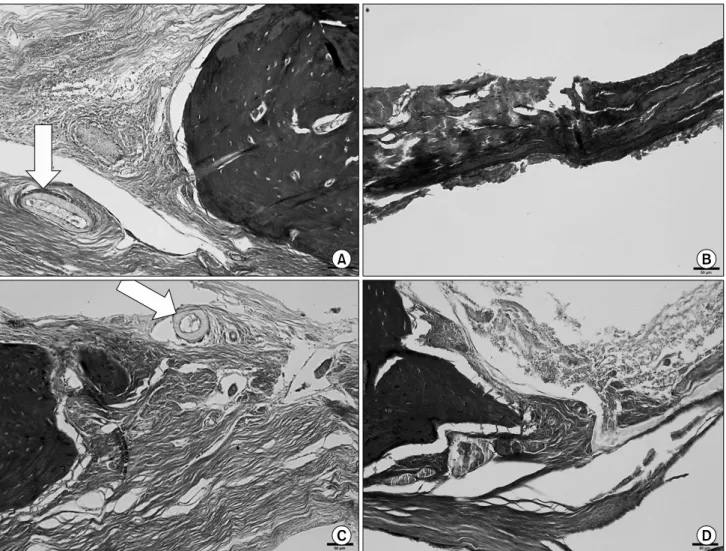
Fig. 2. Histologic section (Masson’s trichrome staining, ×200). New bone formation of each group was found at the border area of bone defect. Residual hydroxyapatite/silk fibroin particle were found in this group (arrow). (A) The experimental group at 4 weeks, (B) The control group at 4 weeks, (C) The expermental group at 8 weeks, (D) The control group at 8 weeks.
Fig. 1. Hydroxyapatite/silk fibroin particle was grafted into the center of parietal bone.
3. 조직학적 검사 및 조직 형태학적 분석
채취한 표본은 10% 중성 formalin 용액에 고정하고 5% Nitric
acid 용액을 이용하여 탈회하였다. 파라핀 블록에 포매한 후 4μm 두께로 절단한 후 Masson’s trichrome (MT) 염색을 시행하였다.
염색한 조직 표본은 디지털 현미경 카메라를 사용하여 이미지를 취득한 후 Sigma Scan Pro 5.0 (Systat Software Inc., San Jose, CA, USA)을 이용하여 신생골의 면적 비율을 측정하였다.
HA/SF군과 아무것도 넣지 않은 대조군의 신생골 면적 비율 차이 의 통계학적 유의성 검사를 위해 SPSS 18.0 (IBM Co., Armonk, NY, USA)를 이용해 independent-samples t-test를 시행하였다.
결 과
MT 염색을 한 조직절편을 현미경으로 관찰한 결과 실험군, 대조군 모두에서 신생골의 형성을 확인할 수 있었다(Fig. 2).
실험군에서는 4주 및 8주에서 분해되지 않고 남아 있는 일부
HA/SF particle을 발견할 수 있었다(Fig. 2, arrow). Particle
주위로 특별한 염증반응 및 감염소견은 발견되지 않았다.
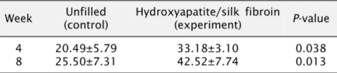
Table 1. Mean new bone formation Week Unfilled
(control)
Hydroxyapatite/silk fibroin
(experiment) P-value
4
8 20.49±5.79
25.50±7.31 33.18±3.10
42.52±7.74 0.038
0.013
염색한 조직 표본의 이미지에서 골결손부에 대한 신생골의 면적 비율을 측정하고 각 그룹에 대한 평균치를 구하였다(Table 1). 4주에서 신생골 면적의 비율은 실험군에서 평균 33.18±3.10%
이고 대조군에서 평균 20.49±5.79%로 실험군에서 유의하게 더 많이 생성되었다( P <0.05). 8주에서 신생골 면적의 비율은 실험 군에서 평균 42.52±7.74%이고 대조군에서 25.50±7.31%로 4주에서와 마찬가지로 통계적으로 유의하게 골이 많이 생성되었 다( P <0.05).
고 찰
구강악안면 영역에서는 악안면부에 발생하는 다양한 낭종, 종 양과 같은 병소 및 외상으로 인해 자연적으로는 치유되기 힘든 골결손부가 발생할 수 있다[1]. 또한 치아상실로 인해 임플란트 식립을 통한 보철치료가 필요할 경우 치조골이 충분치 못하여 추가적인 골이식을 시행해야 하는 경우가 발생할 수 있다[28].
구강악안면 영역에서의 이러한 골결손부는 구강의 저작 및 연하기 능에 영향을 주고 악안면 영역의 심미적 장애를 일으킬 수 있다.
본 연구에서는 HA와 SF의 복합물질을 이용하여 대조군에 비하여 유의할 수준으로 높은 신생골 형성을 관찰하였다( P <0.05). 기 존의 HA/SF 복합이식재의 경우 스폰지 형태나 블록 형태이었던 것에 비하여 이번에 연구한 재료는 입자 형태의 재료로 뼈 이식을 필요로 하는 구강영역의 복잡한 해부학적인 형태를 감안하면 이전 에 개발된 재료에 비하여 임상적으로 다양하게 적용될 수 있을 것으로 생각된다.
본 연구에 사용된 HA/SF particle은 SF 표면에 HA를 도입한 것으로 기존의 알려진 HA와 SF 복합체와 달리 sphere 형태의 SF을 사용했다는 점에서 차이가 있다. 과거 Kye 등[29]은 전기방 사된 silk nano-fiber와 nano-HA 복합체가 가토의 두개골 결손부 모델에서 아무런 처치를 하지 않은 대조군에 비해 신생골 형성량이 유의하게 증가함을 확인하였고, Park 등[30]은 SF/nano-HA/
Corn starch composite가 가토 두개골 결손모델에서 신생골 형성에 효과가 있음을 보고하였다. 이외에도 SF와 platelet-rich fibrin 복합체를 골결손부에 적용 시 신생골 형성에 효과적이었고 [31], 치과용 임플란트 주위의 경미한 골결손부 수복에도 효과적으 로 사용될 수 있음이 보고되었다[32]. 이번 연구에 사용된 이식재 의 경우에는 polyethylene oxide (PEO)를 이용한 상분해를 통해 크기 제어가 가능한 silk sphere를 제조하였고, 균일도가 뛰어난
silk sphere를 제작할 수 있었다[33]. 이렇게 제조한 silk sphere 에 HA를 도입하여 HA의 주형으로 사용하여 HA/SF 복합체의 물리적 강도를 향상시킬 수 있었다.
본 연구에서 사용된 HA/SF particle은 제조과정 중 상분해 시 PEO ratio에 따라 그 입자의 크기가 0.9±0.2에서 5.8±2.9 μm로 다양하게 만들어진다[33]. 만들어진 HA/SF particle은 고운 분말 형태로서 임상적 적용 시 매우 세심한 조작이 필요하며 골결손부에 적용 후 잘 유지될 수 있도록 주의가 필요하다. 골결손 부에서 스며나오는 혈액이나 타액에 의해 particle의 유지력이 조금 향상될 수 있으나 실제 임상에 적용하기에는 어려움이 있다.
새로운 생체 친화적인 재료를 첨가하거나 제조과정의 개선을 통하 여 조작하기 쉬우면서 적절한 강도를 가질 수 있는 HA/SF par- ticle이 필요할 것이다.
본 연구에서는 HA/SF particle이 신생골 형성에 미치는 영향을 알아보기 위해 백서의 두개골에 8 mm 골결손부를 형성하고 아무 처치도 하지 않은 군을 대조군으로 설정하였다. 수술 후 4주가 지났을 때 신생골 형성량이 실험군에서 평균 33.18±3.10%였고 대조군에서 20.49±5.79%로 형성되었고 그 차이도 통계적으로 유의하였다. 수술 8주 후 신생골 형성량은 실험군에서 42.52±7.74%, 대조군에서 25.50±7.31%로 4주에 비해 증가하 였고 4주 결과와 마찬가지로 그 차이가 통계적으로 유의하였다.
결과적으로 HA/SF particle은 골결손부에서의 신생골 형성을 효과적으로 증가시켰다. 이는 SF가 HA 입자의 주형으로 작용하여 HA 입자의 성장을 촉진시키고 유지시켰기 때문이라고 판단된다.
또한 HA/SF particle이 특별한 염증반응 및 감염 없이 분해되어 신생골 형성을 특별한 영향을 주지 않았고 이는 particle이 대부분 분해되고 일부 입자만 실험군 4주와 8주에서 조직에서 관찰되었다 는 사실을 통해 알 수가 있다. 이렇게 대부분 분해되고 일부 입자만 남았다는 사실은 SF의 염증반응에 대한 기존 연구에[17]
비해 긍정적인 결과라 할 수가 있다.
결 론
본 연구에서는 백서의 두개골에 지름 8 mm의 골결손부를
인위적으로 형성한 후 실험군에는 HA/SF particle을, 대조군에는
아무 처치도 하지 않고 수술 후 4주 및 8주에 백서를 희생하여
조직형태학적 분석을 통해 신생골 형성량을 알아보았다. 실험
결과 조직형태학적 분석을 통해 4주 및 8주 모두에서 실험군의
신생골 형성이 대조군에 비해 유의하게 증가하였으며, 특별한
염증반응 없이 대부분의 HA/SF particle 생체 내에서 분해되었
다. HA/SF particle의 물성 및 조작성이 보다 더 개선된다면,
비용이 저렴하면서도 임상적으로 사용 가능한 골이식재로 활용될
수 있을 것이다.
Acknowledgements
이 연구는 농촌진흥청 차세대 바이오그린 21사업(과제번호:
PJ009051052012)에 의해 이루어진 것임.
References