• 검색 결과가 없습니다.

Expression and Clinical Significance of the N-myc Downstream Regulated Gene-1 in Hypopharyngeal Cancer

N/A
N/A
Protected

Academic year: 2021

Share "Expression and Clinical Significance of the N-myc Downstream Regulated Gene-1 in Hypopharyngeal Cancer"

Copied!
5
0
0

로드 중.... (전체 텍스트 보기)

전체 글

(1)

대한 두경부 종양 학회지 제 27 권 제 1 호 2011

Introduction

Head and neck cancer is the sixth most common type of

cancer, representing about 6% of all cancers and accounting for an estimated 650,000 new cancer cases and 350,000 can- cer deaths worldwide every year.

1)

The 5-year survival of

교신저자 : 김철호, 443-751 경기도 수원시 영통구 원천동 산 5 아주대학교 의과대학 이비인후과학교실

전화 : (031) 219-5269 · 전송 : (031) 219-5264 · E-mail : ostium@ajou.ac.kr

Expression and Clinical Significance of the N-myc Downstream Regulated Gene-1 in Hypopharyngeal Cancer

Inkyung Sohn, MD

1

, Nam Soo Han, MD

1

, Yoo Seob Shin, MD

1

, Jang Hee Kim, MD

2

, Chul-Ho Kim, MD

1

Department of Otolaryngology

1

and Pathology,

2

Ajou University School of Medicine, Suwon, Korea

하인두암에서의 N-myc Downstream Regulated Gene-1 발현의 임상적 의의

아주대학교 의과대학 이비인후과학교실,

1

병리학교실

2

손인경1·한남수1·신유섭1·김장희2·김철호1

= 국 문 초 록 =

목 적

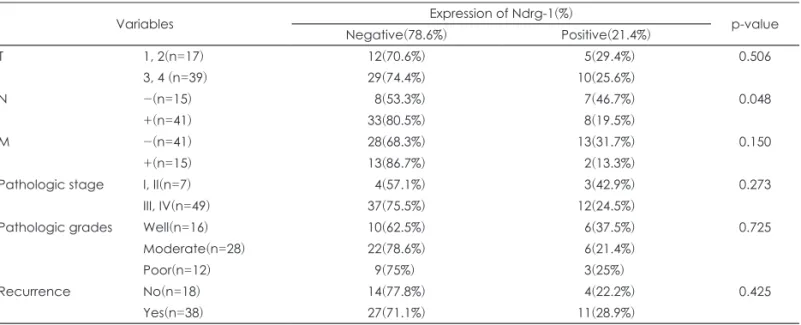
두경부 암은 발생 순위에서 전체 6위에 해당하는 다빈도 암이나 최근 20여년 동안의 노력에도 불구하고 두경부 암의 독톡한 특성상 생존률에서 뚜렷한 향상을 보이지 못하고 있다. 특히, 하인두 암은 원발 부위의 점막하 침윤이 흔하며, 주변 림프절 전이와 원격 전이가 흔하고, 2차 원발 암종 발생이 흔하여 두경부 암 중에서도 가장 불량한 예 후를 보이고 있는 악성 종양이다. 최근에 이러한 암을 치료하고 진단하기 위한 방법으로 분자생물학적 접근법들이 많이 시도 되고 있으며, 그 중 하나로 N-myc downstream regulated gene-1(Ndrg-1)이라는 유전자가 유방, 전 립선, 방광 암 등의 타 악성 종양에서 종양의 전이 및 진행 양상과 관련되어 있다는 보고가 있었다. 이에 본 연구는 하인두 암에서의 Ndrg-1의 발현 양상을 살펴보고 이와 임상 양상과의 연관관계를 살펴보고자 하였다.

방 법

1996년부터 2003년까지 수술 받은 하인두 암 환자 56명을 대상으로 면역조직화학검사를 시행하여 Ndrg-1 발현 을 확인하였고, 3명의 신선 조직을 대상으로 RT-PCR, Western blot을 시행하였다.

결 과

Ndrg-1은 RT-PCR에서 정상 조직과 악성종양 조직 모두에서 비슷한 수준으로 발현되었다. 그러나 Western blot 에서는 정상 조직에서 뚜렷한 증가 양상을 보여 타 연구와 동일한 결과를 보였고, 이는 불필요하며 비효율적인 mRNA수준에서의 발현이 있지만 최종적인 단백 산물 발현에서는 암종의 진행과 연계되어 악성 종양 진행군에서 발현 이 억제되는 결과로 해석된다. 면역조직화학검사에서는 정상 상피조직에서 Ndrg-1 발현이 확인되었으며, 통계적으 로 유의하지는 않으나 불량한 예후를 가진 그룹에서 대체로 발현이 억제되는 악성 종양과의 역 연관 관계를 확인할 수 있었고, 특히 림프절 전이를 보인 그룹과 그렇지 않은 그룹 사이에서는 통계적으로 유의미한 결과가 확인되었다.

결 론

즉, 림프절 전이가 없는 그룹에서 Ndrg-1이 종양의 전이에 관여할 것이라는 타 연구와 일관된 결과로 하인두 암에서도 그 역할이 있음을 나타내는 결과라 할 수 있다.

중심 단어

:Ndrg-1ㆍ하인두암ㆍ경부전이.

online©MLComm

(2)

head and neck cancers for all stages combined on the basis of Surveillance Epidemiology and End Results data is re- mained to be low for several decades, whichs is about 60%

but lower in higher stages(stage III, IV).

2)

Furthermore, of all head and neck cancers, hypopharyngeal squamous cell car- cinoma(HPSCC) has the worst prognosis among the head and neck cancers. The features determining poor survival rate of HPSCC are extensive submucosal infiltration at the primary site, the high incidence of regional lymph node me- tastases or common distant metastasis at presentation or the high incidence of second primaries in the head and neck re- gion, including the lung and esophagus. Despite the aggres- sive multidisciplinary treatment approaches, such as concur- rent chemoradiation or radical surgery followed by postope- rative radiotherapy with or without chemoherapy, 5-year sur- vival rate of hypopharyngeal cancer has not been improved over the past 20 years. Threfore, molecular biological re- searches which enables us to detect the disease and predict the prognosis are strongly required.

Over the past several decades, there has been increasing interest in the growing family of tumor metastasis suppres- sor gene including RKIP, Nm23, KISS1, KAI1, BRMS1, TI- MPs, E-cadherin, MKK4, TXNIP, CRSP3, SSeCKs, RhoG- DI2 and N-myc downstreamregulated gene-1(Ndrg-1).

3)

These recent discoveries have introduced a new approach to treating cancer and preventing metastasis. Ndrg-1, a member of the Ndrg family has been identified as a gene involved in the differentiation of epithelial cells and inhibition of tumor metastasis in colon cancer.

4)

In several clinical studies, a rela- tionship between the expression of Ndrg-1 in serum or in can- cer tissue and the progression of disease has been undisclos- ed. The expression of the Ndrg-1 in patients with prostate, colon and breast cancer is inversely correlated with the clini- cal stage and malignant progression like lymph node or dis- tant metastasis.

4-6)

However, Ndrg-1 expression has not been reported in patients with HPSCC. Therefore, we intend to in- vestigate the expression and distribution of Ndrg-1 in HPSCC and the correlation between Ndrg-1 expression and the sur- vival rate, invasion and metastasis in HPSCC.

Material and Method

1. Patients

56 patients who were treated for HPSCC between 1994 and 2003 were enrolled in this study. We exclude patients who re- ceived prior radiotherapy or chemotherapy before surgical treatment. The patients’ charts were analyzed in terms of age, gender, origin of primary tumor, T stage based on the criteria

of the American Joint Committee on Cancer(2009), and the type of surgery performed. The mean age was 62 years(range 42-77 years). 17 patients had stage I or II disease and 39 had stage III or IV. The histologic type was classified based on the World Health Organization system. Of the 56 tumors, 16 were well, 28 moderately and 12 poorly differentiated. Sur- vival analysis was performed using the Kaplan-Meier meth- od and the statistical significance was evaluated using the log-rank test.

2. Immunohistochemistry

Formalin-fixed, paraffin-embedded tissue was cut into 5- μm sections, then deparaffined and rehydrated via serial pas- sage through xylene and graded ethanol series. The sections were microwaved for 15 minutes in Citrate buffer(pH 6.0) for antigen retrieval. The sections were then treated with 3% hy- drogen peroxide in methanol to quench the endogenous per- oxidase activity followed by incubation with 1% bovine se- rum albumin to block the nonspecific binding. The primary antibodies were goat Ndrg-1 polyclonal antibodies used(Di- luted 1 : 60) at a concentration of 50mg/mL(R&D Systems, Inc., Minneapolis, MN). Primary antibody incubations were carried out for 2 hours at 258C. The sections were extensive- ly washed in Tris-buffered saline(TBS, pH 7.4) and then in- cubated with the appropriate biotinylated secondary antibod- ies followed by avidin-peroxidase. Polink-2 plus HRP anti- goat DAB detection kits(Life science division) were purchas- ed for this purpose and were used in accordance with the ma- nufacturer’s instructions. The chromogenic reaction was car- ried out with 3-3’-diaminobenzidine in a peroxidase substrate solution and lasted for 4 minutes. Hematoxylin was used for nuclear staining. For each experiment, negative controls omit- ting either primary or secondary antibodies were included to examine nonspecific staining. The slides were reviewed by at least two pathologists and scored semiquantitatively as follows : In area, the tumor was defined as point 1 if antigen expression was demonstrated in <30% of the carcinoma cells, as point 2 if demonstrated in <60% and ≥30%, as point 3 if demonstrated in ≥60%. In intensity, as point 1 if antigen expression was demonstrated in weak compared with normal cell, as point 2 if demonstrated in moderate, as point 3 if de- monstrated in strong and same with normal tissue. The tumor was defined as positive if point 3 in intensity and point 2 or 3 in area.

3. Reverse transcriptase polymerase chain reaction

Total RNA was extracted using TRIzol

®

(Invitrogen Gron-

ingen, The Netherlands), and cDNA was prepared using an

Maxime PCR premix kit(Intron Biotech, Korea) according to

(3)

the manufacturer’s instructions. The sequences of the poly- merase chain reaction(PCR) primers were as follows : hu- man Ndrg1(192bp) : sense, 5’-CAT GTC TCG GGA GAT GCA GGA TG-3’, antisense, 5’-ATG GTA GGT GAG GAT GAC AGG-3’ and 5’-AGG TCC ACC ACT GAC ACG TT-3’

for GAPDH. PCR products were separated by electrophore- sis on 1.5% agarose gels and detected under UV light(Bio- Rad, Hercules, CA).

4. Western blot analysis

For Western blot analysis, samples were separated by means of sodium dodecyl sulfate polyacrylamide gel elec- trophoresis and electro-transferred onto a polyvinylidine difluoride membrane(Amersham, Arlington Heights, IL).

The non-specific bindings were blocked with PBS contain- ing Tween 20(0.2%) and skimmed milk. Blots were incubat- ed with primary antibodies Ndrg-1(Santa Cruz Biotechnolo- gy, CA) followed by peroxidase- conjugated donkey anti-goat antibody(Santa Cruz Biotechnology, CA). Bound antibodies were detected using an enhanced chemiluminescence detec- tion system(Pierce).

Result

1. Expression of Ndrg-1 in hypopharyngeal cancer tissue Among the 56 samples, Ndrg-1 was detected in 15 patients (21.4%) by immunohistochemical staining(Table 1). In most of the specimens, Ndrg-1 expression was primarily seen in the epithelial layers of normal cells. In many cases, the cyto- plasm of the epithelial cells were diffusely stained, and the cell membranes were also stained in some cases. Ndrg-1 expres- sion was also detected in some cancer cells and occasionally slightly seen in the basement membrane of the normal cells surrounding the cancer cells(Fig. 1). The rate of Ndrg-1 posi-

tive rate was 29.4%(5 of 17) in patients with T1 or T2 stage tumors(<4cm in diameter), whereas 25.6%(10 of 39) in T3 or T4 tumors(>4cm in diameter). Thus, the expression rate of Ndrg-1 was inversly correlated with tumor size, but it was not statistically significant(Table 1). Ndrg-1 positive rate was 42.9%(3 of 7) in early stage and 24.5%(12 of 49) in advanced- stage carcinoma, but difference was also not statistically sig- nificant. Ndrg-1 postive rate was significantly high in patitents without nodal metastasis(7 of 15, 46.7%) than patients with nodal metastasis(8 of 41, 19.5%)(p=0.048). The Ndrg-1 ex- pression rate was not different between the group with recur- rence(22.2%) and without recurrence(28.9%). Degree of tu- mor differentiation did not affect Ndrg-1 expression(Fig. 2).

Ndrg-1 expression rate was 13.3%(2 of 15) in patients without distant metastasis in contrast to 31.7%(13 of 41) with distant

Table 1. Correlation between the expression pattern of Ndrg-1 and various clinicopathologic factors

Variables Expression of Ndrg-1(%)

p-value Negative(78.6%) Positive(21.4%)

T 1, 2(n=17) 12(70.6%)

05(29.4%)

0.506

3, 4 (n=39) 29(74.4%) 10(25.6%)

N -(n=15)

08(53.3%) 07(46.7%)

0.048

+(n=41) 33(80.5%)

08(19.5%)

M -(n=41) 28(68.3%) 13(31.7%) 0.150

+(n=15) 13(86.7%)

02(13.3%)

Pathologic stage I, II(n=7)

04(57.1%) 03(42.9%)

0.273

III, IV(n=49) 37(75.5%) 12(24.5%)

Pathologic grades Well(n=16) 10(62.5%)

06(37.5%)

0.725

Moderate(n=28) 22(78.6%)

06(21.4%)

Poor(n=12)

09(75%)0. 03(25%)0.

Recurrence No(n=18) 14(77.8%)

04(22.2%)

0.425

Yes(n=38) 27(71.1%) 11(28.9%)

Fig. 1. Immunohistochemical stain for Ndrg-1 protein. Positive re- activity in normal squamous epithelium. Original magnification

×200.

(4)

metastasis. However, there was no significant difference in Ndrg-1 expression rate regarding the distant metastasis(p=

0.194). Of the 56 patients, 36(64.3%) patients died of disease during the follow-up period. The cause of death was local re- currence in 12 patients, distant metastasis in 15 and both in two. There was no statistical difference in survival rate by N- drg-1 positivity.

2. RT-PCR and Western blot analysis in hypopharyngeal cancer tissue

RT-PCR of Ndrg-1 gene was performed with cancer and normal tissue of three hypopharyngeal cancer patients. In all cases, there was no definite difference in amount of mRNA between cancer and normal tissue. Ndrg-1 mRNA was mod- erately expressed in all specimens(Fig. 3). Western blot anal-

ysis was also performed with the specimens from three pa- tients. Ndrg-1 protein was expressed in both the normal and in the cancer tissue. However, Ndrg-1 expression was signif- icantly decreased in the carcinoma tissue(Fig. 4).

Discussion

Overexpression of Ndrg-1 has been reported to inhibit cell growth and promote cell differentiation in various tissues, and its expression might be related to the regulation of cell differentiation and stress.

7)

In various cancers including co- lon, prostate and breast cancer, the expression of Ndrg-1 has found to be significantly down regulated, previously.

5,6,8)

N- drg-1 was originally identified by differential display as be- ing significantly upregulated by induction of differentiation in colon carcinoma cells in vitro.

4)

In addition, the metastasis suppressor activity of Ndrg-1 has also been observed in colon cancer cells in vivo.

8)

Also in prostate cancer, the expression of Ndrg-1 has known to be inversly correlated with the Glea- son grading, Gleason grading, which is the most well-estab- lished pathological criterion for judging the clinical stage and malignant progression in prostate cancer, the overall surviv- al rate and lymph node or bone metastasis.

5)

Bandyopadhyay et al discovered that the expression of the Ndrg-1 protein was significantly reduced in breast tumor cells, particularly in pa- tients with lymph node or bone metastasis as compared to those with localized breast cancer and overexpression of the Ndrg-1 suppresses the invasiveness of breast cancer cells in vitro.

6)

Ndrg-1 expression exhibited significant inverse cor- relation with the disease-free survival rate of patients and em- erged as an independent prognostic factor in breast cancer.

6)

Therefore, these results suggest that Ndrg-1 might have the an- ti-metastatic and anti-progressive property in cancer growth.

Among many head and neck cancers, HPSCC is well known for its high invasion and metastasis rates. These characteris- tics are closely related to the prognosis of the cancer, thus, HP- SCC have one of the worst prognosis between head and neck cancers. By previous researches on Ndrg-1, which is an im- portant component of invasion and metastasis in cancer, we expect that further understanding of the role of Ndrg-1 en-

Fig. 2. Reduced immunohistochemical expression of Ndrg-1 gene in hypopharyngeal carcinoma. Beneath normal squamous epi- thelium, carcinoma cell was not stained. Original magnification

×200.

Fig. 3. RT-PCR of Ndrg-1 in human hypopharyngeal carcinoma tissue. There was no significant difference in the amount mRNA between carcinoma cells and normal cells.

Fig. 4. Western blot analysis of Ndrg-1 in human hypopharyngeal

carcinoma tissue. Ndrg-1 protein expression was significantly de-

creased in cancer tissue.

(5)

ables us to overcome the dire prognosis of HPSCC. Until now, there was no study regarding the Ndrg-1 not only in HPSCC, but also in other head and neck cancers. In our study, we com- pared the clinical features of HPSCC with different Ndrg-l manifestations to analyze the role of Ndrg-1 in HPSCC.

Immunohistochemical staining was performed with spec- imens from 56 HPSCC patients to analyze the expression rate of Ndrg-1. Ndrg-1 was mostly expressed in the epithelium of normal cell, dominantly in the cytoplasm. Ndrg-1 was exp- ressed in only 21.4%(15 of 56) of cases. The patients with nodal metastasis showed significantly low expression rate(8 of 41, 19.5%) compared to the patients without nodal metas- tasis(7 of 15, 46.7%)(p=0.005). This inverse correlation be- tween nodal metastasis and expression of Ndrg-1 are in con- cordance with previous studies regarding other cancers. It might be a foundation of presumption that Ndrg-1 has a im- portant role in nodal metastasis and tumor progression in HP- SCC. Ndrg-1 expression has tendency to be reduced in tumors with distant metastasis or advanced-stage tumors but there was no statistical significance(p=0.150). Furthermore, in tu- mors of positive Ndrg-1 expression, the rates of distant me- tastasis, recurrence and pathologic stage all seemed to have also inverse correlation with Ndrg-1 expression.

On the other hand, Ndrg-1 expression was observed in both normal and cancer specimens with the method of RT-PCR performed on fresh tissue from HPSCC patients in this se- ries. This result does not coincide with the result of immuno- histochemical staining. In spite of repeated RT-PCR, definite expression of Ndrg-1 in cancer cell was detected. To detect protein synthesis, Western blot analysis was performed with the specimens from three HPSCC patients. Ndrg-1 protein was dominantly dectected in the normal cells and slightly in the carcinoma cells. Although mRNA expression in RT-PCR was elevated in the cancer cell, protein synthesis was not el- evated. We assume that aberrant expression of Ndrg-1 at the mRNA level could be the reason fot this result. Ineffective and negative feedback could make this kind of aberrant expres- sion of mRNA, not contributing to protein synthesis.

In summary, immunohistochemical staining of HPSCC tissue showed ubiquitous expression of Ndrg-1, and the level of Ndrg-1 significantly inversly correlated with the nodal me-

tastasis of the tumor. The expression of mRNA of Ndrg-1 was detected in both normal cells and HPSCC cells but the exp- ression of proteins were increased in normal cells compared to cancer cells.

Conclusion

The downregulated expression of Ndrg-1 could be associ- ated with lymph node, distant metastasis which remains to be the most crucial cause of mortality in hypophrynx and the prognosis of patients with HPSCC.

Acknowledgments

This study was supported by CCRB through the GRRC project of the Gyeonggi Provincial Government, Republic of Korea(GRRC Ajou university 2010-A03).

Refernces

1) Parkin DM, Bray F, Ferlay J, Pisani P. Global cancer statistics, 2002. CA Cancer J Clin. 2005;55(2):74-108.

2) Jemal A, Bray F, Center MM, Ferlay J, Ward E, Forman D. Glo- bal cancer statistics. CA cancer J Clin. 200;61:69-90.

3) Welch DR,Hunter KW. A new member of the growing family of metastasis suppressors identified in prostate cancer. J Natl Cancer Inst. 2003;95(12):839-841.

4) van Belzen N, Dinjens WN, Diesveld MP, Groen NA, van der Made AC, Nozawa Y, et al. A novel gene which is up-regulated during colon epithelial cell differentiation and down-regulated in colorectal neoplasms. Lab Invest. 1997;77(1):85-92.

5) Bandyopadhyay S, Pai SK, Gross SC, Hirota S, Hosobe S, Mi- ura K. The Drg-1 gene suppresses tumor metastasis in prostate cancer. Cancer Res. 2003;63(8):1731-1736.

6) Bandyopadhyay S, Pai SK, Hirota S, Hosobe S, Takano Y, Saito K, et al. Role of the putative tumor metastasis suppressor gene Drg-1 in breast cancer progression. Oncogene. 2004;23(33):

5675-5681.

7) Kalaydjieva L, Gresham D, Gooding R, Heather L, Baas F, de Jonge R, et al. N-myc downstream-regulated gene 1 is mutated in hereditary motor and sensory neuropathy-Lom. Am J Hum Genet. 2000;67(1):47-58.

8) Guan RJ, Ford HL, Fu Y, Li Y, Shaw LM, Pardee AB. Drg-1 as

a differentiation-related, putative metastatic suppressor gene

in human colon cancer. Cancer Res.2000;60(3):749-755.

수치

Fig. 1. Immunohistochemical stain for Ndrg-1 protein. Positive re- re-activity in normal squamous epithelium
Fig. 3. RT-PCR of Ndrg-1 in human hypopharyngeal carcinoma  tissue. There was no significant difference in the amount mRNA  between carcinoma cells and normal cells.

참조

관련 문서

In the present study, consistent to that report, up-regulation of VEGF mRNA was observed in the colon cancer cell with the acquired resistance to 5-FU and this

1) Effects of methanol extracts of Capsella bursa-pastoris on cyclooxygenase-2 (COX-2) and Inducible Nitric oxide synthase (iNOS) expression in human prostate cancer cell

The expression of reporter genes driven by the ZAT1 promoter was observed in the tissues undergoing maturation such as border cell of root cap, endodermis,

Inhibitory effects of classified methanol extracts of Smilax china L on the COX-2 and iNOS expression of human colorectal cancer cell lines... Inhibitory

Objective: The purpose of this study was to analyze recent trend in incidence of basal cell carcinoma and squamous cell carcinoma in patients from the Gwangju City

Frequency and clinical significance of the expression of the multidrug resistance proteins MDR1/P-glycoprotein, MRP1, and LRP in acute myeloid leukemia: a

Activated H-Ras expression in human fibroblast cell lines increases the activity of Ku80 to bind injuried DNA, reduces γ-H2AX expression by UV irradiation,

(1) PCR monitoring of bphC gene of Ralstonia eutropha H850 in soil Amount of bphC gene amplification of H850 cells was not generally proportional to the cell density