Korean J Vet Res(2017) 57(4) : 209~214 https://doi.org/10.14405/kjvr.2017.57.4.209
209
<Original Article>
Detection of viral infections in wild Korean raccoon dogs ( Nyctereutes procyonoides koreensis)
Dong-Kun Yang
1,*, Seoug Heon Lee
1, Ha-Hyun Kim
1, Jong-Taek Kim
2, Sangin Ahn
2, In-Soo Cho
11
Animal and Plant Quarantine Agency, Ministry of Agriculture, Food and Rural Affair, Gimcheon 39660, Korea
2
College of Veterinary Medicine, Kangwon National University, Chuncheon 24341, Korea (Received: October 9, 2017; Revised: November 17, 2017; Accepted: December 4, 2017)
Abstract: Wild raccoon dogs ( Nyctereutes procyonoides koreensis) may play a role transmitting several pathogens to humans and pet animals. Information concerning the incidence of rabies, canine distemper virus (CDV), canine parvovirus (CPV), canine adenovirus type 2 (CAdV-2), canine parainfluenza virus type 5 (CPIV-5), and canine herpesvirus (CHV) is needed in wild raccoon dogs. In total, 62 brain samples of raccoon dogs were examined for rabies virus (RABV) and CDV, and 49 lung samples were screened for CDV, CAdV-2, CPIV-5, and CHV. No RABV, CAdV-2, CPIV-5, or CHV was identified, but nine CDV antigens (8.1%, 9/111) were detected. Moreover, 174 serum samples from wild raccoon dogs were screened for antibodies against the five major viral pathogens. The overall sero- surveillance against CDV, CPV, CAdV-2, CPIV-5, and CHV in wild raccoon dogs was 60.3%, 52.9%, 59.8%, 23.6%, and 10.3%, respectively. Comparisons of the sero-surveillance of the five pathogens showed that raccoon dogs of Gyeonggi province have slightly higher sero-positive rates against CDV, CPV, and CHV than those of Gangwon province. These results indicate high incidences of CDV, CPV, and CAdV-2 in wild raccoon dogs of two Korean provinces and a latent risk of pathogen transmission to companion and domestic animals.
Keywords: canine distemper, rabies, raccoon dogs, sero-surveillance
Introduction
Wild raccoon dogs (Nyctereutes procyonoides koreensis) are becoming more likely to invade human residences due to shortages of food during winter, and they may even live in human habitats because of easy access to food. Frequent encounters between wild raccoon dogs and humans increase the incidence of zoonosis. In particular, the raccoon dog is known to be responsible for transmitting many pathogens, including rabies virus (RABV) and Demodex canis in Europe and Asia [4, 18]. A “One Health” concept has been established to prevent the spillover of zoonotic infectious diseases [9].
Contact between raccoon dogs and companion animals can also increase the incidence of infectious diseases such as canine distemper virus (CDV) and kennel cough in dogs and cats.
Rabies is caused by RABV belonging to the genus Lissavi- rus, family Rabdoviridae, and is divided into human and ani- mal rabies. Animal rabies cases have been identified in a variety of animals in the Republic of Korea, including cattle, dogs, raccoon dogs, and cats [21]. According to an epidemi- ological study of rabies in Korea, all animal rabies cases have been linked to rabid raccoon dogs since 1993. There- fore, raccoon dogs have been attracting attention as a carrier
that transmits rabies. Canine distemper caused by CDV belonging to the genus Morbillivirus, family Paramyxoviri- dae, leads to lethal disease in many animal species, includ- ing raccoon dogs. Only a single serotype against CDV is known to exist. Wild Korean raccoon dogs infected with CDV have been reported using a commercial CDV antigen detection kit [4, 6]. Canine parvovirus (CPV) belonging to the genus Parvovirus, family Parvoviridae, causes gastrointesti- nal illness (e.g., acute hemorrhagic diarrhea) in young pup- pies [7]. Animals infected with CPV excrete large amounts of virus in their feces after a natural infection, which is trans- mitted to CPV antibody-negative puppies. The incidence of CPV in raccoon dogs has been reported in Germany and Japan [8]. Moreover, recent advances in molecular technol- ogy such as the next generation sequencing, polymerase chain reaction (PCR), and a variety of enzyme-linked immu- nosorbent assay (ELISA) kits have enabled the detection of many infectious diseases in wild raccoon dogs. Based on advanced techniques, the presence of several infectious dis- eases, including canine adenovirus type 2 (CAdV-2), canine parainfluenza virus type 5 (CPIV-5), and canine herpesvirus type 1 (CHV), representing a problem in raccoon dogs and pet dogs, have been reported in Italy and Norway [3, 13].
*Corresponding author
Tel: +82-54-912-0785, Fax: +82-54-912-0812
E-mail: yangdk@korea.kr
Among infectious pathogens, rabies causes the most cata- strophic damage in all warm-blooded animals. It is neces- sary to identify pathogens in wild raccoon dogs residing in rabies-risk provinces and to continuously monitor major pathogens circulating in wild raccoon dogs to prevent rabies and many other infectious diseases. In this study, we investi- gated the incidence rates of RABV, CDV, CAdV-2, CPIV-5, and CHV from brain or lung samples of raccoon dogs and determined the sero-surveillance of CDV, CPV, CAdV-2, CPIV-5, and CHV in wild Korean raccoon dog sera.
Materials and Methods
Sample collection
In total, 62 brain and 49 lung samples were obtained from the Wild Animal Rescue Center of Kangwon National Uni- versity to determine incidence rates. For sero-surveillance, blood samples were obtained from 174 wild raccoon dogs as part of annual surveillance for the national rabies eradication program. Blood was taken from the cephalic vein. In total, 71 and 103 serum samples were obtained from raccoon dogs liv- ing in Gyeonggi and Gangwon Provinces, respectively from February 2013 to June 2017. Clotted blood samples were centrifuged (3,000 × g, 10 min), and the sera were stored at
−20
oC until use.
Detection of viral pathogens
A direct fluorescent antibody (DFA) test was carried out to detect RABV in 62 brain samples using the procedure described by OIE [20]. Briefly, thin, frozen sections of rac- coon dog brain were placed on a slide and fixed with cold acetone ( −20
oC) for 20 min. The slides with fixed brain tis- sue were subsequently reacted with a specific monoclonal antibody (MEDIAN Diagnostics, Korea) against RABV for 1 h. After washing with phosphate-buffered saline (PBS), the slides were stained with fluorescent isothiocyanate-conju- gated goat-anti mouse IgG+IgM (MEDIAN Diagnostics).
Samples showing specific fluorescence in brain cells were diagnosed as positive. Commercial RT-PCR kits (Canine dis- temper virus detection kit and Canine parainfluenza virus detection kit) manufactured by iNtRON Biotechnology (Korea) were used to detect CDV and CPIV-5 antigens in brain and lung samples according to the manufacturer’s instruc- tions. Primer sets (forward: 5'-TCCCGTTCACCAGCAC- CAGGGCC-3', reverse: 5'-GGTGAGAGGCGGGGGAGGGGT- 3') specific for the CAdV-2 gene were prepared and mixed with PCR premix (Bioneer, Korea) and the extracted lung sample DNA. In brief, PCR was carried out in a PCR pre- mix containing 10 µL of denatured DNA, 1 µL of each primer (50 pmol), and 38 µL of distilled water in a final vol- ume of 50 µL. The cycling profile was denaturation at 95
oC for 5 min followed by 35 cycles of denaturation at 95
oC for 30 sec, annealing at 55
oC for 45 sec, and extension at 72
oC for 1 min, with a final extension at 72
oC for 5 min. The prod- ucts were visualized by 1.8% agarose gel electrophoresis
with RedSafe Nucleic Acid Staining Solution (iNtRON Bio- technology). Lung samples with a 757-bp product were con- sidered positive. All brain and lung samples were inoculated into Vero cells (CRL1586; American Type Culture Collec- tion [ATCC], USA) and MDCK cells (CCL34; ATCC) to isolate CHV. All samples that did not show specific cyto- pathic effects (CPEs) were passaged three times in the same cells.
Detection of antibodies against CDV, CPV and CHV by ELISAs
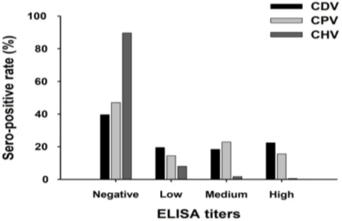
ELISA kits for CDV (INgezim MOQUILLO IgG; Inmu- nología y Genética Aplicada, Spain), CPV (INgezim; Inmu- nología y Genética Aplicada) and CHV (arigo Biolaboratories, Taiwan) were used according to the manufacturer’s instruc- tions to detect antibodies against CDV, CPV, and CHV. In brief, the serum samples were diluted 1:100 (CDV and CPV) or 1:200 (CHV) and added to the ELISA kit. After 10 min of incubation at room temperature, the plates were washed four times with washing solution and then 100 µL of conjugate was added to each well. The sealed plates were incubated for 10 min at room temperature, the conjugate was discarded, and the plates were washed four times. Finally, 100 µL of substrate solution was added and the plates were kept for 5 min at room temperature. After adding 50 or 100 µL of stop solution to each well, the absorbance of each well was read with an ELISA reader (Sunrise; Tecan, Switzerland) at 450 nm. The results were interpreted based on positive or nega- tive samples according to the instructions. The ELISA optical density values were equivalent to each viral neutralization (VN) titer.
Virus neutralization of CAdV-2
The CAdV-2 (strain QIAA1601) used in this study was isolated from a lung sample of naturally infected dogs in 2016. The CAdV-2 strain had been propagated in Vero cells and the CAdV-2 titer was confirmed by CPEs and an indi- rect fluorescent assay with anti-CAdV-2 antibodies (4H1-A7;
VMRD, USA). CAdV-2 was used as the antigen in a VN test. The VN test was performed in 96-well plates in dupli- cate using sera inactivated at 56
oC for 30 min. Then, 50 μL aliquots of two-fold serially diluted serum were mixed with equal volumes of CAdV-2 containing 200 TCID
50/0.1 mL.
After incubating the mixtures at 37
oC for 1 h, 100 μL of a Vero cell suspension containing 20,000 cells was added to each well. The plates were incubated for 3 days in a humidi- fied incubator with 5% CO
2. Each well was examined under a microscope to detect viral-specific CPEs. The virus neutral- izing antibody (VNA) titers were expressed as the reciprocal of the highest serum dilution that completely inhibited CPEs.
Sera showing a VNA titer ≥1:2 were considered positive.
Hemagglutination inhibition (HI) test for CPIV-5
CPIV-5 (strain QIA-B1201) was used as the antigen for the
HI test. The hemagglutination (HA) test was performed by
preparing serial two-fold dilutions of CPIV-5 in 50 µL of PBS (pH 7.2) and adding 50 µL of 0.6% chicken erythro- cytes. The HI test was performed in 96-well microplates as described previously [1, 23]. The HI titer was expressed as the reciprocal of the highest dilution of serum showing com- plete inhibition of HA. Serum samples showing HI titers
≥ 1:10 were considered positive.
Statistical analysis
Chi-square test was used to compare the prevalence rate of each virus between the two different geographic regions. The statistical analysis was performed with SPSS statistics 21.0 software (IBM, USA). A p value < 0.05 was considered sig- nificant.
Results
In total, 62 brain and 49 lung samples were collected from raccoon dogs held at the Wild Animal Rescue Center of Kangwon National University, and RABV, CDV, CAdV-2, CPIV-5, and CHV antigens were detected. As shown in Fig- ure 1, no RABV antigens were detected by the DFA test in the 62 brain samples, and no CAdV-2 or CPIV-5 antigens were detected in the 49 lung samples by PCR or RT-PCR.
However, nine CDV antigens (8.1%, 9/111) in 62 brain and 49 lung samples were detected by RT-PCR. Six brain and three lung samples showed positive reactions against CDV.
No specific CPEs were observed in Vero or MDCK cells inoculated with the 62 brain and 49 lung samples for the iso- lation of CHV.
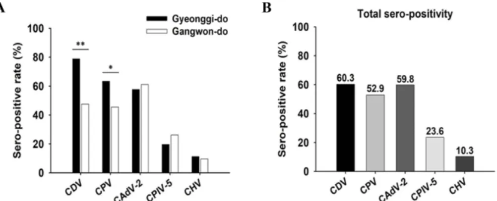
The seroprevalence of five viruses was examined by ELISA, VN, and HI tests in 174 raccoon dog sera collected from Gyeonggi and Gangwon Provinces. As shown in Fig- ure 2, the overall seropositive rates against CDV, CPV, CAdV-2, CPIV-5, and CHV were 60.3% (105/174), 52.9%
(92/174), 59.8% (104/174), 23.6% (41/174), and 10.3% (18/
174), respectively. The regional incidence rates of CDV, CPV, and CAdV-2 were 78.9% (56/71), 63.4% (45/71), and 57.7% (41/71) in Gyeonggi province and 47.0% (49/103), 45.6% (47/103), and 61.2% (63/103) in Gangwon Province, respectively (Fig. 2A). The raccoon dogs captured from Gyeo- nggi Province showed higher incidence rates of CDV (78.9%), CPV (63.4%), and CHV (11.3%) than those from Gangwon Province, and the raccoon dogs from Gangwon Province showed slightly higher incidence rates of CPIV-5 (26.2%) and CAdV-2 (61.2%) than those from Gyeonggi Province.
Fig. 1. Rabies virus (RABV) positive (A) and negative (B) sam- ples by the direct fluorescent antibody test. There was no pos- itive RABV reaction in the 62 raccoon dog brain samples.
Attempts at detecting canine adenovirus type 2 (CAdV-2) (C) and canine parainfluenza virus type 5 (CPIV-5) (D) in lung sam- ples. M, DNA ladder; Lanes 1–4, lung samples; P, positive sam- ple. There were no positive samples against CAdV-2 or CPIV- 5. Detection of canine distemper virus (CDV) in Korean rac- coon dog brain samples (E). Lanes 1–10, brain samples; P, positive sample. Samples 1 and 7 showed positive reactions against CDV.
Fig. 2. Sero-positive rates against canine distemper virus (CDV), canine parvovirus (CPV), canine adenovirus type 2 (CAdV-2), canine parainfluenza virus type 5 (CPIV-5), and canine herpesvirus (CHV) in Korean raccoon dog sera. There were regional differences in the sero-positive rates against CDV and CPV (A). The sero-positive rate (60.3%) of CDV was the highest of the five pathogens (B).
*