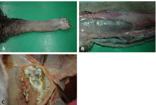
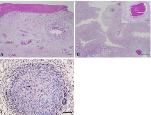
Aspergillus fumigatus infection in an ostrich with an enlarged neck due to respiratory problems
4
0
0
전체 글
(2)
(3)
(4)
수치
관련 문서
Ross: As my lawfully wedded wife, in sickness and in health, until
glen plaids 글렌 플레이드와 캐시미어 카디건, 캐리지 코트, 그리고 케이프 -> 격자무늬의 캐시미어로 된 승마용 바지, 마부용 코트, 말 그림이 수
systemic circulation, in the right ventricle and oxygenated blood from the lungs, or pulmonary circulation, in the left ventricle, as in birds and mammals.. Two vessels,
(A) Interestingly, this has often been the case with English animal expressions.. As you may have observed, they make irregular twists and turns in their
다양한 번역 작품과 번역에 관한 책을 읽는 것은 단순히 다른 시대와 언어, 문화의 교류를 넘어 지구촌이 서로 이해하고 하나가
The index is calculated with the latest 5-year auction data of 400 selected Classic, Modern, and Contemporary Chinese painting artists from major auction houses..
The problems created by welding in industrial site are deformation and residual stress. They not only cause the nonlinear deformation but also have an influence
1. Free radical initiator abstract a hydrogen from polymer chains 2. Through chain transfer of propagating chain with polymer chain 3. Polymer mixtures are mechanically
