pISSN: 2287-4208 / eISSN: 2287-4690
World J Mens Health 2012 December 30(3): 198-201
http://dx.doi.org/10.5534/wjmh.2012.30.3.198
Case Report
Received: Jul 16, 2012; Revised: Nov 18, 2012; Accepted: Nov 26, 2012 Correspondence to: Jae Hyun Bae
Department of Urology, Korea University Ansan Hospital, Korea University College of Medicine, 123, Jeokgeum-ro, Danwon-gu, Ansan 425-707, Korea.
Tel: +82-31-412-5190, Fax: +82-31-412-5194, E-mail: [email protected] Copyright © 2012 Korean Society for Sexual Medicine and Andrology
This is an Open Access article distributed under the terms of the Creative Commons Attribution Non-Commercial License (http://creativecommons.
org/licenses/by-nc/3.0) which permits unrestricted non-commercial use, distribution, and reproduction in any medium, provided the original work is properly cited.
Metastatic Squamous Cell Carcinoma of the Kidney from Cholangiocarcinoma
Hoon Choi1, Tae Il Noh1, Byeong Kuk Ham1, Jae Young Park1, Kang Soo Shim2, Jae Hyun Bae1
Department of Urology, 1Korea University Ansan Hospital, Korea University College of Medicine, Ansan, 2Andong Hospital, Andong, Korea
We present a rare case of a metastatic renal tumor originating from adenosquamous carcinoma of the intrahepatic bile duct. A 64-year-old man treated with bisegmentectomy and extended cholecystectomy for cholangiocarcinoma had a left cystic renal mass, which had irregular wall thickening, heterogeneously low attenuation, and soft tissue infiltration as determined by a computed tomography scan. The first impression was renal abscess. Left nephrectomy was performed and the nonencapsulated mass was gray in color macroscopically. Histological examination of the specimen revealed alveolar proliferation of small cancer cells, which was consistent with the original tumor of the intrahepatic bile duct. The left renal tumor was misdiagnosed as a renal abscess but finally diagnosed as squamous cell carcinoma metastasized from the intrahepatic bile duct. The patient expired because of lung metastasis after 14 months following left nephrectomy. In our opinion, this case would be the first report of a renal metastasis from a cholangiocarcinoma clinically and was treated with nephrectomy.
Key Words: Neoplasm metastasis; Carcinoma, squamous cell; Nephrectomy
Renal metastases are common from disseminated malig- nancies, with a reported rate of 7.6∼12% at autopsy. On the other hand, clinical detection of secondary carcino- mas in the kidney are relatively rare.1-4 Although meta- static renal tumors are the usual findings, it is generally known that aggressive surgical treatment offers minimal survival benefit. Therefore, most cases have been man- aged conservatively.
We report a rare case of renal metastasis from carcino- ma of the intrahepatic bile duct, misdiagnosed as a renal abcess. The patient survived about 14 months after ne- phrectomy.
CASE REPORT
A sixty-four-year old patient presented with complaints of fever, myalgia, and left flank pain lasting for one month.
He had undergone bisegmentectomy and extended chol- ecystectomy for cholangiocarcinoma 110 days earlier and his symptoms had gradually been aggravated during close observation. On the pathologic report after the operation, more than one tumor larger than 5 cm was present and the cancer had spread to the regional lymph nodes. Thus his pathologic stage was T3N1M0.
The nature of his pain was gradual in onset and he had
Hoon Choi, et al: Metastatic Renal Squamous Cell Carcinoma 199
Fig. 1. Left cystic renal mass with irregular wall thickening, heterogeneous attenuation, and soft tissue infiltration.
Fig. 2. Tumor is located in the renal pelvis, and extending to and infiltrating the renal parenchyma.
occasional nausea, minimal radiating pain nature and gen- eralized weakness. There was history of fever without hematuria or pyuria. He was admitted our hospital via the emergency department. His vital signs were in the normal range, except body temperature, which was 38.1oC. A physical exam revealed costovertebral angle tenderness and a palpable left flank mass. Routine hematology and bi- ochemical tests revealed leukocytosis (21,500 mm3), an elevated erythrocyte sedimentation rate (70 mm/hr), and c-reactive protein (13.7 mg/dl). Urinalysis and a chest ra- diograph were normal.
A computed tomography scan of the abdomen and pel- vis showed a large left renal cystic lesion with soft tissue infiltration to the posterior perirenal fascia suggestive of pyonephrosis. The cyst had irregular wall thickening and heterogeneous attenuation (Fig. 1). The left kidney and ureter were normal. There was no ascites or lymphadeno- pathy. He had no history of past radiation exposure or re- nal stones.
We regarded this lesion as a renal abscess. To drain pus from the cyst, a percutaneous drainage tube was inserted under C-arm monitoring. However, the drained material was only blood, and no bacterial growth was found in the culture test. The mild fever persisted and flank pain was not controllable by analgesics. We suspected the cystic re- nal mass could be a malignant lesion. Therefore, we made a decision to perform nephrectomy for his uncomfortable symptoms and oncological management.
A extraperitoneal approach via the flank position was used. An approach to the kidney through the subcostal
route was made and the perinephric space was entered by vertical incision to the Gerota fascia on the lateral aspect of the kidney, revealing underlying perinephric fat. By the guidance of a percutaneous drainage tube, we were able to reach the exact area of the tumor lesion. The kidney was mobilized sharply by developing a plane between the re- nal capsule and the perinephric fat.
Downward traction on the kidney permits the upper pole to be mobilized. With the use of lateral traction on the kidney to expose the hilum, the vascular pedicle is dis- sected free from the surrounding fat and lymphatics. The renal hilum can be approached anteriorly. The mobilized renal vein is retracted to reveal the renal artery located posteriorly. The left renal artery should be differentiated from the superior mesenteric artery by ensuring that the re- nal artery emanates from the lateral aspect of the aorta.
The renal artery is ligated away from the hilum using a 2-0 silk tie. After division of the renal artery, the renal vein is similarly ligated and divided. In the posterior approach the artery is encountered before the renal vein and is ligated and the ureter is doubly clamped and divided. The speci- men is removed. The distal aspect of the transected ureter is suture ligated. A drain is brought out through a separate stab incision.
The specimen of the kidney measuring 12.2×7.0×5.2 cm in size and 440 g in weight was obtained. The whole mass was a distended sac-like structure without any grossly visible renal tissue. On section, the specimen showed an in- filtrative solid mass along the lateral border. The cut surface showed a loculus with necrotic material within it (Fig. 2).
200 World J Mens Health Vol. 30, No. 3, December 2012
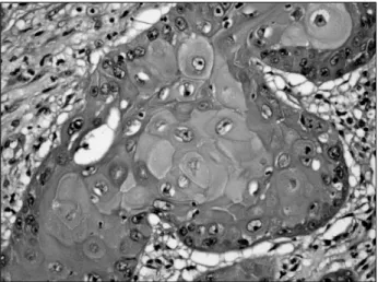
Fig. 3. Moderate-differentiated squamous cell carcinoma. Nests of infiltrating squamous cells with hyperchromatic nuclei and prominent keratin production should be noted (H&E, ×400).
The histology of the cystic mass revealed features of moderately differentiated squamous cell carcinoma. Nests of infiltrating squamous cells with hyperchromatic nuclei and prominent keratin production were noted (Fig. 3).
There were no positive findings on the periodic acid-Schiff stain. The tumor had invaded the renal parenchyma and perirenal soft tissue. There were no tumor emboli in the re- nal artery or veins. The entire tumor showed exclusive squamous differentiation. No transitional element was found within the tumor.
After nephrectomy, the palpable left flank mass with in- termittent pain disappeared and the temperature returned to the normal range. During the follow up, general weak- ness with lung metastasis and multiple systemic dissem- ination were observed about 10 months after neph- rectomy, and the patient expired 14 months after the nephrectomy.
DISCUSSION
Metastatic tumors in the kidney are common. Autopsy studies have shown that 7.6∼12% of patients dying of cancer have renal metastases, making the kidney one of the most common sites for metastatic spreading.1-4 In gen- eral, these metastatic tumors are discovered at autopsy, so only 40 cases have been reported in studies that diag- nosed them before the patient’s death.5
In general, the high blood flow and profuse vascularity
of the kidney make it an abundant growth medium for the deposition and growth of cancer cells. Most renal meta- stases develop through a hematogenous route of spread;
only a small minority are caused by direct invasion of tu- mors derived from adjacent organs such as the pancreas, colon, and adrenal gland.
Primary tumors of the lung (19.8%), breast (12.3%) and stomach (11.1%) are the most common sources of renal metastases.6 Other sources include the cervix, prostate, gallbladder, thyroid, ovary, testis, urinary bladder, con- tralateral kidney, and bone.3
Choyke et al7 found that renal masses in patients with other known primary tumors were 4 times as likely to be metastatic than a primary renal tumor.5 Renal metastases are generally small, bilateral, and multifocal. However, there are no specific radiologic findings to distinguish a secondary renal tumor from primary renal cell carcinoma.4 In our case, a computed tomography scan showed a large renal cystic lesion highly suggestive of pyonephrosis.
Therefore, percutaneous tube drainage with percutaneous fine-needle aspiration cytology could have been an appro- priate next step for diagnosis and treatment in our situation. However, this confusion brought about the de- lay of palliative and therapeutic decision making on the nephrectomy in the end. We are not sure if nephrectomy should be more prompt.
Intrahepatic cholangiocarcinoma is an uncommon neo- plasm and complete resection is the treatment of choice. If completely resected, 3-year survival rates range from 16%
to 61% and 5-year survival rates, 24% to 44%. Factors as- sociated with poor outcome include intrahepatic meta- stases, lymph node metastasis, vascular invasion, and pos- itive margins. There is little evidence for the utility of radia- tion and chemotherapy in intrahepatic cholangiocarcino- ma, and their use is optional.
In this case, the patient expired relatively earlier than we expected. At first, a renal mass biopsy or aspiration could be considered because it may preclude the need for surgical intervention.8 However, we can suspect that de- lay of nephrectomy resulted in shortening of the survival period. Though we had comparatively good control of the original tumor and also of the metastatic renal tumor by nephrectomy and of the intractable cancer-related symp- toms (fever, myalgia, and flank pain), in case of any con-
Hoon Choi, et al: Metastatic Renal Squamous Cell Carcinoma 201
dition suggesting renal metastasis, the metastatic tumor should be the first diagnosis and all the treatment strat- egies should be focused on cancer control, because be- nign disease is rarely related to a survival disadvantage and a cancer-related event can lead to a regrettable result.
REFERENCE
1. Abrams HL, Spiro R, Goldstein N. Metastases in carcinoma;
analysis of 1000 autopsied cases. Cancer 1950;3:74-85 2. Bracken RB, Chica G, Johnson DE, Luna M. Secondary renal
neoplasms: an autopsy study. South Med J 1979;72:806-7 3. Mayer RJ. Infiltrative and metastatic disease of the kidney. In:
Rieselbach RE, Garnick MB, editors. Cancer and the kidney.
Philadelphia: Lea & Febiger; 1982;707.
4. Sánchez-Ortiz RF, Madsen LT, Bermejo CE, Wen S, Shen Y, Swanson DA, et al. A renal mass in the setting of a nonrenal malignancy: When is a renal tumor biopsy appropriate? Cancer 2004;101:2195-201
5. Peterson RO. Urologic pathology. Philadelphia: Lippincott;
1992;127.
6. Wagle DG, Moore RH, Murphy GP. Secondary carcinomas of the kidney. J Urol 1975;114:30-2
7. Choyke PL, White EM, Zeman RK, Jaffe MH, Clark LR. Renal metastases: clinicopathologic and radiologic correlation.
Radiology 1987;162:359-63
8. Rybicki FJ, Shu KM, Cibas ES, Fielding JR, vanSonnenberg E, Silverman SG. Percutaneous biopsy of renal masses: sensi- tivity and negative predictive value stratified by clinical set- ting and size of masses. AJR Am J Roentgenol 2003;180:
1281-7