• 검색 결과가 없습니다.

Interaction between epidermal growth factor receptor– and cyclooxygenase 2–mediated pathways and its implications for the chemoprevention of head and neck cancer

N/A
N/A
Protected

Academic year: 2021

Share "Interaction between epidermal growth factor receptor– and cyclooxygenase 2–mediated pathways and its implications for the chemoprevention of head and neck cancer"

Copied!
9
0
0

로드 중.... (전체 텍스트 보기)

전체 글

(1)

Interaction between epidermal growth factor receptor–

and cyclooxygenase 2–mediated pathways and its implications for the chemoprevention

of head and neck cancer

Mi Sun Choe,

1

Xin Zhang,

1

Hyung Ju C. Shin,

2

Dong M. Shin,

1

and Zhuo (Georgia) Chen

1

1

Department of Hematology/Oncology, Winship Cancer Institute, Emory University and

2

Quest Diagnostics, Atlanta, Georgia

Abstract

Head and neck squamous cell carcinoma is a well-known model for chemoprevention studies because of its field cancerization effect, its multistep carcinogenesis pro- cess, and the easy accessibility of biopsies to target lesions. With new understandings of head and neck carcinogenesis and the development of molecular targeted therapy, chemoprevention trials for head and neck squamous cell carcinoma have been rapidly updated. Cyclooxygenase-2 (COX-2) and epidermal growth factor receptor (EGFR) tyrosine kinase inhibitors are gaining significant attention as potential chemo- preventive agents. Both COX-2 and EGFR are involved in head and neck carcinogenesis. Targeting COX-2 and EGFR separately has shown promising antitumor activity.

Recently, combinations of COX-2 and EGFR tyrosine kinase inhibitors have been reported to show synergistic/

additive effects in preclinical studies. Because COX-2 and EGFR tyrosine kinase inhibitors are toxic as single agents in clinical trials, the combination of COX-2 and EGFR tyrosine kinase inhibitors used at lower doses seems more promising than monotherapy with either as a novel strategy in head and neck cancer chemoprevention. [Mol Cancer Ther 2005;4(9):1448 – 55]

Introduction

Approximately 40,000 cases of new head and neck cancer (HNC) develop annually, accounting for f3% of all new cancers in the United States (1), and 90% of these are squamous cell carcinoma [head and neck squamous cell carcinoma (HNSCC)]. Due to its location and anatomic complexity, HNC causes almost inevitable functional and social impairment even before it becomes life threatening.

Despite great advances in therapy, the overall survival rate for patients with HNC has not improved significantly, emphasizing the importance of preventive intervention (2 – 4).

Chemoprevention can be defined as the use of specific agents to suppress, reverse, or prevent carcinogenesis, thus stopping the progression to invasive cancer by modulating the carcinogenic process or by removal (apoptosis) of premalignant cells (5, 6). Recently, the concept of chemo- prevention has been substantially incorporated into cancer treatment goals. Progress in clinical trials has shown that the combined preventive approach is more effective than single-agent chemoprevention.

This review will briefly summarize chemopreventive approaches in HNSCC before highlighting a novel and promising chemopreventive modality combining cyclo- oxygenase-2 (COX-2) and epidermal growth factor receptor (EGFR) tyrosine kinase inhibitors.

Chemoprevention for HNC

HNSCC is an excellent model for the chemopreventive approach in several aspects. First, HNSCC is notorious for its high tendency to develop second primary tumors, one of the main reasons for the typical dismal outcome of this cancer. The lifetime cumulative risk of second primary tumor is >20% and has been reported in up to 47% in patients with previously treated laryngeal cancer (2, 7). This multi- centricity is best explained by the fact that the whole mucosa of the upper aerodigestive tract is affected by the same carcinogens, most likely tobacco and alcohol (field cancer- ization; ref. 8). Second, HNSCC well exemplifies multistep carcinogenesis with stepwise accumulations of genetic alterations (9, 10). Pathologically, the course follows from normal epithelium, hyperplasia, and dysplasia to invasive HNSCC. Multicentricity and multistep carcinogenesis pro- vides the rational background for a chemopreventive

Received 9/20/04; revised 7/13/05; accepted 7/15/05.

Grant support: NIH grant U01 CA101244.

The costs of publication of this article were defrayed in part by the payment of page charges. This article must therefore be hereby marked advertisement in accordance with 18 U.S.C. Section 1734 solely to indicate this fact.

Requests for reprints: Zhuo (Georgia) Chen, Department of Hematology/

Oncology, Winship Cancer Institute, Emory University, Room 3086, 1365-C Clifton Road, Atlanta, GA 30322.

E-mail: georgia_chen@emoryhealthcare.org

Copyright C 2005 American Association for Cancer Research.

doi:10.1158/1535-7163.MCT-04-0251

Minireview

(2)

approach. In addition, HNC exhibits clinically well defined premalignant lesions, such as oral leukoplakia and eryth- roplakia, to be targeted. These can be easily identified compared with premalignant lesions in other organ sites and provide accessibility for macroscopic and microscopic evaluation or biomarker evaluation of lesions.

HNSCC has been one of the main models for chemo- preventive approaches and important issues have been learned from chemopreventive clinical trials. The most extensively studied chemopreventive agents in HNC areretinoids (11). The first randomized clinical trial was conducted by Hong et al. (11) using a high dose of 13-cis- retinoic acid (13-cRA) for a short period in patients with oral premalignant lesions, which showed significant clinical and pathologic responses. In spite of the encouraging results, this trial raised two important issues common in chemo- preventive clinical trials. Dose-related toxicity was an important obstacle to treating patients on a long-term basis, and the remission induced by short-term treatment did not last long after cessation of treatment. This observation led to a subsequent low-dose maintenance trial. Lippman et al. (5) treated patients with oral premalignant lesions with high- dose 13-cRA for 3 months and switched to either low-dose 13-cRA or h-carotene for maintenance. Although the results showed that using low-dose 13-cRA for maintenance could effectively repress disease progression, the long-term follow-up failed to show a difference in the cancer development rate in both groups (11). In terms of pre- vention of second primary tumor, the first phase III clinical trial using 13-cRA showed a statistically significant sup- pression of second primary tumor, although there was no significant difference in survival. Subsequent randomized clinical trials using retinoids have failed to show a difference in the development of second primary tumor or survival (11).

Obtaining long-lasting efficacy with low toxicity has been an important issue, provoking combinations of retinoids with other agents or searches for new chemopreventive agents. Other chemopreventive agents that have shown preventive effects include selenium and vitamin E. Recently, Papadimitrakopoulou et al. (12) reported that a combination of 13-cRA, vitamin E, and IFN-a restored advanced laryngeal premalignant lesions. Shin et al. (13, 14) showed that the same combination suppressed the development of second primary tumor and/or recurrence, and achieved an excellent survival rate in stage III/IV HNSCC, making this a promising combination chemopreventive approach. With a better understanding of relevant molecular changes in each carcinogenic step of HNC and increased availability of specific molecular targeting agents, chemoprevention in HNC has evolved to a new era. New agents in this context include COX inhibitors, EGFR tyrosine kinase inhibitors, farnesyl transferase inhibitors, and others (11).

Rationale for Blocking COX-2 and EGFR Path- ways in Chemoprevention of HNC

Selection of chemopreventive agents requires consider- ation of several criteria. Biological efficacy impeding the

carcinogenic process, selectivity for transformed tissue, minimal toxicity, and the ability to obtain good patient compliance are essential. As new potential agents, COX-2 inhibitor and EGFR inhibitor show promise for chemo- prevention of HNC. Both the COX-2 and EGFR signaling pathways play major roles in head and neck carcinogen- esis (15, 16). Both COX-2 and EGFR are overexpressed in premalignant and malignant tissues of the head and neck compared with normal tissue (17 – 20). Blocking these pathways has already shown promising antitumor activity. In addition, both the COX-2 and EGFR inhibitors used in clinics are orally bioavailable and their side effects are well tolerated at clinically relevant dosages (21 – 23).

COX-2 Pathway in Carcinogenesis

COX-1 and COX-2 catalyze prostanoid synthesis from arachidonic acid. COX-1 is constitutively expressed in nearly all normal tissues and has a beneficial housekeep- ing role. COX-2 is undetectable (or at low levels) in most normal tissues but is rapidly induced in response to inflammatory or mitogenic stimuli, including cytokines, growth factors, and tumor promoters (24, 25).

COX-2 has been implicated in carcinogenesis ever since the discovery that intake of nonsteroidal anti-inflammatory drugs that inhibit COX activity also decrease the relative risk for development of colorectal cancer (26). There is also direct evidence for the carcinogenic role of COX-2. Oshima et al. (27) showed that selective genetic elimination of COX- 2 protected APC tumor suppressor gene – deleted mice from developing intestinal tumors. In humans, COX-2 overexpression has been found in premalignant and malignant lesions in several tumors (17, 18, 25; for review, see 28). In HNC, there is a stepwise increase in COX-2 expression through the normal, hyperplastic, dysplastic, and invasive carcinoma stages, suggesting its carcinogenic role in HNC (17, 18, 29).

COX-2 overexpression contributes to many aspects of carcinogenesis, such as inhibition of apoptosis, promotion of cell proliferation, induction of angiogenesis, and increasing invasiveness, mainly through increasing the amount of prostaglandins, including prostaglandin E

2

(PGE

2

), PGF

2a

, PGD

2

, TXA

2

, PGI

2

, and PGJ

2

(28, 30).

Prostaglandins exert their effect mainly by binding to G-protein – coupled receptors, including EP-1, EP-2, EP-3, and EP-4, on cell surfaces (for review, see ref. 31). Each is specifically recognized by certain prostaglandins and activates signaling transduction from extracellular signal regulated kinase (32), phosphatidyl inositol 3-kinase/AKT, and cyclic AMP/protein kinase A (32 – 35). Alternatively, cyclopentenone prostaglandins, such as PGJ

2

, can bind to the nuclear transcription factor peroxisome proliferator- activated receptor, which regulates expression of several genes involved in cell proliferation (36).

EGFR Signaling Pathway in Cancer Progression

EGFR, a surface receptor with intrinsic tyrosine kinase

activity, is one of several known pivotal intermediates in

many epithelial malignancies (37). It belongs to the erbB

growth factor receptor family. The ligands binding to the

(3)

extracellular domain of EGFR induce homodimerization of EGFR or heterodimerization with other members of the erbB growth factor receptor family, activating the intrinsic tyrosine kinase and its downstream signaling molecules (38). Potential EGFR downstream signaling pathways include Ras/mitogen-activated protein kinase, phosphati- dyl inositol 3-kinase, phospholipase Cg, the Src kinase family, Janus kinase, signal transducers, activators of transcription, and others (39). Activation of EGFR is involved in pertinent pleiotropic cellular processes, such as cell proliferation, apoptosis, differentiation, angiogene- sis, and motility (37 – 43).

Overexpression of EGFR has been frequently reported in human malignant neoplasms (44). In HNC, expres- sion of EGFR and its ligands, transforming growth factor-a or epidermal growth factor, are up-regulated in histologically normal epithelium adjacent to invasive cancer compared with control normal epithelium in indi- viduals without cancer. Dysplastic lesions overexpress EGFR, and invasive cancer displays a more jumped-up pattern of overexpression (19, 20). All this implicates the EGFR signaling pathway in the early stages of head and neck carcinogenesis and progression. EGFR overexpres- sion is also associated with a poorer prognosis in HNC patients (45).

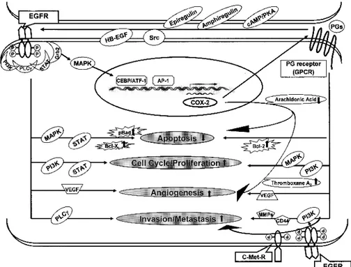
Interaction between EGFR and COX-2 Pathways Whereas the pathways by which EGFR and COX-2 contribute to carcinogenesis have been separately consid- ered and targeted, increasing evidence indicates a tight connection between these two pathways (Fig. 1).

EGFR and COX-2 signaling pathways form a positive feedback loop. Activation of EGFR has been shown to

induce increased COX-2 expression in various normal and tumor cell lines, including HNSCC cell lines (46–54).

Both transforming growth factor-a and epidermal growth factor, ligands of EGFR, were found to induce COX-2 expression (46 – 50). Expression of COX-2 is regulated at both the transcriptional and posttranscrip- tional level. The signaling pathway involved in COX-2 induction via EGFR varies depending on the type of cells and inducers, but the ras/raf/mitogen-activated protein kinase signaling pathways mainly contribute to both increased transcriptional and posttranscriptional control. One explanation for the linkage between EGFR/mitogen-activated protein kinase and transcrip- tional activation of COX-2 may be the activation of transcription factors such as cyclic AMP response element-binding protein/activating transcription factor and activator protein-1 by mitogen-activated protein kinase signaling (25, 46, 51, 54). Binding sites for these transcription activators have been identified in the COX-2 promoter region.

On the other hand, COX-2 induces transactivation or increased expression of EGFR (55, 56). Transactivation of EGFR by PGE

2

, a major prostaglandin involved in carci- nogenesis, has been well documented but the process seems to be quite complex and cell type dependent. Pai et al. (56) showed that PGE

2

transactivated EGFR and triggered the activation of extracellular signal regulated kinase-2 pathways in normal gastric epithelial cells and colon cancer cells, inducing cell proliferation in vitro and in vivo. The G-protein coupled receptor, to which the major prostanoids receptors belong, is involved in EGFR trans- activation. The mechanism by which G-protein coupled

Figure 1. Interaction between the EGFR and COX-2 pathways. Activation of EGFR induces COX-2 mainly via the mitogen-activated protein kinase pathway.

On the other hand, prostaglandins, a

product of COX-2, can stimulate EGFR

activation. Both EGFR and COX-2 path-

ways act in common on several aspects of

carcinogenesis.

(4)

receptor mediates the transactivation of EGFR has not been clearly defined but the release of EGFR ligand by Src- activated transmembrane metalloproteinase has been sug- gested (31, 57). Consistent with these findings, Pai et al.

(56) observed that PGE

2

-mediated transactivation of EGFR also involved transforming growth factor-a, likely re- leased by Src-activated metalloproteinase. Complicating these findings, Buchanan et al. (58) reported that the transactivation of EGFR by PGE

2

occurred via an intracellular Src-mediated event but not through the release of an extracellular epidermal growth factor-like ligand in colon cancer cell lines. On the other hand, Shao et al. (34) showed that PGE

2

activated EGFR through the induction of increased amphiregulin expression, one of the EGFR ligands. They showed that PGE

2

activated the cyclic AMP/protein kinase A pathway, which induced expression of amphiregulin in a colon cancer cell line.

Induction of EGFR expression by overexpression of COX-1 or COX-2 was reported by Kinoshita et al. (55) in a human colon cancer cell line. There is a recent evidence for EGFR and proliferator-activated receptor transactivation through an Src-dependent pathway (59).

A direct collaborative effect between PGE

2

and EGFR on tumor cell phenotypes, such as invasion and proliferation, is also well documented. Shao et al. showed this collabo- ration between COX-2/PGE

2

and EGFR pathways (34, 60), observing synergistic induction of amphiregulin by PGE

2

and transforming growth factor-a (34). In a follow-up study, they showed that activation of both PGE

2

and EGFR signaling pathways synergistically promoted the growth and migration of colon cancer cells (60). Pai et al. (61) reported that PGE

2

enhancement of invasiveness in a colon cancer cell line was mediated by transactivation of c-Met-R (hepatocyte growth factor receptor), partly through the transactivation of EGFR. Buchanan et al. (58) made a similar observation, showing that PGE

2

-induced cell migration was mediated by the transactivation of EGFR, and associated with intracellular Src activation in colon cancer cells.

It is worth noting that COX inhibitor repressed the EGFR-related pathway and in turn, EGFR inhibitor repressed COX-2 expression, indirectly suggesting their interaction (62, 63). COX inhibitors were reported to block the cell proliferation induced by epidermal growth factor in NIH 3T3 cells, which could be reversed by adding exo- genous PGE

2

(62). Gefitinib, an EGFR inhibitor, showed inhibition of COX-2 expression in a HNSCC cell line (63).

In addition, celecoxib, a selective COX-2 inhibitor, showed a protective effect against HER-2/neu-induced experimen- tal breast cancer, indirectly suggesting a relationship between the epithelial growth factor receptor family and COX-2 (64).

On top of their known interactions, both the EGFR and COX-2 pathways affect the same aspects of carcinogenesis, such as inhibition of apoptosis and induction of angiogen- esis. The conclusion that the EGFR and COX-2 pathways directly or indirectly collaborate in pertinent carcinogenic pathways seems justified.

Therapeutic Implication of Targeting COX-2 and EGFR-Mediated Pathways in Chemopre- vention

COX-2 as a Target of Therapeutic and Chemopreven- tive Agents

Selective COX-2 inhibitors have been developed to avoid interrupting the biosynthesis of prostaglandins by COX-1.

Studies in preclinical models have clearly showed that COX-2 inhibitors repressed tumor growth (65). Further- more, COX inhibitors reduce tumor cell migration and tumor invasiveness as well as inhibit angiogenesis in various cell lines and in a xenograft animal model (66 – 69). These inhibitory effects have been reported in HNC as well as by in vivo and in vitro experiments (70 – 74).

In terms of a chemopreventive effect in HNC, Wang et al.

(75) reported a significant delay of tumor cell growth and reduced angiogenesis using a COX-2 inhibitor, celecoxib, in a xenograft mouse model. In a head and neck carci- nogenesis model, Shiotani et al. (76) pretreated rats with a carcinogen, 4-nitroquinoline-1-oxide, followed by a selec- tive COX-2 inhibitor, nimesulide, at the postinitiation stage. They found that the ingestion of carcinogen induced COX-2 expression in premalignant tongue lesions and squamous cell carcinoma. Subsequent COX-2 inhibitor treatment significantly reduced the development of inva- sive squamous cell carcinoma. The antitumorigenic prop- erties of these inhibitors are both COX dependent and independent (77, 78).

Based on these promising results, many clinical trials of chemoprevention using various COX-2 inhibitors have been conducted in various organs, although mostly in colon. The first chemopreventive trial using a selective COX-2 inhibitor in humans was conducted on familial adenomatous polyposis patients. Familial adenomatous polyposis patients were treated with celecoxib 400 mg bid for 6 months and were compared with a placebo- treated group for their polyp burden. A significant reduction in polyp burden was observed in the cele- coxib-treated group (21). Observing the data from this study, the Food and Drug Administration approved celecoxib as an adjuvant therapy for familial adenoma- tous polyposis patients. Currently, phase II clinical trials are under way to evaluate COX-2 inhibitors for the prevention of recurrence or development of second primary tumor in early-stage HNC patients and the prevention of cancer in patients with oral leukoplakia or dysplasia, using celecoxib.

However, notable cardiovascular toxicity was reported for specific COX-2 inhibitors recently, which resulted in reevaluation of the clinical use of COX-2 inhibitors (79).

3

Studies of COX-2 – specific inhibitors, including celecoxib, showed that COX-2 inhibitors increased the thromboem- bolic cardiovascular risks. A hypothetical explanation is

3

Department of Health and Human Services. NIH halts use of COX-2 inhibitor in large cancer prevention trial. NIH News, December 17, 2004.

Available at http://www.nih.gov/news/pr/dec2004.

(5)

that, unlike nonselective nonsteroidal anti-inflammatory drugs, COX-2 inhibitors could not inhibit platelet aggre- gation (80, 81). Due to concerns about cardiovascular complications associated with long-term use of COX-2 inhibitors, in December 2004, the National Cancer Institute announced the early cessation of a large colorectal cancer prevention clinical trial with celecoxib.

3

But in April 2005, the Food and Drug Administration announced restricted use of nonsteroidal anti-inflammatory drugs, including celecoxib, by providing revised labels to include more specific information about the potential cardiovascular and gastrointestinal risks.

4

EGFR as a Target of Therapeutic or Chemopreventive Agents

A variety of strategies have been developed to block EGFR specifically, including monoclonal antibodies, tyro- sine kinase inhibitors, ligand-linked immunotoxins, and antisense approaches (37, 82, 83). Among those strategies, monoclonal antibodies, such as IMC-225 (Cetuximab), and tyrosine kinase inhibitors, such as ZD1839 (Iressa or Gefitinib), and OSI-774 (Tarceva or Erlotinib) have shown promising efficacy and are currently being used in clinical studies singly or in combination with other chemothera- peutic agents or radiotherapy. The antineoplastic effects of EGFR inhibitors include inhibition of cell cycle progression, induction of apoptosis, inhibition of angiogenesis, and decreasing metastasis (40, 84). In HNC, EGFR inhibition also showed growth inhibition and inhibition of metastasis in in vitro and in vivo experiments (84 – 89).

Like other anticancer agents, EGFR inhibitor toxicity was also reported. The main toxic effects of EGFR tyrosine kinase inhibitors, such as OSI-774 and ZD1839, include headache, diarrhea, and skin rash (22, 90, 91). Rare asso- ciation with interstitial lung disease was also observed using ZD1839 (92, 93).

Combined Chemopreventive Therapy Using COX-2 and EGFR Inhibitors

Because the underlying genetic mechanisms of many malignant neoplasms are possibly multipath processes combined with complex cross-talk between pathways, specific blocking of single molecular targets would not outwit the variability and complexity of genetic alterations in cancer. Combined treatments using appropriate multi- agents may be more effective than single agent treatments.

In terms of chemoprevention, combining low doses of drugs too toxic for single use may result in negligible toxicity while eluding drug resistance, resulting in an elegant strategy that is both effective and safe.

Considering the tight connection of the COX-2 and EGFR pathways, the combination of their particular inhibitors may block both pathways in a synergistic or additive manner. This idea has been supported by in vitro and in vivo studies combining COX-2 and EGFR inhibitors (Table 1). Tortora et al. (94) showed a supra-additive inhibitory effect on tumor growth and angiogenesis by combined treatment with SC-236 (COX-2 inhibitor), ZD1839, and protein kinase A antisense in human colon and breast cancer cell lines and a colon cancer xenograft.

We also observed a synergistic growth-inhibitory effect by combining celecoxib and ZD1839 in HNSCC in vitro (95) and in vivo (96). The combination of celecoxib and ZD1839 augmented G

1

cell cycle arrest and further suppressed phosphorylation of EGFR downstream signaling mole- cules, such as EGFR, extracellular signal regulated kinase, and AKT. This synergistic growth inhibitory effect was also observed in a breast cancer cell line. De Luca et al. (97) found a synergistic growth inhibitory effect on breast cancer cell lines with a combination of rofecoxib and ZD1839. As with our observation, this effect was associated with significant further inactivation of AKT and extracel- lular signal regulated kinase.

A synergistic growth inhibitory effect also has been achieved in chemoprevention models. Torrance et al. (98) showed that a combination of sulindac, an inhibitor of both COX-1 and COX-2, and EKB-569, an EGFR tyrosine kinase inhibitor, protected APC

Min/+

mice remarkably Table 1. Preclinical and clinical studies of combined therapy using COX and EGFR inhibitors

Author (reference) COX inhibitor EGFR inhibitor In vitro/ in vivo/clinical

Tortora et al. (94) SC-236 ZD1839 (plus protein kinase A

antisense)

In vitro/vivo

Chen et al. (95) Celecoxib ZD1839 In vitro

Zhang et al. (96) Celecoxib ZD1839 In vivo

Luca et al. (97) Refecoxib ZD1839 In vitro

Torrance et al. (98) Sulindac EKB-569 In vivo

Krysan et al. (102) Celecoxib OSI-774 In vitro

Reckamp et al. (100) Celecoxib ZD1839 Clinical

Writh et al. (101) Celecoxib ZD1839 Clinical

Dannenberg et al. (28) Celecoxib EKB-569 Clinical*

Choe et al. (this review) Celecoxib OSI-774 Clinical*

*

HNSCC chemoprevention trials.

4

The Food and Drug Administration announces series of changes to the class of marketed nonsteroidal anti-inflammatory drugs. Food and Drug Administration News, April 7, 2005. Available at http://www.fda.gov/

bbs/topics/news/2005.

(6)

from the development of intestinal neoplasia, compared with the use of single agents alone. Although 100% of the untreated APC

Min/+

mice developed f20 polyps, nearly half the mice treated with both agents developed no polyps at all. In HNC, we observed that pretreatment with celecoxib and ZD1839 before tumor injection results in significant tumor growth delay and inhibition compared with control and single-drug – treated groups (96). The same effect could usually be achieved by at least more than double dose of ZD1839 alone (50 mg/kg in the combina- tion versus 150 mg/kg as a single drug; refs. 96, 99).

Combining COX-2 and EGFR inhibitors seems to impede or avert several carcinogenic steps with lower toxicity. As observed previously, cellular proliferation is also more efficiently repressed by this combinational strategy. It may inhibit further progression of genetic instability and make abnormal cells more vulnerable to apoptotic cell death.

Synergistic inhibition of angiogenesis has also been achieved. It is rational to expect that inhibiting both pathways with a low dose of each inhibitor would reduce toxicity, because the toxicity from COX-2 inhibitor and EGFR inhibitor have unrelated mechanisms.

Based on preclinical studies, several clinical trials using a combination of COX-2 and EGFR inhibitors are either ongoing or pending activation. Two phase I trials using COX-2 and EGFR tyrosine kinase inhibitors were reported in the American Society of Clinical Oncology meetings of last year and this year (100, 101). The first trial enrolled 12 patients with advanced metastatic and recurrent HNSCC (100). Three of nine evaluable patients showed partial response with no dose-limiting toxicities up to the reporting date. The second trial involved 15 patients with advanced NSCLC (101). The combined drugs were well tolerated. Four of 12 evaluable patients showed partial response and three showed stable disease with no unanticipated toxicities. At present, a randomized multicenter trial using celecoxib and EKB-569 (EGFR tyrosine kinase inhibitor) for preventing oral cancer in patients with oral leukoplakia is pending at the M. D. Anderson Cancer Center (28). We are also planning phase I/II chemopreventive clinical trials using a combina- tion of OSI-774 (Tarceva) and celecoxib for former smokers with premalignant lesions and patients with early stage (stage I/II) HNSCC to prevent HNSCC and second primary and recurrent tumors, respectively. Once launched, these clinical trials will provide valuable information on toxicity, response rate, and appropriate biomarkers for using the combination of COX-2 and EGFR inhibitors in cancer clinics.

Future Prospects and Conclusions

In spite of extensive research in EGFR and COX-2 inhibitors, their combined use for chemoprevention is still in its developmental stage. Further investigations are indeed necessary to develop appropriate chemopreventive strategies in HNC. First, the mechanism by which EGFR and COX-2 pathways are deregulated, interact with each other, or contribute to head and neck carcinogenesis must be clarified. Second, more preclinical studies are critical to evaluate the effectiveness of these combinations and to

understand their mechanisms of action as single agents and in combination in HNC specifically. The generation of a good animal model for HNC chemoprevention would be very helpful. Possible drug toxicity from combined use must be evaluated over the long term, although low doses of each drug will be used. Also, substantial biomarkers that can reflect the effect of COX-2 and EGFR combinations need to be identified. Eventually, we may hope to identify the patient population who would derive the most benefit from this combinational chemopreventive approach.

In conclusion, an expanding body of evidence shows tight interaction between the EGFR and COX-2 pathways, which may provide a target for a synergistic inhibitory effect on cancer cell growth using EGFR and COX-2 inhibitors in combination. This combination approach is a promising novel strategy for the chemoprevention of HNC, which may achieve more effective cancer prevention with less drug toxicity or drug resistance than monotherapy with either drug.

References

1. Jemal A, Tiwari RC, Murray T, et al. American Cancer Society. Cancer statistics, 2004. CA Cancer J Clin 2004;54:8 – 29.

2. Vokes EE, Weichselbaum RR, Lippman SM, Hong WK. Head and neck cancer. N Engl J Med 1993;328:184 – 94.

3. Vokes EE, Stenson KM. Therapeutic options for laryngeal cancer.

N Engl J Med 2003;349:2087 – 9.

4. Argiris A, Brockstein BE, Haraf DJ, et al. Competing causes of death and second primary tumors in patients with locoregionally advanced head and neck cancer treated with chemoradiotherapy. Clin Cancer Res 2004;

10:1956 – 62.

5. Lippman SM, Batsakis JG, Toth BB, et al. Comparison of low-dose isotretinoin with beta carotene to prevent oral carcinogenesis. N Engl J Med 1993;328:15 – 20.

6. Sun SY, Hail N, Lotan R. Apoptosis as a novel target for cancer chemoprevention. J Natl Cancer Inst 2004;96:662 – 72.

7. Gao X, Fisher SG, Mohideen N, Emami B. Second primary cancer patients with laryngeal cancer: a population-based study. Int J Radiat Oncol Biol Phys 2003;56:427 – 35.

8. Slaughter DP, Southwick HW, Smejkal W. Field cancerization in oral stratified squamous epithelium. Cancer 1953;6:963 – 8.

9. Hunter KD, Parkinson EK, Harrison PR. Profiling early head and neck cancer. Nat Rev Cancer 2005;5:127 – 35.

10. O’Shaughnessy JA, Kelloff GJ, Gordon GB, et al. Treatment and prevention of intraepithelial neoplasia: an important target for accelerated new agent development. Clin Cancer Res 2002;8:314 – 46.

11. Papadimitrakopoulou VA, Shin DM, Hong WK. Chemoprevention of head and neck cancer. In: Harrison LB, Sessions RB, Hong WK, editors.

HNC, a multidisciplinary approach. Philadelphia (PA): Lippincott Williams &

Wilkins; 2004. p. 985 – 1000.

12. Papadimitrakopoulou VA, Clayman GL, Shin DM, et al. Biochemopre- vention for dysplastic lesions of the upper aerodigestive tract. Arch Otolaryngol Head Neck Surg 1999;125:1083 – 9.

13. Shin DM, Khuri FR, Murphy B, et al. Combined interferon-a, 13-cis - retinoic acid, and a-tocopherol in locally advanced head and neck squamous cell carcinoma: novel bioadjuvant phase II trial. J Clin Oncol 2001;19:3010 – 7.

14. Shin DM, Richards TJ, Seixas-Silva JA, et al. Phase II trial of bioadjuvant therapy with interferon-a2a (IFN-a2a), 13-cis -retinoic acid (13-cRA), and a-tocopherol (a-TF) for locally advanced squamous cell carcinoma of the head and neck (SCCHN): long term follow-up. Proc Am Soc Clin Oncol 2003;22:496.

15. Lin DT, Subbaramaiah K, Shah JP, Dannenberg AJ, Boyle JO.

Cyclooxygenase-2: a novel molecular target for the prevention and

treatment of HNC. Head Neck 2002;24:792 – 9.

(7)

16. Ford AC, Grandis JR. Targeting epidermal growth factor receptor in HNC. Head Neck 2003;25:67 – 73.

17. Chan G, Boyle JO, Yang EK, et al. Cyclooxygenase-2 expression is up-regulated in squamous cell carcinoma of the head and neck. Cancer Res 1999;9:991 – 4.

18. Renkonen J, Wolff H, Paavonen T. Expression of cyclooxygenase-2 in human tongue carcinoma and its precursor lesions. Virchows Arch 2002;440:594 – 7.

19. Shin DM, Ro JY, Hong WK, Hittleman WN. Dysregulation of epi- dermal growth factor receptor expression in premalignant lesions during head and neck tumorigenesis. Cancer Res 1994;54:3153 – 9.

20. Grandis JR, Tweardy DJ, Melhem MF. Asynchronous modulation of transforming growth factor a and epidermal growth factor receptor protein expression in progression of premalignant lesions to head and neck squamous cell carcinoma. Clin Cancer Res 1998;4:13 – 20.

21. Steinbach G, Lynch PM, Phillips RK, et al. The effect of celecoxib, a cyclooxygenase-2 inhibitor, in familial adenomatous polyposis. N Engl J Med 2000;342:1946 – 52.

22. Soulieres D, Senzer NN, Vokes EE, Hidalgo M, Agarwala SS, Siu LL.

Multicenter phase II study of erlotinib, an oral epidermal growth factor receptor tyrosine kinase inhibitor, in patients with recurrent or metastatic squamous cell cancer of the head and neck. J Clin Oncol 2004;22:77 – 85.

23. Cohen EE, Rosen F, Stadler WM, et al. Phase II trial of ZD1839 in recurrent or metastatic squamous cell carcinoma of the head and neck.

J Clin Oncol 2003;21:1980 – 7.

24. Turini ME, Dubois RN. Cyclooxygenase-2: a therapeutic target. Annu Rev Med 2002;53:35 – 57.

25. Dannenberg AJ, Subbaramaiah K. Targeting cyclooxygenase-2 in human neoplasia: rationale and promise. Cancer Cell 2003;4:431 – 6.

26. Thun MJ, Namboodiri MM, Heath CW, Jr. Aspirin use and reduced risk of fatal colon cancer. N Engl J Med 1991;325:1593 – 6.

27. Oshima M, Dinchuk JE, Kargman SL, et al. Suppression of intestinal polyposis in Apc

D716

knockout mice by inhibition of cyclooxygenase 2 (COX-2). Cell 1996;87:803 – 9.

28. Dannenberg AJ, Lippman SM, Mann JR, Subbaramaiah K, DuBois RN.

Cyclooxygenase-2 and epidermal growth factor receptor: pharmacologic targets for chemoprevention. J Clin Oncol 2005;23:254 – 66.

29. Choe MS, Rogatko A, Tighiouart M, et al. Role of cyclooxygenase-2 (Cox-2) expression in tumor progression and survival in the squamous cell carcinoma of the head and neck (HNSCC). Proc Am Soc Clin Oncol 2005;23:502s.

30. Cao Y, Prescott SM. Many actions of cyclooxygenase-2 in cell dynamics and in cancer. J Cell Physiol 2002;190:279 – 86.

31. Narumiya S, Sugimoto Y, Ushikubi F. Prostanoid receptors: structure, properties, and functions. Physiol Rev 1999;79:1193 – 225.

32. Sheng H, Shao J, Washington MK, Dubois RN. Prostaglandin E

2

increases growth and motility of colorectal carcinoma cells. J Biol Chem 2001;276:18075 – 81.

33. Sonoshita M, Takaku K, Sasaki N, et al. Acceleration of intestinal polyposis through prostaglandin receptor EP2 in Apc

D716

knockout mice.

Nat Med 2001;7:1048 – 51.

34. Shao J, Lee SB, Guo H, Evers BM, Sheng H. Prostaglandin E

2

stimulates the growth of colon cancer cells via induction of amphiregulin.

Cancer Res 2003;63:5218 – 23.

35. Dohadwala M, Batra RK, Luo J, et al. Autocrine/paracrine prostaglan- din E

2

production by non-small cell lung cancer cells regulates matrix metalloproteinase-2 and CD44 in cyclooxygenase-2-dependent invasion.

J Biol Chem 2002;277:50828 – 33.

36. Eibl G, Reber HA, Hines OJ, Go VLW. COX and PPAR: possible interactions in pancreatic cancer. Pancreas 2004;29:247 – 53.

37. Raymond E, Faivre S, Armand JP. Epidermal growth factor receptor tyrosine kinase as a target for anticancer therapy. Drugs 2000;60:15 – 23.

38. Jorissen RN, Walker F, Puoliot N, Garrett TPJ, Ward CW, Burgess AW. Epidermal growth factor receptor: mechanisms of activation and signaling. Exp Cell Res 2003;284:31 – 53.

39. Holbro T, Civenni G, Hynes NE. The erbB receptors and their role in cancer progression. Exp Cell Res 2003;284:99 – 110.

40. Kari C, Chan TO, Quadros MR, Rodeck U. Targeting the epidermal growth factor receptor in cancer: apoptosis takes center stage. Cancer Res 2003;63:1 – 5.

41. Buettner R, Mora LB, Jove R. Activated STAT signaling in human tumors provides novel molecular targets for therapeutic intervention. Clin Cancer Res 2003;8:945 – 54.

42. Busse D, Doughty RS, Ramsey TT, et al. Reversible G

1

arrest induced by inhibition of the epidermal growth factor receptor tyrosine kinase requires up-regulation of p27

KIP1

independent of MAPK activity. J Biol Chem 2000;275:6987 – 95.

43. Kong M, Mounier C, Wu J, Posner BI. Epidermal growth factor- induced phophatidylinositol 3-kinase activation and DNA synthesis:

identification of Grb2-associated binder 2 as the major mediator in rat hepatocytes. J Biol Chem 2000;275:36035 – 42.

44. Herbst RS, Shin DM. Monoclonal antibodies to target epidermal growth factor receptor-positive tumors: a new paradigm for cancer therapy. Cancer 2002;94:1593 – 611.

45. Dassonville O, Formento JL, Francoual M, et al. Expression of epidermal growth factor receptor and survival in upper aerodigestive tract cancer. J Clin Oncol 1993;11:1873 – 8.

46. Matsuura H, Sakaue M, Subbaramaiah K, et al. Regulation of cyclooxygenase-2 by interferon g and transforming growth factor a in normal human epidermal keratinocytes and squamous carcinoma cells: role of mitogen-activated protein kinases. J Biol Chem 1999;274:29138 – 48.

47. Coffey RJ, Hawkey CJ, Damstrup L, et al. Epidermal growth factor receptor activation induces nuclear targeting of cyclooxygenase-2, baso- lateral release of prostaglandins and mitogenesis in polarizing colon cancer cells. Proc Natl Acad Sci U S A 1997;94:657 – 62.

48. Zakar T, Mijovic JE, Eyster KM, Bhardwaj D, Olson DM. Regulation of prostaglandin H

2

synthase-2 expression in primary human amnion cells by tyrosine kinase dependent mechanisms. Biochim Biophys Acta 1998;1391:37 – 51.

49. Mestre JR, Chan G, Zhang F, et al. Inhibition of cyclooxygenase-2 expression. An approach to preventing HNC. Ann N Y Acad Sci 1999;

889:62 – 71.

50. Saha D, Datta PK, Sheng H, et al. Synergistic induction of cyclo- oxygenase-2 by transforming growth factor-h1 and epidermal growth factor inhibits apoptosis in epithelial cells. Neoplasia 1999;1:508 – 17.

51. Huh Y, Kim S, Chun J. Differentiation status-dependent regulation of cyclooxygenase-2 expression and prostaglandin E

2

production by epidermal growth factor via mitogen-activated protein kinase in articular chondrocytes. J Biol Chem 2003;278:9691 – 7.

52. Guo Y, Cheng J, Jin G, Gutkind JS, Hellmich MR, Townsend CM, Jr.

Gastrin stimulates cyclooxygenase-2 expression in intestinal epithelial cells through multiple signaling pathways: evidence for involvement of ERK5 kinase and transactivation of the epidermal growth factor receptor.

J Biol Chem 2002;277:48755 – 63.

53. DuBois RN, Awad J, Morrow J, Roberts LJ, Bishop PR. Regulation of eicosanoid production and mitogenesis in rat intestinal epithelial cells by transforming growth factor-a and phorbol ester. J Clin Invest 1994;93:

493 – 8.

54. Chen LC, Chen BK, Chang JM, Chang WC. Essential role of c-Jun induction and coactivator p300 in epidermal growth factor-induced gene expression of cyclooxygenase-2 in human epidermoid carcinoma A431 cells. Biochim Biophys Acta 2004;1683:38 – 48.

55. Kinoshita T, Takahashi Y, Sakashita T, Inoue H, Tanabe T, Yoshimoto T. Growth stimulation and induction of epidermal growth factor receptor by overproduction of cyclooxygenase 1 and 2 in human colon carcinoma cells. Biochim Biophys Acta 1999;438:120 – 30.

56. Pai R, Soreghan B, Szabo IL, Pavelka M, Baatar D, Tarnawski AS.

Prostaglandin E

2

transactivates EGF receptor: a novel mechanism for promoting colon cancer growth and gastrointestinal hypertrophy. Nat Med 2002;8:289 – 93.

57. Luttrell LM, Daaka Y, Lefkowitz RJ. Regulation of tyrosine kinase cascades by G-protein coupled receptors. Curr Opin Cell Biol 1999;11:

177 – 83.

58. Buchanan FG, Wang D, Bargiacchi F, DuBois RN. Prostaglandin E

2

regulates cell migration via the intracellular activation of the epidermal growth factor receptor. J Biol Chem 2003;278:35451 – 7.

59. Slomiany BL, Slomiany A. Role of epidermal growth factor trans- activation in PPAR[g]-dependent suppression of helicobacter pyliori interference with gastric mucin synthesis. Inflammopharmacology 2004;

12:177 – 88.

60. Shao J, Evers BM, Sheng H. Prostaglandin E

2

synergistically

enhances receptor tyrosine kinase-dependent signaling system in colon

cancer cells. J Biol Chem 2004;279:14287 – 93.

(8)

61. Pai R, Nakamura T, Moon WS, Tarnawski AS. Prostaglandins promote colon cancer cell invasion; signaling by cross-talk between two distinct growth factor receptors. FASEB J 2003;17:1640 – 7.

62. Handler JA, Danilowicz RM, Eling TE. Mitogenic signaling by epidermal growth factor (EGF), but not platelet-derived growth factor, requires arachnoid acid metabolism in BALB/c 3T3 cells: modulation of EGF-dependent c-myc expression by prostaglandins. J Biol Chem 1990;

265:3669 – 73.

63. De Lorenzo MS, Chang M, Du B, et al. Gefitinib (‘‘Iressa’’, ZD1839) inhibits the expression of Cox-2 and the growth of experimental head and neck squamous-cell carcinoma. Proc Am Assoc Cancer Res 2004;45:1072.

64. Howe LR, Subbaramaiah K, Patel J, et al. Celecoxib, a selective cyclooxygenase 2 inhibitor, protects against human epidermal growth factor receptor 2 (HER-2)/neu-induced breast cancer. Cancer Res 2002;

62:5405 – 7.

65. Subbaramaiah K, Dannenberg AJ. Cyclooxygenase 2: a molecular target for cancer prevention and treatment. Trends Pharmacol Sci 2003;24:96 – 102.

66. Attiga FA, Fernandez PM, Weeraratna AT, Manyak MJ, Patierno SR.

Inhibitors of prostaglandin synthesis inhibit human prostate tumor cell invasiveness and reduce the release of matrix metalloproteinases. Cancer Res 2000;60:4629 – 37.

67. Chen W, Wei S, Liu JM, Hsiao M, Kou-Lin J, Yang WK. Tumor invasiveness and liver metastasis of colon cancer cells correlates with cyclooxygenase-2 (COX-2) expression and inhibited by a COX-2-selective inhibitor, etodolac. Int J Cancer 2001;91:894 – 9.

68. Rozic JG, Chakraborty C, Lala PK. Cyclooxygenase inhibitors retard murine mammary tumor progression by reducing tumor cell migration, invasiveness and angiogenesis. Int J Cancer 2001;93:497 – 506.

69. Masferrer JL, Leahy KM, Koki AT, et al. Antiangiogenic and antitumor activities of cyclooxygenase-2 inhibitors. Cancer Res 2000;60:1306 – 11.

70. Peng JP, Liu LT, Chang HC, Hung WC. Enhancement of chemother- apeutic drug-induced apoptosis by a cyclooxygenase-2 inhibitor in hypopharyngeal carcinoma cells. Cancer Lett 2003;201:157 – 63.

71. Hashitani S, Urade M, Nishimura N, et al. Apoptosis induction and enhancement of cytotoxicity of anticancer drugs by celecoxib, a selective cyclooxygenase-2 inhibitor, in human head and neck carcinoma cell lines.

Int J Oncol 2003;23:665 – 72.

72. Minte HA, Eveson JW, Huntley S, Elder DJ, Hague A. The cyclooxygenase 2-selective inhibitor NS398 inhibits proliferation of oral carcinoma cell lines by mechanisms dependent and independent of reduced prostaglandin E

2

synthesis. Clin Cancer Res 2003;9:

1885 – 97.

73. Zweifel BS, Davis TW, Ornberg RL, Masferrer JL. Direct evidence for a role of cyclooxygenase 2-derived prostaglandin E

2

in human head and neck xenograft tumors. Cancer Res 2002;62:6706 – 11.

74. Sumitani K, Kamijo R, Toyoshima T, et al. Specific inhibition of cyclooxygenase-2 results in inhibition of proliferation of oral cancer cell lines via suppression of prostaglandin E

2

production. J Oral Pathol Med 2001;30:41 – 7.

75. Wang Z, Fuentes CF, Shapshay SM. Antiangiogenic and chemo- preventive activities of celecoxib in oral carcinoma cell. Laryngoscope 2002;112:839 – 43.

76. Shiotani H, Denda A, Yamamoto K, et al. Increased expression of cyclooxygenase-2 protein in 4-nitroquinoline-1-oxide-induced rat tongue carcinomas and chemopreventive efficacy of a specific inhibitor, nimesu- lide. Cancer Res 2001;61:1451 – 6.

77. Hwang DH, Fung V, Dannenberg AJ. National cancer institute workshop on chemopreventive properties of nonsteroidal anti-inflamma- tory drugs: role of cox-dependent and -independent mechanisms.

Neoplasia 2002;4:91 – 7.

78. Song X, Lin H, Johnson AJ, et al. Cyclooxygenase-2, player or spectator in cyclooxygenase-2 inhibitor-induced apoptosis in prostatic cancer cells. J Natl Cancer Inst 2002;94:585 – 91.

79. Bannwarth B. Do selective cyclo-oxxygenase-2 inhibitors have a future? Drug Safety 2005;28:183 – 9.

80. FitzGerald GA. Coxibs and cardiovascular disease. N Engl J Med 2004;351:1709 – 11.

81. White WB, Faich G, Borer JS, Makuch RW. Cardiovascular thrombotic events in arthritis trials of the cyclooxygenase-2 inhibitors celecoxib. Am J Cardiol 2003;92:411 – 8.

82. Khalil MY, Grandis JR, Shin DM. Targeting epidermal growth factor

receptor: novel therapeutics in the management of cancer. Expert Rev Anticancer Ther 2003;3:367 – 80.

83. Grunwald V, Hidalgo M. Developing inhibitors of the epidermal growth factor receptor for cancer treatment. J Natl Cancer Inst 2003;95:851 – 67.

84. Matheny KE, Barbieri CE, Sniezek JC, Arteaga CL, Pietenpol JA.

Inhibition of epidermal growth factor receptor signaling decreases p63 expression in head and neck squamous carcinoma cells. Laryngoscope 2003;113:936 – 9.

85. Di Gennaro E, Barbarino M, Bruzzese F, et al. Critical role of both p27KIP1 and p21CIP1/WAF1 in the antiproliferative effect of ZD1839 (‘‘Iressa’’), an epidermal growth factor receptor tyrosine kinase inhibitor, in head and neck squamous carcinoma cells. J Cell Physiol 2003;195:

139 – 50.

86. Shintani S, Li C, Mihara M, et al. Gefitinib (‘‘Iressa’’, ZD1839), an epidermal growth factor receptor tyrosine kinase inhibitor, up-regulates p27KIP1 and induces G

1

arrest in oral squamous cell carcinoma cell lines.

Oral Oncol 2004;40:43 – 51.

87. Shintani S, Li C, Mihara M, Nakashiro K, Hamakawa H. Gefitinib (‘‘Iressa’’), an epidermal growth factor receptor tyrosine kinase inhibitor, mediates the inhibition of lymph node metastasis in oral cancer cells.

Cancer Lett 2003;201:149 – 55.

88. Faust RA, Tawfic S, Davis AT, Ahmed K. Apoptosis and growth inhibition of head and neck tumor cell line induced by epidermal growth factor receptor tyrosine kinase inhibitor. Oral Oncol 1999;35:290 – 5.

89. Modjtahedi H, Affleck K, Stubberfield C, Dean C. EGFR blockade by tyrosine kinase inhibitor or monoclonal antibody inhibits growth, directs terminal differentiation and induces apoptosis in the human squamous cell carcinoma HN5. Int J Oncol 1998;13:335 – 42.

90. Barlesi FA, Tchouhadjian CA, Doddoli C, et al. Gefitinib [(ZD1839), Iressa (R)] in non-small-cell lung cancer: a review of clinical trials from daily practice perspective. Fund Clin Pharm 2005;19:385 – 93.

91. Hidalgo M, Siu LL, Nemunaitis J, et al. Phase I and pharmacologic study of OSI-774, an epidermal growth factor receptor tyrosine kinase inhibitor, in patents with advanced solid malignancies. J Clin Oncol 2001;

19:3267 – 79.

92. Inoue A, Saijo Y, Maemondo M, et al. Severe acute interstitial pneumonia and gefitinib. Lancet 2003;361:137 – 9.

93. Forsythe B, Faulkner K. Overview of the tolerability of gefitinib (IRESSA

2

) monotherapy. Drug Safety 2004;27:1081 – 92.

94. Tortora G, Caputo R, Damiano V, et al. Combination of a selective cyclooxygenase-2 inhibitor with epidermal growth factor receptor tyrosine kinase inhibitor ZD1839 and protein kinase A antisense causes cooperative antitumor and antiangiogenic effect. Clin Cancer Res 2003;9:1566 – 72.

95. Chen Z, Zhang X, Li M, et al. Simultaneously targeting epidermal growth factor receptor tyrosine kinase and cyclooxygenase-2, an efficient approach to inhibition of squamous cell carcinoma of the head and neck.

Clin Cancer Res 2004;10:5930 – 9.

96. Zhang X, Chen Z, Choe MS, et al. Tumor growth inhibition by simultaneously blocking EGFR a cyclooxygenase-2 in a xenograft model.

Clin Cancer Res. In press. 2005.

97. De Luca A, Maiello MR, Perrone F, et al. Cooperative inhibitory effect of gefitinib (‘‘Iressa’’, ZD1839) in combination with rofecoxib on human breast cancer cell. Proc Am Assoc Cancer Res 2004;45:894.

98. Torrance CJ, Jackson PE, Montgomery E, et al. Combinatorial chemoprevention of intestinal neoplasia. Nat Med 2000;6:1024 – 8.

99. Sirotnak FM, Zakowski MF, Miller VA, Scher HI, Kris MG. Efficacy of cytotoxic agents against human tumor xenografts is markedly enhanced by coadministration of ZD1839 (Iressa), an inhibitor of EGFR tyrosine kinase. Clin Cancer Res 2000;6:4885 – 92.

100. Writh LJ, Haddad RI, Wieczorek TJ, et al. Phase I study of gefitinib plus celecoxib in patients with metastatic and/or locally recurrent squamous cell carcinoma of the head and neck (SCCHN). Proc Am Soc Clin Oncol 2004;40:496.

101. Reckamp KL, Dubinett SM, Krysan K, Figlin RA. A phase I trial of targeted COX-2 and EGFR TK inhibition in advanced NSCLC. Proc Am Soc Clin Oncol 2005;41:648s.

102. Krysan K, Reckamp KL, Escuadro B, Bando JM, Dubinett SM. PGE

2

-

sitimulated proliferation of NSCLC cells is epidermal growth factor

receptor-independent and resistant to erlotinib. Proc Am Assoc Cancer

Res 2005;46:36.

(9)

2005;4:1448-1455.

Mol Cancer Ther

Mi Sun Choe, Xin Zhang, Hyung Ju C. Shin, et al.

for the chemoprevention of head and neck cancer

Updated version

http://mct.aacrjournals.org/content/4/9/1448 Access the most recent version of this article at:

Cited articles

http://mct.aacrjournals.org/content/4/9/1448.full.html#ref-list-1 This article cites 98 articles, 37 of which you can access for free at:

Citing articles

http://mct.aacrjournals.org/content/4/9/1448.full.html#related-urls

This article has been cited by 17 HighWire-hosted articles. Access the articles at:

E-mail alerts Sign up to receive free email-alerts related to this article or journal.

Subscriptions

Reprints and

. pubs@aacr.org Department at

To order reprints of this article or to subscribe to the journal, contact the AACR Publications

Permissions

. permissions@aacr.org Department at

To request permission to re-use all or part of this article, contact the AACR Publications

수치

Figure 1. Interaction between the EGFR and COX-2 pathways. Activation of EGFR induces COX-2 mainly via the mitogen-activated protein kinase pathway.

참조

관련 문서

1 John Owen, Justification by Faith Alone, in The Works of John Owen, ed. John Bolt, trans. Scott Clark, "Do This and Live: Christ's Active Obedience as the

Patients with breast cancer; ovarian cancer; renal cell carcinoma; pancreatic neuroendocrine cancer; colorectal cancer; head and neck cancer; non-small cell lung

Different composition between parent and product (Ex. The growth of the B-rich phase into the A-rich α-phase) Growth of the new phase will require long-range diffusion

Results: 44 of 1048 patients with gastric cancer(4.1%) had synchronous and metachronous cancers. The average time interval between gastric cancer and secondary primary cancer

Caffeic acid phenethyl ester induces E2F-1-mediated growth inhibition and cell-cycle arrest in human cervical cancer cells.

In this study, the expression profiles of miRNAs were compared and analyzed for establishment of miRNAs related cancer cell growth inhibition in normal human oral

Concentration-Dependent inhibition of cell viability by adenosine in human oral fibroblast and FaDu human head and neck squamous cell carcinoma · · ·

Objective :::: The neovascularization is an essential a factor for the growth of solid organ cancer, and especially vascular endothelial growth factor (VEGF)