비스포스포네이트 연관 악골괴사증(BRONJ)
선플러스 치과, 강릉원주대학교 치과대학 치과보철학교실
김현묵․박찬진
근래에 비스포스포테이트(bisphosphonate) 투약의 부작용으로 악골괴사에 관한 보고들이 점차적으로 증가하고 있다. 최근에 보고된 유병율은 대략 0.8% - 12 % 정도이다. 하악은 상악보다 2배 정도 호발하는 것으로 보고되었 고 이의 60 – 70 %는 치과진료와 연관성이 거론되었다. 임상적으로 골괴사 현상이 일어나기 전에 건강한 치주조 직에 변화가 일어나는데 치유지연 점막궤양, 치아동요와 원인을 알 수 없는 연조직 감염등이 관찰되었다. 치아발 거가 치과영역에서 가장 흔한 사전 처치행위로 알려져 있다. 장기적인 비스포스포네이트 투약시 세심한 예방적 치과진료가 이러한 심각한 괴사로 이어지지 않도록 하는데 절대적이다.
주요어: 비스포스포네이트, 치과진료, 악골괴사, BRONJ (
구강회복응용과학지 2011:27(4):449~454)
서 론
골조직 전이는 진행된 암병소에서 가장 흔한 현상이고 고칼슘혈증, 골동통, 병적인 파절, 활동 제한 및 척수압박 등의 심각한 임상적 합병증을 유발한다. 질소 함유 비스포스포네이트 (정맥주 사형 졸레드로닉산 혹은 파미드로닉산)은 다른 치료옵션과 함께 이러한 골격성 질환처치에 널 리 사용되어지고 있다
1). 2003년 Wang 등
2)이 악 골에 발생하는 비정상적인 골수염 증상에 대해 보고한 이래로 많은 보고들
3-9)이 주로 정맥주사 형 졸레드로닉산 혹은 파미드로닉산과 같은 비 스포스포네이트의 영향에 대해 언급하고 이를 비스포스포네이트 연관 악골괴사증(BRONJ)라 명명하였다. Yarom 등
10)은 경구투여 비스포스포 네이트 환자중 BRONJ가 발현된 11증례를 발표 하기도 하였다. 비스포스포네이트는 파골세포가
교신저자:
박찬진
강원도 강릉시 죽헌길 7, 강릉원주대학교 치과대학, 210-702
Tel: +82-33-640-3153, Fax: +82-33-640-3113, E-mail: [email protected]
원고접수일: 2011년 11월 10일, 원고수정일: 2011년 12월 03일, 원고채택일: 2011년 12월 25일
매개되는 골의 turnover와 혈관세포기능을 저해 하는 작용을 한다. 즉, 비스포스포네이트에 영향 을 받은 세포는 증식이 감소되고 사멸이 증가하 며 모세혈관 형성의 감소를 보이게 된다
6,11). 또 한, vascular endothelial growth factor(VEGF)의 순 환농도를 현저히 감소시킴으로써 혈관형성을 저 해하는 것으로 알려져 있다
12).
비록 확실치 않지만, 몇 증례와 함께 비스포스 포네이트 연관 악골괴사증의 임상적 특징, 방사 선학적 양상, 그리고 치과적인 예방처치 등에 관 해 고찰해 보고자 한다.
증례 및 고찰
BRONJ의 임상적 양상과 형태는 방사선악골
괴사증에서와 같은 일반적인 수술적 방법에 쉽
게 반응하지 않는 골 및 부골의 노출 혹은 발치
Fig. 1. Swelling and exposed sequestrum-like lesion is shown in anterior area of mandible.
후 연조직이나 경조직의 치유지연 등이 특징적 이다(Fig. 1).
임상적 증상으로 이환부의 동통, 치아 동요, 점막부종, 궤양등을 보인다. 또한, 일부 환자에서 는 이환부의 감각변화를 동반하기도 한다
13). 구 강외 누공의 형성과 피부를 통한 부골의 노출을 보이며 때때로 환자 이환부의 부골이 일부 탈락 되기조차 한다. 방사선학적 변화가 확실한 골의 변화가 있을 때까지는 거의 변화가 없다는 것이 특징적이다. BRONJ의 초기상태는 일반적인 파 노라마와 치근단방사선사진으로는 발견하기가 어려운 경우가 많으며 진행 상태라 하더라도 일 반적인 치과방사선사진에서 볼 수 있는 치근단
Fig. 2. After tooth extraction, unhealed lesion of that was prolonged during 2 months.
염증 병소 혹은 골수염의 형태와 아주 유사할 수 있다. 또한, 치유가 되지 않은 발치와, 치주인대 강의 확대, 골경화성 lamina dura등도 발견될 수 있다(Fi.g 2와 3). 광범위하게 침범한 골파괴병소 의 경우 골수염의 그것과 유사한 mottled 골소견 을 보이기도 한다.
Ruggiero 등
6)은 비스포스포네이트 투여 환자 군의 8%에서 상악 혹은 하악에 병적파절이 발생 하였음과 양측성 골절이 드물게 존재함을 보고 하였다.
하치조신경관으로의 병소 확장은 하순의 이상 감각을 유발하고 Tc-99m 메틸렌 디포스포네이트 를 이용한 골스캔은 종종 병소부위내와 주위의 집중적인 증가양상을 나타내는데 이는 이 병소 부위가 활동적인 골 turnover 지역임을 나타내며, 병리학적으로 손상된 파골세포 기능은 정상적인 골대사와 골흡수 기능에 영향을 주고 있음을 나 타낸다. Reid와 Bolland
14)는 최근에 골내 축적된 비스포스포네이트의 농도에 주목하여 일정 농도 가 구강상피에 직접적인 독소로 작용한다는 병
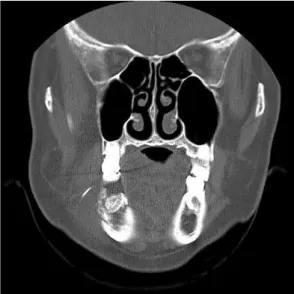
Fig. 3. CT image reveals separated lamina dura
on socket of Fig. 2. and buccal soft
tissue swelling.
Fig. 4. Dental implants treatment was done on edentulous space.
리적 가설을 주장하였다. 이로 인해 마치 의치에 의한 만성적 외상에서처럼 연조직 병소의 치유 가 지연된다고 하였다. 일련의 연구에서
15-20)BRONJ와 연관된 몇가지 위험요소에 관해 언급 된 것들이 있으며 이는 발치기왕력, 비스포스포 네이트 투약 기간, 비스포스포네이트 약제의 유 형등의 연관성들이다. 가장 주된 것은 최근의 치 과에서의 수술과 연관성이다(Fig. 4와 5). Fig. 4 의 환자는 임플란트 식립과 보철수복을 비교적 최근에 받은 환자로 비스포스포네이트를 장기간 복용중이었다. 연조직의 부종, 동통과 구강내 누 공의 형성으로 수술적인 처치를 하였음에도 장 기간 치유가 되지 않았으며 골수염과 같은 증상 으로 발전되었다. 경부의 누공이 발생되었고 하 악우측 입술 및 턱부위의 마비가 있었다. 4차례 수술과 투약으로 해당부위의 임플란트와 부골 (Fig. 6)이 제거되었다. 병리학적 소견에서는 괴 사된 골조직, 염증세포의 침윤 등이 관찰되었다.
괴사된 골은 정상적인 골세포가 사라지고 주변 부 흡수와 세균증식이 동반되었다.
조직학적으로 노출된 골조직에 괴사골과 육아 조직뿐 아니라 세균 집락중 Actinomyces의 증가, 괴사골 주변으로 유사상피 과증식 등이 보고되 었으나
10)골수염자체가 비스포스포네이트와 관 련된 골괴사에 의해 이차적으로 발생하는지 아 니면 일차감염이 골괴사를 유발하는 것인지는
Fig. 5. Osteomyelitis-like symptoms and signs were evoked and surgical intervention was conducted.
Fig. 6. Removed sequestra.
확실하지 않은 것으로 알려져 있다.
2007년 미국 구강악안면외과학회에서는 이러
한 BRONJ의 임상적 상황과 치료에 있어서 가이
드라인을 정비하였다
21). 비스포스포네이트 투약
중인 환자군에 있어서 치과적 특히 관혈적 시술
을 받아야 하는 환자는 명백한 골변화가 없더라
도 BRONJ의 발현성에 대해 교육이 되어져야 한
다고 하였고, 제 1 형인 증상이 없으나 골노출,
부골이 발견되고 염증의 증거가 없는 경우에는
항균구강세정요법을 동반한 매 4개월마다 주기
적 내원을 권고하였다. 감염을 동반하고 동통과
부종이 있는 경우인 제 2 형은 광범위 항생제 투
약, 항균구강세정요법, 동통조절, 연조직을 자극 하는 골 표층부 제거등을 시행하고 누공형성, 파 절 등을 포함하는 광범위한 질환의 진행기인 제 3 형은 위의 보존적 요법과 함께 수술적인 절제 도 고려해야 함을 권고하였다. 현재 추천되고 있 는 것은 과도한 외과적 처치를 피하라는 것이다.
즉 외과적 수술의 결과를 예측하기 어려울 뿐만 아니라 오히려 상태의 악화를 초래할 수도 있다 는 이유 때문인데 이는 치료전에 환자와 의사가 모두 인식해야 하는 것으로 치료의 목적이 반드 시 BRONJ를 없애고 완치를 하는 것만은 아니라 는 것이다. 상태에 따라 통증과 감염을 조절하고, 골괴사 진행을 억제함으로써 보다 장기적이고 보존적 관점을 갖도록 하는 것이다. 질환자체의 조절이 중요하다는 개념이다. 그렇지만, 치과적 처치시 필요한 특별한 예방적 조치는 일반적인 상식수준을 넘지 않는다. 구강위생에 철저하고 BRONJ의 발현가능성에 관한 사전고지정도이다.
비스포스포네이트 투약을 미리 예단하여 중지한 다 하더라도 어느 정도의 기간이 유효한지도 밝 혀져 있지 않은 실정이다. 아직 병리학적 기전과 임상적 증상의 확실한 연관성 등이 규명되지 않 았으므로 이를 극복하기 위한 많은 연구가 필요 하다.
결 론
비스포스포네이트는 파골세포의 골대사기전 및 작용에 관여하는 강력한 골흡수억제제로 가 장 보편적으로 사용되고 있으나 최근에 일부 환 자에서 악골괴사가 발생하는 증례보고가 증가하 고 있다. 하지만, 아직 정확한 발생기전은 모르 는 실정이므로 관혈적 치과시술을 계획하는 치 과의사들은 환자병력, 기왕력, 복용약물 등에 대 한 점검이 필요하고 필요한 술전 검사를 시행하 며 최대한의 위생적인 구강환경을 만드는 것이 중요하다. 또한 환자에게 충분한 설명과 내과의 와의 협진도 수행하여 예방을 하려는 노력이 필 요하다.
참 고 문 헌
1. Wu S, Dahut WL, Gulley JL. The use of bisphos- phonates in cancer patients. Acta Oncol 2007;46:581- 91
2. Wang J, Goodger NM, Pogrel MA. Osteonecrosis of the jaws associated with cancer chemotherapy. J Oral Maxillofac Surg 2003;61:1104-7
3. Rosenberg TJ, Ruggiero S. Osteonecrosis of the jaws associated with the use of bisphosphonates. J Oral Maxillofac Surg 2003;61:60 (letter)
4. Marx RE. Pamidronate (Aredia) and zoledronate (Zometa) induced avascular necrosis of the jaws: a growing epidemic. J Oral Maxillofac Surg 2003; 61:
1115-7
5. Migliorati CA. Bisphosphonates and oral cavity avascular bone necrosis. New Engl J Med 2003;21:
4253-4 (letter)
6. Ruggiero SL, Mehrotra B, Rosenberg TJ, et al.
Osteonecrosis of the jaws associated with the use of bisphosphonates: a review of 63 cases. J Oral Maxillofac Surg 2004;62:527-34
7. Lugassy G, Shaham R, Nemets A, et al. Severe osteomyelitis of the jaw in long-term survivors of multiple myeloma: a new clinical entity. Am J Med 2004;117:440-1
8. Bagan JV, Murillo J, Poveda R, et al. Avascular jaw osteonecrosis in association with cancer chemotherapy: series of 10 cases. J Oral Pathol Med 2005;34:120-3
9. Vannucchi AM, Ficarra G, Antonioli E, et al.
Osteonecrosis of the jaw associated with zoledronate therapy in a patient with multiple myeloma. Br J Haematol 2005;128:738
10. Yarom N, Yahalom Y, Shoshani, et al. Osteonecrosis of the jaw induced by orally administered bisphos- phonates: incidence, clinical features, predisposing factors and treatment outcome. Osteoporos Int 2007;18(10):1363-70
11. Lehrer S, Montazem A, Ramanathan L, Pessin-
Minsley M, Pfail J, Stock RG, et al. Normal serum
bone markers in bisphosphonate-induced
osteonecrosis of the jaws. Oral Surg Oral Med Oral
Pathol Oral Radiol Endod 2008; 106 : 389-91.12.
Ficarra G, Beninati F. Bisphosphonate-related osteonecrosis of the jaws: an update on clinical, pathological and management aspects. Head Neck Pathol 2007; 1 : 132-40.
13. Kos M, Kuebler JF, Luczak K, Engelke W.
Bisphosphonate related osteonecrosis of the jaws: a review of 34 cases and evaluation of risk. J Craniomaxillofac Surg 2010; 38 : 255-9.14. Reid IR, Bolland MJ. Is bisphosphonates-associated osteonecrosis of the jaw caused by soft tissue toxicity? Bone 2007;41:318-20
15. Migliorati CA, Schubert MM, Peterson DE, et al.
Bisphosphonate-associated osteonecrosis of mandibular and maxillary bone: an emerging oral complication of supportive cancer therapy. Cancer 2005;104:83-93
16. Merigo E, Manfredi M, Meleti M, et al. Jaw bone necrosis without previous dental extractions associated with the use of bisphosphonates (pamidronate and zoledronate): a four- case report. J Oral Pathol Med 2005;34:613-7
17. Olson KB, Hellie CM, Pienta KJ. Osteonecrosis of jaw in patient with hormone-refractory prostate cancer treated with zoledronic acid. Urology 2005;66:658
18. Zervas K, Verrou E, Teleioudis Z, et al. Incidence, risk factors and management of osteonecrosis of the jaw in patients with multiple myeloma: a single- centre experience in 303 patients. Br J Haematol 2006;134:620-3
19. Sanna G, Preda L, Bruschini R, et al. Bisphos- phonates and jaw osteonecrosis in patients with advanced breast cancer. Ann Oncol 2006;17:1512–6 20. Badros A, Weikel D, Salama A, et al. Osteonecrosis of the jaw in multiple myeloma patients: clinical features and risk factors. J Clin Oncol 2006;24:945- 52
21. American Association of Oral and Maxillofacial
Surgeons position paper on bisphosphonates-related
osteonecrosis of the jaws. J Oral Maxillofac Surg
2007;65:369-76
Bisphophonate-Related Osteonecrosis of the Jaw (BRONJ)
Hyeon-Mook Kim*, Chan-Jin Park
*Sunplus Dental Clinic,
Department of Prosthodontics and Institute of Oral Science, College of Dentistry, Gangneung-Wonju National University Recently, jawbone osteonecrosis has been largely reported as a potential adverse effect of bisphosphonate (BP)administration. Currently available published incidence data for BRONJ are based on retrospective studies and estimates of cumulative incidence range from 0.8 to 12%. The mandible is more commonly affected than the maxilla (2:1 ratio), and 60–70% of cases are preceded by a dental surgical procedure. The signs and symptoms that may occur before the appearance of clinical evident osteonecrosis include changes in the health of periodontal tissues, non-healing mucosal ulcers, loose teeth and unexplained soft-tissue infection. Tooth extraction as a precipitating event is a common observation. The significant benefits that bisphosphonates offer to patients clearly surpass the risk of potential side effects; however, any patient for whom prolonged bisphosphonate therapy is indicated, should be provided with preventive dental care in order to minimize the risk of developing this severe condition.
Key words: bisphosphonate, dental treatment, osteonecrosis, BRONJ
Correspondence to : Chan-Jin Park
Department of Prosthodontics, College of Dentistry, Gangneung-Wonju National University, 7 Jukheongil, Gangneung city, Gangwon-do, South Korea, 210-702
Tel: +82-33-640-3153, Fax: +82-33-640-3113, E-mail: [email protected]
Received: November 10, 2011, Last Revision: December 03, 2011, Accepted: December 25, 2011