Effects of the Applications of Chitin and Chitosan on Soil Organisms
Jinu Eo, Myung-Hyun Kim, Soon-Kun Choi, Hea-Son Bang*, and Kee-Choon Park1 Climate Change and Agroecology Division, National Academy of Agricultural Science
1National Institute of Horticultural & Herbal Science, Rural Development Administration
(Received: November 3 2014, Revised: February 3 2015, Accepted: April 21 2015)
Effects of chitin and chitosan treatments on soil microorganisms and the mesofauna were investigated in a microcosm and a fumigated field experiment. Responses of microorganisms were determined using microbial phospholipid fatty acid (PLFA) analysis, whereas responses of the mesofauna were measured in terms of the abundances of nematodes and microarthropods. Soil nitrate concentration increased on the application of chitin. Overall, chitin promoted bacterial and fungal abundance, leading to an increase in abundance of free-living soil nematodes that feed on decomposers. The ratio of saturated to unsaturated fatty acids was highest in the chitin-treated soil. Chitosan had a minimal effect on the abundance of microorganisms;
however, it reduced the abundance of collembolans in the microcosm experiment. These results indicate that the application of chitin has beneficial effects on the supply of nutrients and promotion of the abundance of soil organisms.
Key words: Microarthropod, Nematode, PLFA
Changes in soil biota as affected by the applications of chitin and chitosan.
Microorganisms Nematodes Microarthropods
Bacteria Fungi Bacterivorous Fungivorous Collembolans Mites ---- PLFA ng g-1 ---- --- ind g-1 --- --- ind 100 mL-1 ---
Control 11.3b 2.3b 0.57b 0.18ab 1.18a 1.29b
Chitin 19.1a 6.4a 3.78a 0.34a 0.97a 4.41a
Chitosan 10.7b 1.3b 0.35b 0.06b 0.43a 0.43b
1)
*Corresponding author : Phone: +82632382525, E-mail: [email protected]
§Acknowledgement: This study was carried out with the support of “Research Program for Agricultural Science & Technology Development (Project No. PJ008608)”, National Academy of Agricultural Science, Rural Development Administration, Republic of Korea.
Introduction
키틴은 N-acetyl-D-glucosamine이 연결된 구조로 되어 있으며, 생태계에서 다당류로는 셀루로스 다음으로 많이 존 재한다. 갑각류와 곰팡이의 세포벽을 구성하는 물질이며 (Nicol, 1991), 새우나 게와 같은 갑각류로부터 대량으로 분 리한다 (Abdou et al., 2008). 키토산은 키틴의 탈아세틸화 에 의해 생성되며, 곰팡이나 접합균류의 세포벽에 포함되어 있다 (Somashekar and Joseph, 1996). 자연생태계에서는 키틴보다 적게 존재하며 산업적으로 농업, 식품 및 약품 등 에 사용된다 (Gooday, 1990). 일반적으로는 키틴에 강염기 를 첨가하여 키토산을 생산하고, 곰팡이로부터 직접 추출하거 나 미생물과 효소를 이용하여 제조하는 방법도 있다 (Abdou et al., 2008).
키틴과 키토산은 토양생물에게 먹이원이 되며, 이차적으 로는 분해된 물질이 식물의 양분으로 이용되기 때문에 농업 의 토양관리 측면에서 활용할 수 있다. 세균처럼 키틴을 포 함하고 있지 않는 생물들도 키틴분해효소를 생성하여, 유기 물을 분해하거나 자신을 보호하는데 이용한다 (Oranusi and Trinci, 1985). 일부 응애류는 소화기관에 미생물이 공생하 면서 토양의 키틴을 분해한다 (Smrz and Catska, 2010). 키 토산을 분해하는 효소는 세균, 곰팡이 및 식물에 존재하며, 대 부분의 세균은 이 효소를 생산한다 (Somashekar and Joseph, 1996).
이들 유기물은 작물의 생육 증진과 토양 병해충에 의한 피해를 경감시키는 목적으로도 활용된다 (Bell et al., 1998).
키틴을 토양에 처리할 경우 작물의 생육이 증진하거나 그 구성성분이 변하는 효과도 있다 (Liopa-Tsakalidi et al., 2010). 키틴은 토양 미생물상을 변화시켜 토양 병원균을 억 제하고 (Cretoiu et al., 2014), 키틴분해효소를 분비하는 미 생물을 촉진하여 식물기생성 선충을 감소시키는 효과도 있 다 (Sarathchandra et al., 1996). 키토산도 식물병원성 세 균과 곰팡이를 억제하고 (Cheah et al., 1997; Rabea and Steurbaut, 2010), 식물기생성 선충의 피해를 감소시킨다 (Khalil and Badawi, 2012). 또한, 키토산을 식물에 처리하 면 병원균을 억제하는 물질의 생성을 유도하는 효과가 있다 (Walker et al., 2003).
유기물 투입을 통해 양분의 공급, 토양 이화학성 개선 및 토양유래 병원균 억제 등을 촉진하기 위해서는 토양 미생물 뿐만 아니라 미소동물의 역할도 중요하다 (Wickings and Grandy, 2013). 자활성 선충은 분해자인 세균이나 곰팡이를 섭식하여 물질순환에 참여하고, 병원균을 직접적으로 억제 하는 기능을 갖고 있다 (Mikola and Sulkava, 2001). 미소 절지동물 중에 톡토기나 응애도 물리적으로 유기물 분해를 촉진하고, 토양에 서식하는 병원균을 섭식하여 병억제력에 영향을 미친다 (Filser, 2002). 또한, 미생물과 미소동물은
먹이망 구조를 통해 상호작용하기 때문에, 유기물 시용의 전체적인 효과를 알기 위해서는 이들을 동시에 연구할 필요 가 있다.
본 연구에서는 키틴과 키토산이 토양이화학성과 생물상 에 미치는 영향을 알아보기 위하여 두 가지 실험으로 구분 하여 수행하였다. 마이크로코즘 실험에서는 유기물이 국지 적으로 미치는 영향을 경시적으로 조사하였으며, 훈증포장 실험에서는 이들의 단기적 영향을 구명하는 것을 목표로 하 였다.
Materials and Methods
마이크로코즘 실험 상하가 개방된 원통모양의 용기 (지름 8 cm, 높이 10 cm)를 이용하여 토양을 채워 넣고 포 장에 매설하는 방법으로 마이크로코즘을 설치하였다. 키틴 은 4 Mg ha-1로 토양과 혼합하여 매설 전에 처리하였고, 키 토산은 매설 후 2L ha-1의 농도로 토양표면에 2주 간격으로 5회 살포하였다. 실험은 난괴법 5반복으로 수행하였다. 토 양이화학성과 생물상의 변화를 알아보기 위해 4, 10, 18 주 후 용기 안의 토양을 모두 수거하여 이화학성과 생물상을 조사하였다.
훈증포장 실험 훈증을 하기 위해 다조멧을 400 kg ha-1 으로 처리한 후 비닐을 피복하였고 3주 후에 피복을 제거하 였다. 키틴은 4 Mg ha-1로 투입한 후 표토에서 약 30 cm 깊 이로 로터리로 경운하여 토양에 혼입하였다. 키토산은 2L ha-2의 농도로 토양 표면에 2주 간격으로 3회 살포하였다.
처리구는 난괴법 3반복 (각 5 × 6 m)으로 배치하였다. 토양 시료는 처리시부터 8주 후에 각 처리구별로 0-10 cm 깊이 에서 토양을 채취하여 조사하였다.
토양화학성 및 미생물체량 분석 상온에서 건조하여 보관한 토양 시료를 이용하여 이화학성을 분석하였다. pH와 EC는 초자전극법으로 측정하였고, 유효인산함량은 Lancaster 법으로 측정하였다. 토양내 질산태질소는 2 M KCl로 추출후 켈달분석법에 의해 질소분석기 (K-314, Büchi, Swizerland) 로 분석하였다. 치환성 양이온함량은 1 N NH4OAc (pH 7.0) 으로 침출시킨 후 그 여과액을 ICP (Integra XL DUAL, GBC Scientific Equipment, USA)를 이용하여 측정하였다.
토양미생물 분석 토양 미생물의 인지질지방산 (PLFA, phospholipid fatty acid)은 Li et al. (2006)의 방법을 따라 분석하였다. 냉동 건조한 토양시료 5 g에 chloroform (4 mL), methanol (8 mL), buffer solution (3.2 mL, pH 7.4)을 혼 합하여 지질을 추출한 후 silicic acid column으로 neutral-, glyco- 및 phospho-lipid로 분리하였다. MIDI Sherlock
Table 1. Chemical properties of soil as affected by the applications of chitin and chitosan in the microcosm experiment.
pH EC OM NO3- Av. P2O5 K+ Mg2+ Ca2+
ds m-1 g kg-1 mg kg-1 mg kg-1 --- Ex. cation (cmol+ kg-1) ---
Control 5.9a 0.2b 4.5a 2.4b 65.2a 0.2a 0.6b 5.6a
Chitin 5.7b 0.3a 4.3a 21.2a 47.0b 0.2a 0.6a 4.4b
Chitosan 5.9a 0.2b 4.5a 3.6b 64.9a 0.2a 0.6b 5.3a
Values indicated by the same letter are not significantly different according to Fisher’s LSD test (P < 0.05). Data are average of three sampling dates.
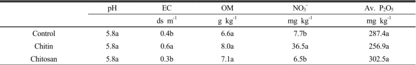
Table 2. Chemical properties of soil as affected by the applications of chitin and chitosan in the fumigated field experiment.
pH EC OM NO3- Av. P2O5
ds m-1 g kg-1 mg kg-1 mg kg-1
Control 5.8a 0.4b 6.6a 7.7b 287.4a
Chitin 5.8a 0.6a 8.0a 36.5a 256.9a
Chitosan 5.8a 0.3b 7.1a 6.5b 302.5a
Values indicated by the same letter are not significantly different according to Fisher’s LSD test (P < 0.05).
Microbial Identification System (MIDI Inc., Newark, DE) 으로 지방산을 정성 및 정량 하였다. 각 지방산의 값은 19:00 지방산을 표준으로 이용하여 계산하였다. 단불포화 지방산 은 16:1 ω5c, 17:1 ω8c, 18:1 ω7c, 포화지방산은 14:00, 15:00, 16:00, 17:00, 18:00, 20:00을 지표로 사용하였다. 호기성균 은 16:1 ω7t, 혐기성균은 cy19:0을 사용하였다. 그램 음성균 의 지표 지방산은 18:1 ω7c, 19:0cy ω8c, 17:1 ω8c, 그램양 성균은 i14:0, i15:0, a15:0, i16:0, i17:0, a17:0, 세균은 그 램 음성균과 양성균을 모두 이용하였다. 곰팡이는 18:2 ω 6,9c, 방선균은 10Me16:0, 10Me17:0, TBSA10Me18:0, 균근 균은 16:1 ω5c을 지표로 이용하였다.
미소동물 조사 토양 미소동물은 자활성 선충과 미소절 지동물로 나누어 조사하였다. 자활성 선충의 밀도를 측정하기 위해 20 g의 토양을 이용하여 베르만깔대기법에 의해 48시간 동안 추출 후 TAF (2% triethanolamine, 2.8% formaldehyde) 용액에 시료를 보관하였다. 보관하였던 선충을 광학현미경 으로 관찰하면서 식성에 따라 세균섭식성과 곰팡이섭식성 으로 분류하였다 (Okada, 2002). 미소절지동물은 350 mL 의 토양으로 툴그렌 장치를 이용하여 72시간 동안 추출하였 다. 추출된 시료는 광학현미경으로 관찰하면서 톡토기와 응 애로 분류하였다.
통계분석 유기물이 미생물 PLFA 및 미소동물에 미치 는 영향은 Fisher’s LSD 검정법으로 분석하였다. 마이크로 코즘 실험에서 토양 이화학성과 PLFA지표는 세 번 조사한 결과의 경향이 비슷하였기 때문에 이들의 평균값으로 나타 냈다. 모든 통계과정은 XLSTAT (Addinsoft, Brooklyn, NY, USA)를 이용하여 수행하였다.
Results and Discussion
토양 이화학성 마이크로코즘실험과 훈증포장실험에서 유사한 경향이 나타났다 (Table 1, Table 2). 마이크로코즘 실험에서 대부분의 토양화학성의 경시적 변화는 시기별로 비슷한 경향이었기 때문에 평균값만을 나타냈다 (Table 1).
다만, 질산태질소의 농도는 4주 후에 55.3 mg kg-1로 증가 하였다가 10주 후부터는 대조구와 비슷하였다. 토양 EC와 질산태질소의 함량이 키틴처리구에서 증가하였고, 유효태 인산은 마이크로코즘 실험에서 키틴처리구에서 감소하였 다. 키틴 투입이 토양 화학성에 미치는 영향에 대한 보고는 미미하나, 키틴과 그 부산물들은 질소 함량이 6.1~8.3%로 비교적 높기 때문에 (Yen and Mau, 2007) 미생물에 의해 분해될 경우 질소질 양분 공급에 유용하다. 키틴은 미생물 밀도를 증가시키며, 호기적 호흡에 의해 분해되어 암모니아 화와 질산화 과정을 거쳐서 이산화탄소와 질산으로 변한다 (Wieczorek et al., 2014). 본 연구결과에서도 질산태질소 농도 증가가 뚜렷하였기 때문에 이러한 추측을 뒷받침하는 근거가 된다. 한편, 유효태인산의 감소는 작물의 생육에 영 향을 줄 수 있기 때문에 이에 대해서는 유의할 필요가 있다.
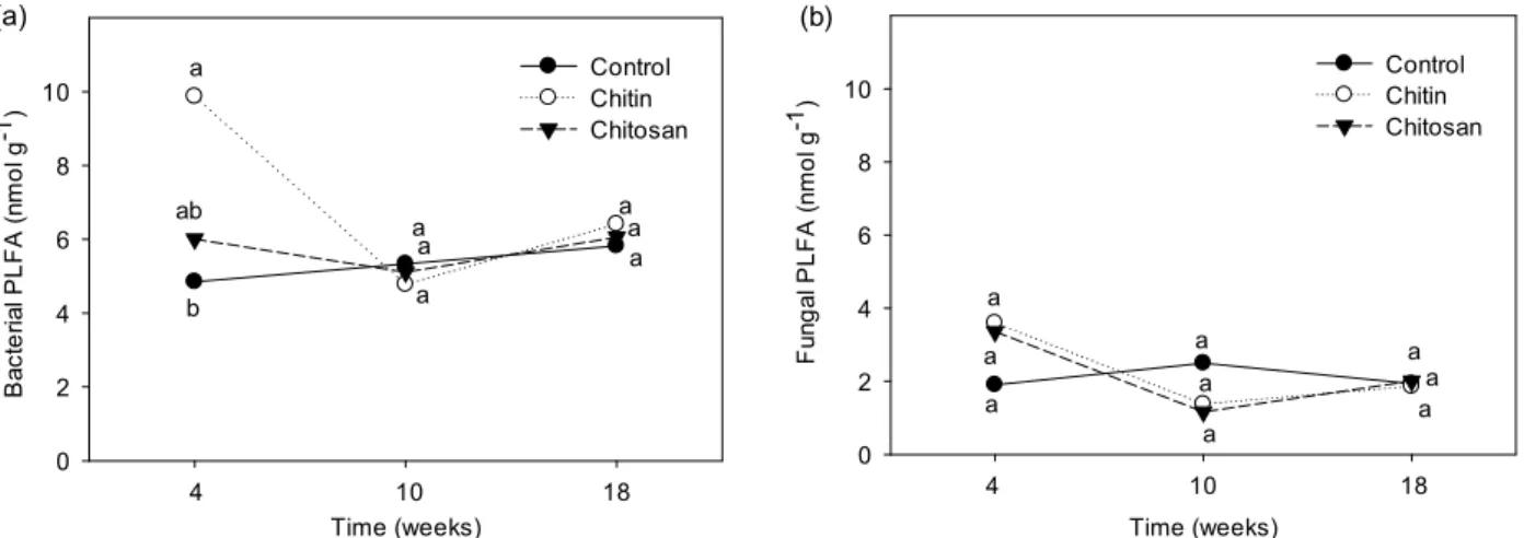
키틴이 미생물에 미치는 영향 마이크로코즘 실험의 키틴처리구에서 세균 밀도가 4주 후에 증가하였으나, 그 이 후로는 차이가 없었다 (Fig. 1). Russell (2014)은 키틴이 토 양에서 쉽게 분해될 수 있으며, 투입 후 수 일 안에 토양호 흡이 64% 증가하고, 암모늄이 32% 증가하였다고 보고하였 다. 또한, 적절한 토양 환경에서는 일반 토양유기물보다 분 해되는 속도가 3배 빠르다 (Vorobev et al., 2007). 따라서, 키틴처리구에서 10주 후에 차이가 없었던 이유는 투입된 키
Time (weeks) Bacterial PLFA (nmol g-1)
0 2 4 6 8
10 Control
Chitin Chitosan
4 10 18 a
ab
b (a)
a a a
a aa
Time (weeks) Fungal PLFA (nmol g-1)
0 2 4 6 8
10 Control
Chitin Chitosan
4 10 18 (b)
a a a
a a a
a a a
Fig. 1. Changes in microbial PLFA as affected by chitin and chitosan in the microcosm experiment. (a) bacterial PLFA; (b) fungal PLFA. Values indicated by the same letter are not significantly different according to Fisher’s LSD test (P < 0.05).
Table 3. Indicators of microbial phospholipid fatty acid (PLFA) in the microcosm experiment.
Gram -/
Gram +
Aerobic/
anaerobic
Saturated/
unsaturated
Cyclo/
precursor
Control 0.9ab 3.1a 1.2ab 0.4a
Chitin 0.7b 2.6a 1.3a 0.4a
Chitosan 1.1a 3.9a 1.1b 0.3a
Values indicated by the same letter are not significantly different according to Fisher’s LSD test (P < 0.05). Data are average of three sampling dates.
Table 4. Changes in soil biota as affected by chitin and chitosan in the fumigated field experiment.
Microorganisms Nematodes Microarthropods
Bacteria Fungi Bacterivorous Fungivorous Collembolans Mites --- PLFA ng g-1 --- --- ind g-1 --- --- ind 100 mL-1 ---
Control 11.3b 2.3b 0.57b 0.18ab 1.18a 1.29b
Chitin 19.1a 6.4a 3.78a 0.34a 0.97a 4.41a
Chitosan 10.7b 1.3b 0.35b 0.06b 0.43a 0.43b
Values indicated by the same letter are not significantly different according to Fisher’s LSD test (P < 0.05).
틴이 미생물에 의해 조기에 분해되어 단기적인 효과에 국한 되었던 것으로 보인다.
일반적으로 미생물 PLFA 지표 중에서 그램 음성균/양성 균의 비율이 증가하면 양분이 많아지는 상태를 나타낸다 (Kaur et al., 2005). 키틴처리구에서는 기질의 증가에도 불 구하고 키토산처리구보다 이 비율이 낮았는데 (Table 3), 토 양이나 유기물의 구성성분에 따라 토양미생물의 반응이 다 를 가능성이 있다. 포화/불포화 지방산 비율은 환경스트레 스의 증가에 따라 높아지며, 키토산처리구보다 키틴처리구 에서 이 비율이 높았다. 이것은 토양 pH 저하 등에 의한 환 경스트레스 차이 때문인 것으로 생각된다.
훈증포장 실험에서도 키틴 처리구에서 미생물 밀도가 뚜렷
하게 증가하였다 (Table 4). 키틴은 세균과 방선균의 밀도를 증 가시킨다 (Bell et al., 1998). 특히 키틴에 의해 Streptomyces와 같은 방선균이나 Oxalobacteraceae 등의 세균이 증가하는 것으로 보고되었다 (Cretoiu et al., 2014; Jacquiod et al., 2013; Ueno and Miyashita, 2000). 본 실험에서는 곰팡이의 밀도도 증가하였으며 (Table 4), 곰팡이 중에서는 Mortierella 나 Fusarium 등이 키틴을 분해한다고 알려져 있다 (De Boer et al., 1998).
곰팡이 밀도에 대해서는 두 실험의 결과가 서로 달랐는 데 (Table 4, Fig. 1), 훈증한 토양에서는 대부분의 생물이 소멸하므로 복원하는 과정에서 유기물 투입에 대한 토양 미 생물의 반응에 차이가 있었던 것으로 추측된다. 또한, 미생 물에 의한 키틴의 분해는 토성이나 온도에 따라 다르며 (Manucharova, 2009), 토양함수량에도 영향을 받으므로 (Vorobev et al., 2007) 시기별로 차이가 나타날 수 있다.
키토산이 미생물에 미치는 영향 키토산이 세균과 곰 팡이에 미치는 영향은 두 실험 모두에서 처리간 차이가 없 었다 (Fig. 1, Table 4). 키토산이 키틴과 비교하여 영향이 적 었다는 결과는 기존의 발표와 일치한다 (Bell et al., 1998).
또한, 키토산은 유기물분해에 대해서도 영향을 미치지 않았 을 수 있다 (Wieczorek et al., 2014). 키틴은 대부분의 곰팡 이에 존재하지만 키토산은 접합균류에서만 발견되는 것에
Time (weeks) Bacterivorous nematode ( ind g-1)
0 2 4 6 8 10
Control Chitin Chitosan
4 10 18 a
ab b (a)
a a
a
a a a
Time (weeks) Fungivorous nematode ( ind g-1)
0 2 4 6 8 10
Control Chitin Chitosan
4 10 18 a
b ab (b)
a a a
a a a
Time (weeks) Collembolans ( ind 100 mL-1)
0 2 4 6 8 10
Control Chitin Chitosan
4 10 18 a
a
ab (c)
a a a
a a a
Time (weeks) Mites ( ind 100 mL-1)
0 2 4 6 8 10
Control Chitin Chitosan
4 10 18 (d)
aa a
a a
a aaa
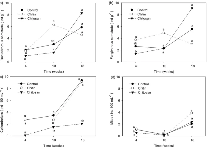
Fig. 2. Changes in abundances of mesofauna as affected by chitin and chitosan in the microcosm experiment. (a) bacterivorous nematode; (b) fungivorous nematode; (c) collembolans; (d) mites. Values indicated by the same letter are not significantly different according to Fisher’s LSD test (P < 0.05).
서 알 수 있듯이 키틴은 일반적으로 키토산보다 자연생태계 에서 많이 존재하기 때문에 토양에 투입할 경우 토양 미생 물은 키틴에 더 쉽게 반응할 수 있다 (Wieczorek et al., 2014).
키틴이 미소동물에 미치는 영향 마이크로코즘 실험 에서 키틴처리구는 대조구와 비교하여 세균섭식성 선충 밀 도는 10주차에 높았고, 곰팡이섭식성 선충의 밀도는 4주차 에 높았다 (Fig. 2). 키틴이 식물 기생성 선충에 대해 억제 효과가 있다는 보고는 많이 있으나 (Chen et al., 1999), 자 활성 선충에 대한 보고는 미미하다. 세균섭식성 선충의 경 우 이들의 먹이가 되는 세균의 밀도가 4주차에 높았기 때문 에, 먹이원의 증가에 의한 연쇄적인 반응으로 생각할 수 있 다. 또한, 훈증포장 실험의 키틴 처리구에서도 동일한 결과 가 관찰되었다 (Table 4).
키토산이 미소동물에 미치는 영향 마이크로코즘 실 험의 키토산 처리에서 톡토기의 밀도가 실험기간 중 계속적 으로 가장 낮았다 (Fig. 2). Mitschunas et al. (2006)은 키 토산이 곰팡이 생장 억제를 통한 먹이원 감소로 톡토기의 밀도를 간접적으로 줄이거나 또는 직접적으로 억제하는 기
작이 있다고 추측하였다. 하지만 본 연구에서는 토양 곰팡 이의 밀도는 대조구와 비교하여 차이가 없었기 때문에 먹이 량 변화에 따른 이차적인 반응이라기 보다는 직접적인 영향 이 있었던 것으로 추측된다.
Conclusion
키틴과 키토산이 토양이화학성과 토양생물에 미치는 영 향을 알아보기 위하여 마이크로코즘과 훈증포장에서 실험 을 수행하였다. 두 실험에서 유기물 처리에 대한 토양미생 물과 미소동물의 반응은 비슷하게 나타났다. 대체로 키틴은 토양생물의 밀도를 증가시켰으나, 이에 비해 키토산의 영향 은 미미하였다. 키틴의 투입으로 유기물을 분해하는 미생물 의 증가와 함께 이를 섭식하는 미소동물이 증가한 것으로 추측된다. 또한, 키틴에 의해 토양 화학성 중 질산태질소 농 도와 EC가 증가하였다. 따라서 키틴은 토양관리측면에서 양분공급뿐만 아니라 토양생물 밀도를 증진시키는 목적으 로 활용할 수 있을 것으로 판단된다. 한편, 키틴은 근권 미 생물상도 변화시키기 때문에 (Hallmann et al., 1999) 작물 생육과 관련하여도 추가적인 연구가 필요하다.
References
Abdou, E.S., K.S.A. Nagy, and M.Z. Elsabee. 2008. Extraction and characterization of chitin and chitosan from local sources.
Bioresour. Technol. 99:1359-1367.
Bell, A.A., J.C. Hubbard, L. Liu, R.M. Davis, and K.V.
Subbarao. 1998. Effects of chitin and chitosan on the incidence and severity of Fusarium yellows of celery. Plant Disease 82:
322-328.
Cheah, L.H., B.B.C. Page, and R. Shepherd. 1997. Chitosan coating for inhibition of Sclerotinia rot of carrots. N.Z. J. Crop and Hort. Sci. 25:89-92.
Chen, J., G.S. Abawi, and B.M. Zuckerman. 1999. Suppression of Meloidogyne hapla and its damage to lettuce grown in a mineral soil amended with chitin and biocontrol organisms. J.
Nematology 31(4S):719-725.
Cretoiu, M.S., A.M. Kielak, A. Schluter, and J.D. Van Elsas.
2014. Bacterial communities in chitin-amended soil as revealed by 16S rRNA gene based pyrosequencing. Soil Biol. Biochem.
76:5-11.
De Boer, W., P.J.A. Klein Gunnewiek, P. Lafeber, J.D. Janse, B.E. Spit, and J.W. Woldendorp. 1998. Antifungal properties of chitinolytic dune soil bacteria. Soil Biol. Biochem. 30:
193-203.
Filser, J. 2002. The role of collembola in carbon and nitrogen cycling in soil. Pedobiologia 46:234-245.
Gooday, G.W. 1990. The ecology of chitin degradation. Adv.
Microb. Ecol. 11:387-430.
Hallmann, J., R. Rodriguez-Kabana, and J.W. Kloepper. 1999.
Chitin-mediated changes in bacterial communities of the soil, rhizosphere and within roots of cotton in relation to nematode control. Soil Biol. Biochem. 31:551-560.
Jacquiod, S., L. Franqueville, S. Cecillon, T.M. Vogel, and P.
Simonet. 2013. Soil bacterial community shifts after chitin enrichment: An integrative metagenomic approach. PLOS 11:e79699.
Kaur, A., A. Chaudhary, A. Kaur, R. Choudhary, and R. Kaushik.
2005. Phospholipid fatty acid - a bioindicator of environment monitoring and assessment in soil ecosystem. Cur. Sci.
89:1103-1112.
Khalil, M.S., and E.I. Badawi. 2012. Nematicidal activity of a biopolymer chitosan at different molecular weights against root-knot nematode. Plant Prot. Sci. 48:170-178.
Li, W.H., C.B. Zhang, H.B. Jiang, G.R. Xin, Z.Y. Yang. 2006.
Changes in soil microbial community associated with invasion of the exotic weed, Mikania micrantha HBK. Plant Soil 281, 309-324.
Liopa-Tsakalidi, A., D. Chalikiopoulos, and A. Papasavvas.
2010. Effect of chitin on growth and chlorophyll content of two medicinal plants. J. Med. Plants Res. 4:499-508.
Manucharova, N.A. 2009. The microbial destruction of chitin, pectin, and cellulose in soils. Eurasian Soil Sci. 42:1526-1532.
Mikola, J., and P. Sulkava. 2001. Responses of microbial-feeding nematodes to organic matter distribution and predation in experimental soil habitat. Soil Biol. Biochem. 33:811-817.
Mitschunas, N., M. Wagner, and J. Filser. 2006. Evidence for a positive influence of fungivorous soil invertebrates on the seed bank persistence of grassland species. J. Ecol. 94:791-800.
Nicol, S. 1991. Life after death for empty shells. New Scientist 129:46-48.
Okada, H., 2002. Role of nematodes in soil ecosystems-effects on dynamics of inorganic nitrogen. Root Res. 11:3-6.
Oranusi, N.A., A.P.J. Trinci. 1985. Growth of bacteria on chitin fungal cell walls and fungal biomass, and the effect of extracellular enzymes produced by these cultures on the antifungal activity of amphotercin B. Microbios 43:17-30.
Rabea, E.I., and W. Steurbaut. 2010. Chemically modified chitosans as antimicrobial agents against some plant pathogenic bacteria and fungi. Plant Prot. Sci. 46:149-158.
Russell, A.E. 2014. Unexpected effects of chitin, cellulose, and lignin addition on soil dynamics in a wet tropical forest.
Ecosystem 17:918-930.
Sarathchandra, S.U., R.N. Watson, N.R. Cox, M.E. Di Menna, J.A. Brown, G. Burch, and F.J. Neville. 1996. Effects of chitin amendment of soil on microorganisms, nematodes, and growth of white clover (Trifolium repens L.) and perennial ryegrass (Lolium perenne L.). Biol. Fert. Soils 22:221-226.
Smrz, J., and V. Catska. 2010. Mycophagous mites and their internal associated bacteria cooperate to digest chitin in soil.
Symbiosis 52:33-40.
Somashekar, D., and R. Joseph. 1996. Chitosanases – Properties and applications: a review. Bioresour. Technol. 55:35-45.
Ueno, H., and K. Miyashita. 2000. Inductive production of chitinolytic enzymes in soil microcosms using chitin, other carbon-sources, and chitinase-producing streptomyces. Soil Sci. Plant Nutr. 46:863-871.
Vorobev, A.V., N.A. Manucharova, A.M. Yaroslavtsev, E.V.
Belova, D.G. Zvyagintsev, and I.I. Sudnitsyn. 2007. The composition of the chitinolytic microbial complex and its effect on chitin decomposition at various humidity levels. Microbiology 76:557-562.
Walker, T.S., H.P. Bais, K.M. Halligen, F.R. Stermitz, and J.M.
Vivanco. 2003. Metabolic profiling of root exudates of Arabidopsis thaliana. J. Agri. Food Chem. 51:2548-2554.
Wickings, K., and A.S. Grandy. 2013. Management intensity interacts with litter chemistry and climate to drive temporal patterns in arthropod communities during decomposition.
Pedobiology 56:105-112.
Wieczorek, A.S., S.A. Hetz, and S. Kolb. 2014. Microbial responses to chitin and chitosan in oxic and anoxic agricultural soil slurries. Biogeosciences 11:3339-3352.
Yen, M.T., and J.L. Mau. 2007. Selected physical properties of chitin prepared from shiitake stipes. Food Sci. Technol. 40:
558-563.