국민건강보험공단 일산병원 내과, 병리과, 영상진단 의학과, 연세대학교 의과대학 내과학교실 나형중, 김승업1, 김도현, 남동혁, 김정주, 이선민, 기정혜2, 홍용국3
A Case of Bronchiolitis Obliterans Organizing Pneumonia from Epstein-Barr Virus
Hyoung Jung Na, M.D., Sueng Up Kim, M.D.1, Do Hyun Kim, M.D., Dong Hyug Nam, M.D., Sun Min Lee, M.D., Chong Ju Kim, M.D., Jeong-Hae Kie, M.D.2, Yong Kug Hong, M.D.3
Department of Internal Medicine and Pathology2, Radiology3, National Health Iinsurance Corporation Ilsan Hospital, Koyang, Korea Department of Internal Medicine, Yonsei University College of Medicine, Seoul, Korea1
In the average adult with a normal immune state, Epstein-Barr virus pneumonia is very rare, especially in the form of interstitial lung disease. According to recent studies, the Epstein-Barr virus is also associated with lymphocytic interstitial pneumonia, AIDS and Langerhans cell histiocytosis, but not with sarcoidosis.
BOOP is caused by lung injury due to an infection or drug intoxication, and is related to connective tissue disease or bone marrow transplantation, but is sometimes idiopathic. We experienced a patient with symptoms and signs of interstitial lung disease, with confirmed BOOP and EBV ingection from an open lung biopsy and serologic examination, respectively Herein, this case is reported, with a review of the literature. (Tuberc Respir Dis 2007; 62: 51-55) Key words: Bronchiolitis obliterans organizing pneumonia, Epstein-barr virus.
Address for correspondence: Chong Ju Kim, M.D.
Department of Internal Medicine, National Health Insurance Corporation Ilsan Hospital 1232, Paeksok- dong, Ilsan-donggu, Koyang-shi, Kyunggi-do, 410-719, Korea
Phone: 82-31-900-0237 Fax: 82-31-900-0343 E-mail: [email protected]
Received: Oct. 26. 2006 Accepted: Dec. 7. 2006
서 론
기질화 폐렴을 동반한 폐쇄성 기관지염(bronchio- litis obliterans organizing pneumonia: BOOP)은 다 양한 비특이성 폐손상에 대해 말초 세기관지와 폐포 관 내에 과도한 육아조직의 증식과 더불어 염증세포 의 간질내 침윤 및 주위 폐포관 및 폐포내로 기질화 폐렴 형성등의 만성 염증 소견을 보이는, 폐렴과 유사 한 특징을 갖는 질병으로, 임상적병리적인 증후군이 다1. BOOP은 감염증, 약물독성에 의한 폐손상에 기인 하거나, 결체조직 질환, 폐나 골수이식2과 동반되지만 원인을 모르는 경우도 많으며 이러한 경우를 특발성 간질성 폐렴(cryptogenic organizing pneumonia:
COP)이라 한다. BOOP을 일으키는 감염증 중 신이식 환자에서 Pneumocystis carinii 감염증3,4, capsule-
deficient cryptococcosis5 에 의한 발생 보고가 있고 바이러스로는 파라인플루엔자바이러스(parainfluenza virus) 또는 아데노바이러스(adenovirus)가 원인이 될 수 있는 것으로 알려져 있으나6 Epstein-Barr virus 에 대해서는 아직 보고된 경우가 없다.
정상 면역 상태의 성인에서 Epstein-Barr virus (EBV)에 의한 폐렴은 드물며, 특히 간질성 폐질환의 양상으로 오는 경우 또한 매우 드물다. EBV 감염과 관련된 간질성 폐질환은 특발성 폐섬유증(Idiopathic pulmonary fibrosis: IPF), 림프구성 간질성 폐렴 (Lymphocytic interstitial pneumonia: LIP), AIDS, 및 호산구성 육아종 등이며 유육종증과는 관련이 없다7. 이에 저자들은 간질성 폐렴의 양상으로 내원하여 개흉 폐생검을 통하여 BOOP 양상이 확인되고 이의 원인으로 혈청학적 검사를 통해 EBV 감염증을 확인 한 환자를 경험하였기에 문헌고찰과 함께 보고하는 바이다.
증 례 환 자: 김OO, 여자 51세 주 소: 기침, 가래, 전신 쇠약감
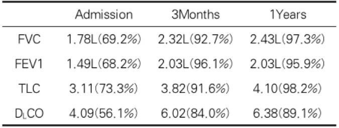
Admission 3Months 1Years FVC 1.78L(69.2%) 2.32L(92.7%) 2.43L(97.3%) FEV1 1.49L(68.2%) 2.03L(96.1%) 2.03L(95.9%) TLC 3.11(73.3%) 3.82(91.6%) 4.10(98.2%) DLCO 4.09(56.1%) 6.02(84.0%) 6.38(89.1%) Table 1. Follow up findings of PFT values
A B
Figure 1. On H-E section, loose branching fibroblastic proliferations are observed within the terminal airway and alveolar spaces (A, H-E, ×200) with relatively mild interstitial inflammatory cell infiltration (B, H-E, ×400).
현병력: 평소 건강하게 지내던 51세 여자가 내원 2 개월 전부터 시작된 기침, 가래, 전신 쇠약감, 피로 및 호흡곤란을 주소로 내원하였다.
과거력 및 가족력: 당뇨, 결핵등 특이 과거력은 없 었고 흡연력, 직업력 및 가족력상 특이 소견은 없었 다.
신체검사 소견: 내원 당시 신체 검진상 혈압 110/70 mmHg, 맥박수 80회/분, 호흡수 20회/분, 체온은 36.
2℃ 이었다. 흉부 청진상 양측 폐 하부에서 흡기말에 가는 수포음이 청진되었으며 심장 청진에서는 특이 소견은 없었다. 복부 팽만은 없었으며 장음도 정상이 었으며 목이나 액와부에 만져지는 림프절은 없었다.
검사실 소견: 혈액학 검사에서 백혈구 8,600/mm3, 혈색소 13.8 g/dL, 헤마토크릿 40.7%, 혈소판 289,000/
mm3 이었다. 혈청 생화학 검사에서 나트륨 140 mEq/L, 칼륨 4.1 mEq/L, 염소 111.0 mEq/L, 총이산 화탄소 21.9 mEq/L, 혈액요소질소 18 mg/dL, 크레아 티닌 0.7 mg/dL이었다. 동맥혈 검사에서 pH 7.39,
Pco2 41.1 mmHg, Po2 77.6 mmHg, HCO3- 24.6 mmol/L, 산소 포화도 95.6%였다. 객담 AFB 도말 및 배양검사는 음성이었고 ANA 및ANCA(antineutro- phil cytoplasm antibody), 류마티스 인자는 음성이었 고 HIV 항체도 음성이었다.
방사선학적 소견: 흉부 방사선검사상 양측 폐야에 반점형 경화성 병변을 보이는 폐렴 소견이었고, 흉부 전산화 단층촬영상 양측 폐야에 간유리 음영과 폐경 결 소견, 망상형 혼탁과 경도의 기관지 확장 소견이 관찰되었다.
폐기능 검사 소견: 폐기능 검사는 FVC 1.78L (69.2%), FEV1 1.49L(68.25%), FEV1/FVC 83.5%, DLCO 56.1% 로 경증 제한성 환기장애와 증등도의 폐확산능 감소 소견을 보였다(Table 1).
치료 및 경과: 정확한 진단을 위하여 8일째 개흉 폐 생검 시행하였고 비 특이적인 BOOP 소견을 보였다 (Figure 1). 혈청 생화학적 검사상 EBV-EA IgM 항 체 15 U/mL(정상 < 8 U/mL), EBV-VCA IgM 항체 15 U/mL(정상 < 8 U/mL) 로 양성 소견을 보였고 EBV-VCA IgG 항체 124 U/mL로 양성(정상 < 8 U/mL), EBNA 항체 7 U/mL로 음성(정상 < 8 U/mL) 소견을 보였다. 단순포진바이러스(herpes simplex virus: HSV), 풍진바이러스(rubella virus), 수두대상 포진바이러스(varicella zoster vrus: VZV), 아데노바 이러스, 및 거대세포바이러스(cytomegalovirus: CMV) 에 대한 혈청검사는 모두 음성이었고, 다른 감염증의
Admission
EBV-EAlgM Positive(15)
EBV-VCAlgM Positive(15)
Table 2. Follow up findings of serum anti EBV immunoglobulin(U/mL)
A B
Figure 2. Chest radiograph shows diffuse ground glass opacity and reticular opacity in the both lungs (A). Chest PA after 5 months shows resolution of diffuse ground glass and reticular opacity in the both lungs (B).
A B C
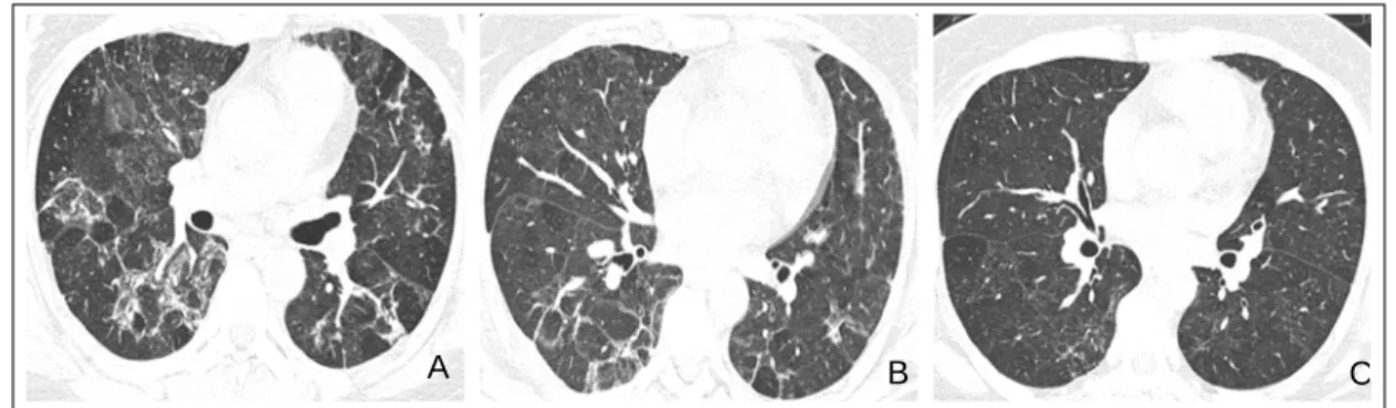
Figure 3. Axial CT image shows diffuse patch distributed ground glass opacity and consolidation along the bronchovascular bundles (A). Three week follow-up Chest CT shows improvement of ground glass opacity and consolidation in the both lungs (B). Follow-up Chest CT after 1 year demonstrates resolution of diffuse pulmonary infiltration. Mild ground glass opacity and reticular opacity are remained (C).
증거는 발견되지 않았다. 따라서, EBV 감염증에 의한 BOOP으로 진단하고, 프레드니솔론 30mg부터 치료 시작하여 2개월 간 투약 후 추적 흉부 방사선 검사상 호전되는 양상 보였고 폐기능 검사상 정상소견으로 회복하였다(Table 1). 이후 추적 흉부 방사선 검사, 흉 부 CT 촬영 상 명확한 호전이 관찰되었으며(Figure 2, 3), 6개월 추적 검사에서 EBV-EA IgM항체,
EBV-VCA IgM 항체는 borderline 양성이었고, 1년 후 추적검사는 모두 음전되었다(Table 2).
고 찰
기질화 폐렴을 동반한 폐쇄성 기관지염(bronchio- litis obliterans organizing pneumonia : BOOP)은 대 개 40~60대에서 호발하며 남녀의 차이는 없고 흡연 력과는 무관하다. 대부분이 2~3개월 전에 발열, 기침, 무력감 등의 감기 몸살 증상이나 피로-쇠약감, 체중 감소 등의 증상으로 시작되며, 마른기침이 지속되며 진찰상 흡기성 수포음이 들리고, 폐기능 검사상으로 는 제한성 병변과 함께 저산소증이 관찰된다. 방사선
학적으로 양 폐야의 주변 부위에 미만성 폐포성 침윤 이 대부분에서 관찰되며 흉부 전산화 단층촬영 (HRCT)상에서도 폐 주변부, 기관지 중심성으로 경결 성 음영이나 간유리 음영이 산발적으로 관찰된다. 말 초 폐야의 반상 음영은 삼각형 모양을 하고 있는데, 삼각형의 기저부는 흉막에 접해 있으며 이것이 BOOP의 특징적인 전산화 단층촬영 소견이 된다8. 이 러한 BOOP은 감염증, 약물독성에 의한 폐 손상에 기 인하거나, 결체조직 질환, 폐나 골수이식과 동반되지 만 원인을 모르는 경우도 많다. 감염증으로는 Chlamydia, Legionella, Mycoplasma pneumoniae 등 이 원인이 될 수 있고, 특히 바이러스로는 파라인플루 엔자바이러스 및 아데노바이러스가 알려져 있다6. BOOP의 진단은 임상적 증상과 방사선학적 소견, 병 리학적으로 기질화된 미만성 폐포 손상의 소견이 보 일 때 가능하다. BOOP은 환자의 1/3정도가 재발하거 나 반응이 없으며 사망률은 5%정도로 알려져 있고 UIP와는 달리 스테로이드 치료로 환자의 2/3에서 완 전 치유를 보이기 때문에 반드시 정확한 진단이 필요 하다.
일반적으로 EBV에 의한 감염증은 여려 종류의 양 성 또는 악성 종양처럼 면역 억제 상태에서 잘 발생하 며 EBV-EA IgM 항체, EBV-VCA IgM 항체 소견을 참고로 진단을 내리며 최근에는 EB viral load를 이용 하는 진단법이 사용되고 있다. 가장 흔하게 이용되는 두 가지 기법으로는 quantitative PCR과 real-time PCR을 들 수 있으며 이들 검사법이 환자의 혈액 내에 서 EB virus의 DNA를 검출하는데 있어서 감수성과 특이성이 뛰어난 방법임이 입증되었다. 본 증례에서 처럼 정상 면역 상태의 성인에서 EBV에 의한 폐렴은 드물며, 특히 간질성 폐질환의 양상으로 오는 경우는 매우 드물다. EBV 감염과 관련된 간질성 폐질환은 특 발성 폐섬유증, 림프구성 간질성 폐렴, AIDS, 및 호산 구성 육아종 등이며 유육종증과는 관련이 없다7. 양측 폐야의 경화나 미만성 질환을 보인는 환자들은 감염 의 다른 원인을 배제하기 위해 기관지 내시경을 통한 세포학적 검사, 진균배양 검사 등 세균학적 검사가 행 해져야 한다.
본 증례에서는 흉부 방사선검사상 양측 폐야에 반
점형 경화성 병변을 보이는 폐렴소견이었고, 흉부 전 산화 단층촬영상 양측 폐야에 간유리 음영과 폐경결 소견, 망상형 혼탁과 경도의 기관지 확장 소견이 관찰 되어 특발성 간질성 폐렴의 감별진단을 위하여 개흉 폐생검을 시행하였다. H-E 염색상 느슨한 가지의 섬 유 아세포 증식이 말초 기도와 폐포에서 관찰되었고 간질성 세포 침윤이 보여 BOOP으로 진단할 수 있었 으며(Figure 1) 다른 원인이 의심되는 소견은 없었다.
다른 감염증의 소견이 없으면서 혈청 생화학적 검사 상 EBV 감염증을 보였고 추적 검사상 호전 소견 보 여 EBV 감염증에 의한 BOOP으로 판단되었다. 건강 한 사람에게서 EBV 감염에 의한 간질성 폐렴 양상은 매우 드물고 특히 BOOP형태로 확인된 경우는 없어 보고하는 바이다.
요 약
임상적으로 BOOP을 시사하는 경우 여러가지 감염 증, 그 중에서도 EBV에 의한 감염증도 고려하여야 한 다고 생각한다.
참 고 문 헌
1. Kim KS, Lee YM, Choi YS, Shin JH, Han GJ. Moon SH, et al. 2 case of idiopathic BOOP associated with rare radiologic finding. Tuberc Respir Dis 1996;43:
228-35.
2. Dodd JD, Muller NL. Bronchiolitis obliterans organi- zing pneumonia after bone marrow transplantation. J Comput Assist Tomogr 2005;29:540-3.
3. Ahn MS, Koh YM, Shin J, Jeong HB, Lee SE, Chung YT. A case of Pneumoncystis carinii pneumonia with histopathologic finding of bronchiolitis obliterans organizing pneumonia in patient with AIDS. Tuberc Respir Dis 1998;45:444-50.
4. Verma N, Soans B. Cryptogenic organizing pneu- monia associated with Pneumocystis carinii infection and sirolimus therapy in a renal transplant patient.
Australas Radiol 2006;50:68-70.
5. Chantraumat C, Sittipunt C. Bronchiolitis obliterans organizing pneumonia caused by capsule-deficient cryptococcosis. Southeast Asian J Trop Med Public Health 2005;36:174-7.
6. Epler GR. Bronchiolitis obliterans organizing pneu-
monia. Arch Intern Med2001;161:158-64.
7. Marzouk K, Corate L, Saleh S, Sharma OP. Epstein- Barr-virus-induced interstitial lung disease. Curr Opin Pulm Med 2005;11:456-60.
8. Costabel U, Teschler H, Schoenfeld B, Hartung W, Nusch A, Guzman J, et. al. BOOP in Europe. Chest 1992;102(Suppl):14S-20S.