Ⅰ. 서 론
타액선 종양은 전체 타액선 질환의 약 20%를 차지하며 타액 상피 실질(parenchymal) 또는 주위의 간질(간엽조직, mesenchy-
mal)에서 유래된다. 다형성선종(Pleomorphic adenoma)은 실질
기원 종양으로 가장 호발 하는 타액선 양성 종양이며 현미경 적 소견에서 주로 도관구조를 구성하는 상피 세포와 소성의 점액양 간질에 높은 비율로 존재하는 근상피 세포 및 간엽조직 성분을 포함하는 매우 다양한 소견을 보여준다1,2)
.
선양낭성 암종(Adenoid cystic carcinoma)은 대, 소타액선에서 발생하는 악성 타액선 종양으로, 침습성이 매우 강하고, 신경 주위의 침윤이 특히 심한 악성종양으로서, 전체 악성 종양 중, 대타액선에서는 4.4%, 소타액선에서는 1.2%의 빈도를 보이며 외과적 절제술의 치료 후 높은 재발률을 보인다. 조직병리학 적으로 주로 사상체(cribriform)형을 이루고 있으며, 도관형
(tubular form), 충실소(solid nest)형 등 3가지 형태로 구성되어 있
고 세포구조는 기저양세포(basaloid cell), 또는 근상피세포(myoepithelial cell)로 구성되어 있다
3,4,5). 또한 선양낭성 암종의
임상적 양상이나 예후는 그 암종의 조직학적 형태와 관련있다 고 알려져 왔다6,7).
상피성장인자(Epidermal growth factor, EGF)는 53 amino-acids 로 구성된 single-chain polypeptide로써, 세포 표면의 수용체
(EGFR)와 결합하여 다양한 형태의 세포들의 증식을 유도할 수
다형성 선종과 선양낭성 암종에서 상피성장인자 발현에 관한 연구
박승구∙한세진∙김철환∙김경욱 단국대학교 치과대학 구강악안면외과학교실
Abstract (J. Kor. Oral Maxillofac. Surg. 2008;34:245-249)
김 경 욱
330-716
충남 천안시 안서동29
단국대학교 치과대학 부속병원 구강외과 Kyung-Wook KimDept. of OMFS, College of Dentistry, Dankook University, 29 Amseodong, Choenan, Chungnam, 330-716, Korea Tel: 82-41-550-1994 Fax: 82-41-550-8988 E-mail: [email protected]
THE STUDY OF EGF EXPRESSION BETWEEN HUMAN PLEOMORPHIC ADENOMA AND ADENOID CYSTIC CARCINOMA
Seung-Gu Park, Se-Jin Han, Chul-Hwan Kim, Kyung-Wook Kim Dept. of Oral and Maxillofacial Surgery, College of Dentistry, Dankook University
Epidermal growth factor is a single-chain polypeptide consisting of 53 amino acids and has a potent mitogenic activity that stimulates proliferation of various normal and neoplastic cells through the interaction with its specific receptor(epidermal growth factor receptor, EGFR).
Pleomorphic adenoma is the most common salivary benign tumor and histologically, it contains the epithelial cell, the myo-epithelial cell and mes- enchymal ingredient, which is various aspect. Adenoid cystic carcinoma is an infiltrative malignant salivary gland tumor with three different histologi- cal patterns: cribriform, tubular or solid. The tumor cell structure composed of modified myoepithelial cell, and basaloid cell. In this study, we used an immunohistochemical technique to investigate the expression of EGF in 6 specimens of adenoid cystic carcinoma and 10 specimens of pleomorphic adenoma taken from patients treated at Dept. of Oral and Maxillofacial Surgery, Dankook University.
The results were as follows.
1. In pleomorphic adenoma, ductal structure and scattered spindle cells in hyalinized stroma, disclosing myxoid stroma and hyalin, cartilage forma- tion were observed. Immunohistologically, weak EGF expression in ductal structure and negative in stromal area were observed.
2. Cribriform type of adenoid cystic carcinoma showed numerous pseudocyst surrounded by dark small neoplastic cells in the back-ground of fibrous connective tissue and moderate EGF expression of dark cells adjacent to pseudo lumen in cribriform pattern, while weak expression in other most cells.
3. Tubular type of adenoid cystic carcinoma showed numerous ductal pattern surrounded by two layered neoplastic cells in the back-ground of fibrous connective tissue and strong EGF expression in luminal cells of ductal structure, while weak expression in outer cells.
From the results obtained, we suggest that EGF is mainly biosynthesized in cells forming duct like structures of tubulo-ductal type or cribriform adenoid cystic carcinoma and it may play a role, as a cell mitogen in adenoid cystic carcinoma growth.
Key words: Epidermal growth factor, Pleomorphic adenoma, Adenoid cystic carcinoma
있는 강력한 분열 유발성 능력을 갖고 있는 단백질이다.
Cohen(1962) 등이 생쥐의 악하선으로부터 처음으로 추출하였
으며 이후 사람의 urine, 타액, 세포외액에서도 추출되게 되었 고 많은 관련 연구가 시행되었다8,9).
상피성장인자수용체(epidermal growth factor receptor, EGFR)는 다양한 세포의 세포막에 존재하는 분자량 170 kDa의 막투과성 당단백질이다10)
. 상피성장인자수용체는 상피의 증식이 빠르
게 일어나는 여러 종류의 상피세포와 상피조직에서 관찰된다11).
상피성장인자와 세포의 세포막에 존재하는 상피성장인자수 용체와의 결합은 수용체의 세포 내 domain의 tyrosinekinase를 활성화시킨다. 이와 같이 활성화된 tyrosine kinase는 downstream 기질을 인산화시킴으로써 궁극적으로는 DNA의 복제와 세포 분열을 야기 시키며, 이런 특성이 종양 세포에서 증식 및 발암 기전에 중요한 역할을 하는 것으로 생각되고 있다. 이에 저자 는 사람의 다형성 선종과 선양낭성 암종에서 상피성장인자(EGF) 발현 양상을 관찰하고 종양형성에 어떠한 관계가 있는
지 알아보고자 하였다.Ⅱ. 연구 재료 및 방법 1. 연구 재료
연구 대상으로는 단국대학교 구강악안면외과에서 수술 시 행한 타액선 종양 중 조직병리학적으로 확진된 다형성 선종 10 례와 선양낭성암종 6례의 조직표본을 대상으로 하였으며 그 중, 선양낭성암종은 조직병리학적 형태에 따라 사상형 3례, 도 관형 3례로 분류하여 실험하였다.
2. 연구 방법
(1) H & E 염색
채취된 종양을 절취하여 10% 중성 포르말린에 고정 후 4㎛ 파 라핀 절편을 제작한 후, 통법에 따라 H & E 염색을 시행하고 광 학현미경하에서 각 표본의 조직병리학적 특징을 관찰하였다.
(2) 면역조직화학적 검사
절취한 조직을 고정한 후 Poly-L-Lysine으로 처리된 슬라이드 에 4~8㎛ 파라핀 절편을 제작한 후, 면역조직화학적 염색을 시 행하기 위하여 절편을 0.3% H2
O
2에 5분 간 부란시키고 10mMPBS 로 세척하여 제 1차 항체인 Polyclonal anti-EGF Ab(Oncogene Science, U.S.A.)를 사용하였다.
PBS로 세척하고 LSAB(labelled streptavidine biotin)법으로 처리
후, 발색제는 Diaminobenzidine을 이용하였으며 Mayer’sHematoxylin으로 대조염색 시행하였다.
염색된 각 조직표본을 광학현미경으로 관찰 후, 각각의 상피 성장인자(EGF) 발현 양상을 관찰하여 다음과 같이 기록하였 다(Table 1).
Ⅲ. 연구 결과 1. H & E 염색 소견
다형성 선종은 2가지의 우세한 분화 형태인 도관과 근상피 형태가 특징이며, 관구조의 내강은 입방세포로 이장되어 있으 며, 방추형의 신장된 근상피세포가 결합 조직의 유리질화 된 간질에 넓게 퍼져있는 양상을 나타내었다(Fig. 1).
Table 1.
EGF Staining Nomenclature(-) Negative
(+) Weak : positive staining of 10% or less of tumor cells
(++) Moderate : positive staining of 11-50%
(+++) Strong : positive staining of more than 50%
Table 2.
Immunohistochemical Staining of EGF in Normal Salivary GlandCell type Striated duct Intercalated duct Excretory duct Acinus cells
EGF + + - -
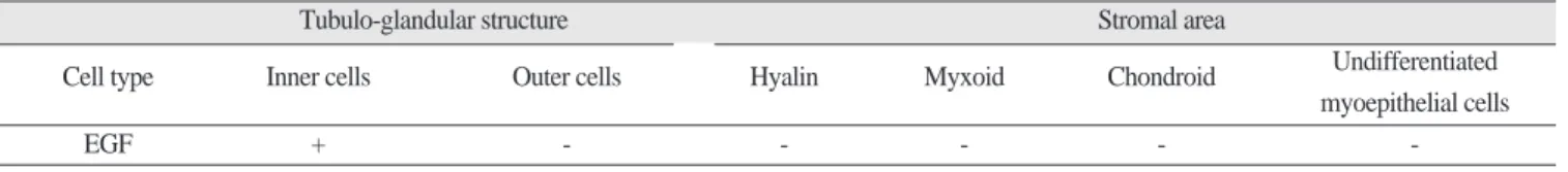
Table 3.
Immunohistochemical Staining of EGF in Pleomorphic AdenomaTubulo-glandular structure Stromal area
Cell type Inner cells Outer cells Hyalin Myxoid Chondroid Undifferentiated
myoepithelial cells
EGF + - - - - -
Table 4.
Immunohistochemical Staining of EGF in Adenoid Cystic CarcinomaCribriform type Tubulo-ductal type
Cell type Pseudocystic area cells Other area cells Inner cells Outer cells
EGF ++ + +++ +
Fig. 1. H & E staining findings in pleomorphic adenoma (×100).
Fig. 2. H & E staining findings in cribriform type of adenoid cystic carcinoma(×100).
Fig. 3. H & E staining findings in tubulo-ductal type of adenoid cystic carcinoma(×100).
Fig. 4. EGF staining immunohistochemical findings in pleomorphic adenoma(×200).
Fig. 5. EGF staining immunohistochemical findings in cribriform type of adenoid cystic carcinoma(×200).
Fig. 6. EGF staining immunohistochemical findings in tubulo- ductal type of adenoid cystic carcinoma(×200).
선양낭성 암종의 사상형(cribriform type)은 다수의 소낭이 수 많은 원주로 분리되어 스위스 치즈 양상을 보이며, 소낭 주위 로 둘러싸는 세포들을 관찰할 수 있었다. 관상형(tubulo-ductal
type)에서는 유리질화 된 기질 사이로 1-3층의 기저양 세포로
둘러싸여 있는 다수의 작은 관 구조를 관찰하였다(Fig. 2, 3).2. 면역조직화학적 검사 소견
1) 정상 타액선
정상 타액선의 선조도관 및 개재도관에서 EGF 양성반응을 보였으나, 분비도관 및 도관내세포에서는 음성반응을 보였 다(Table 2).
2) 다형성 선종
다형성 선종에서는 도관 내세포와 도관을 이루는 세포에 양 성 반응을, 산재하는 근상피세포, 점액성 조직, 초자양 물질 및 초자양 지역에서는 음성반응을 보였다(Fig. 4, Table 3).
3) 선양낭성 암종
선양낭성 암종의 사상형에서는 가성낭 조직을 둘러싸는 세 포에서 EGF 양성반응을 보였고 사상체를 이루는 종양세포에 서는 산재되어 미약한 양성반응을 나타냈으며, 도관형에서는 두 층의 종양세포 중 도관 내세포에서 강한 EGF 양성반응을 보였다(Fig. 5, 6, Table 4).
Ⅳ. 총괄 및 고찰
포유류의 세포 성장과 분화는 다양한 성장인자들에 의해 조
절 된다11)
. 성장인자는 세포의 특정한 수용기와 상호작용 하는
단백질로서, 표현형의 조절, 세포의 운동성과 세포골격구조의 변화, 세포 증식 속도의 변화 등을 포함하는 다양한 생물학적 반응들을 야기한다. 성장인자는 특정 세포에서만 생산되는 것 이 아니라 여러 종류의 세포에서 생산 되며, 다양한 범위의 서 로 중복되는 생물학적 기능을 가지고 있고 일반적으로 비교적 짧은 거리에서 작용한다. 대부분 포유류 세포들은 여러 가지 성장인자를 분비 한다12-14)
.
상피성장인자(Epidermal growth factor, EGF)는 53 amino-acids 로 구성된 single-chain polypeptide로써, 세포 표면의 수용체
(EGFR)와 결합하여 다양한 형태의 세포들의 증식을 유도할 수
있는 강력한 분열 유발성 능력을 갖고 있는 단백질이다15,16). Cohen등이 생쥐의 악하선으로부터 처음으로 추출하였으며 이
후 사람의 urine, 타액, 세포외액에서도 추출되게 되었고 많은 관련 연구가 시행되었다8,9,17,18).
상피성장인자수용체(epidermal growth factor receptor, EGFR)는 다양한 세포의 세포막에 존재하는 분자량 170 kDa의 막투과성 당단백질이다10)
. 상피성장인자수용체는 상피의 증식이 빠르
게 일어나는 여러 종류의 상피세포와 상피조직에서 관찰 된다11,19)
. 상피성장인자와 세포의 세포막에 존재하는 상피성장인
자수용체와의 결합은 수용체의 세포 내 domain의 tyrosinekinase 를 활성화시킨다. 이와 같이 활성화된 tyrosine kinase는 down-
stream 기질을 인산화시킴으로써 궁극적으로는 DNA의 복제
와 세포 분열을 야기 시킨다. 상피성장인자는 상피성장인자 수용체와의 결합으로 세포의 증식을 촉진시키는데 중요한 역 할을 한다. 이러한 성장인자 생산의 증가는 구강에서 발생하 는 암종의 발암기전 및 암종의 성장에 중요한 역할을 할 것으 로 생각 된다20).
구강 편평상피 세포암종12)
, 위암
21), 타액선 종양
22)등에서 상피 성장인자의 발현이 관찰되어왔다. Tahara 등은 위암에서의 상 피성장인자 발현 연구를 통해 상피성장인자 발현 양성인 위암 환자가 음성인 환자보다 그 예후가 더 좋지 않았다고 하였으 며 상피성장인자가 위암의 침습적 성장에 중요한 역할을 하며위암 환자의 악성도를 판단하는 생체 인지자(biologic marker)로 서 기여할 수 있다고 주장하였다21)
.
Mori 등은 타액선에 발생한 선양낭성 암종에서의 상피성장
인자 발현을 처음으로 보고하였다22). 선양낭성 암종(Adenoid cystic carcinoma)은 대, 소타액선에서 발생하는 악성 타액선 종
양이다. 침습성이 매우 강하고, 신경주위의 침윤이 특히 심한 악성종양으로서, 주로 50대 후반에 호발하며 전체 악성 종양 중, 대타액선에서는 4.4%, 소타액선에서는 1.2%의 빈도를 보 이며 이하선, 악하선에서 가장 흔한 발병을 나타내고, 구강내 의 구개부, 구강저의 소타액선에서도 발생한다23). 외과적 절제
술 후, 임상적으로 5년 생존율은 비교적 높지만, 초기 치료 후10~15년 내에 국소전이와 혈류전이로 인하여 약 40%이상 재
발하는 낮은 완치율을 가진 암종이다3-5).
선양낭성암종은 조직병리학적으로 주로 사상체(cribriform) 형을 이루고 있으며, 도관형(tubular form), 충실소(solid nest)형 등으로 구성되어 있다. 세포구조는 기저양세포(basaloid cell), 또는 근상피세포(myoepithelial cell)로 구성되어 있다. 각각의 암 종세포군 내에서 수많은 위낭 공간을 형성하여 사상체 형태를 가지는 것이 특징적인데, 가성낭은 근상피세포 형태의 암종 세포에 둘러싸인 모양을 하고 있고 기저막 성분을 포함하는 세포외 기질로 채워져 있다24-28)
. 가성낭 내부에는 거의 세포성
분은 없으나 암종 세포에서 세포 간극으로 유리되는 세포외기 질 성분들로 구성되어 있다29-30).
이번 연구에서는 선양낭성 암종의 상피성장인자 발현이 암 종의 가성낭 또는 도관형 구조에서 양성 반응을 나타내었는데 이들 구조물은 암종의 조직병리학적 특성을 나타내는 것들로 써 상피성장인자가 이들을 만들어내는 세포들로부터 생성되 었다고 생각된다. 대표적 타액선 양성 종양인 다형성 선종의 경우, 도관을 형성하는 세포에서만 미약한 상피성장인자 발현 양성 반응을 나타내었다는 사실과 비교했을 때, 선양낭성 암 종의 상피성장인자를 만들어내는 가성낭, 도관형 구조 생성 세포들이 암종의 분화와 성장에 중요한 역할을 하리라 여겨 진다.
Ⅴ. 결 론
선양낭성 암종은 침습적이고 재발 경향이 높은 타액선 악성 종양으로 상피성장인자(EGF) 발현이 종양의 특성과 연관되어 있다고 추정되며, 이에 사람의 다형성 선종과 선양낭성 암종 에서 상피성장인자(EGF) 발현 양상을 관찰, 비교하기 위해 단 국대학교 구강악안면외과에서 수술 시행한 타액선 종양 중 조 직병리학적으로 확진된 다형성 선종 10례와 선양낭성 암종 6 례의 조직표본을 대상으로 면역조직화학적 검사를 시행하여 다음과 같은 결과를 얻었다.
1. 다형성 선종에서는 도관 내세포와 도관을 이루는 세포에
양성 반응을, 산재하는 근상피세포, 점액성 조직, 초자양 물질 및 초자양 지역에서는 음성반응을 보였다.2. 선양낭성 암종의 사상형에서 가성낭 조직을 둘러싸는 세
포에서 EGF 양성반응을 보였고 사상체를 이루는 종양세 포에서는 산재되어 미약한 양성반응을 나타냈다.
3. 선양낭성 암종의 도관형에서는 두 층의 종양세포 중 도관
내세포에서 강한 EGF 양성반응을 보였다.이상의 소견으로 다형성 선종에서는 EGF가 도관을 형성하 는데 국한되어 있지만, 선양낭성 암종의 사상형에서는 비록 근상피세포 기원 종양세포의 형성에는 무관하였으나 가성낭 을 만드는데 관여하며 도관형에서는 도관 형성에 관여하는 것 으로 생각되었다. 이에 EGF 발현은 타액선에 발생하는 선양낭 성 암종의 형성과 성장에 밀접한 관계가 있으리라 사료되 었다.
참고문헌