2020 Keimyung University School of Medicine
Tumor Size is Associated with Long-term Outcomes after Resection of Gastric
Gastrointestinal Stromal Tumors
Kyung In Shin, Ju Yup Lee, Yoo Jin Lee, Kyung Sik Park
Department of Internal Medicine, Keimyung University School of Medicine, Daegu, Korea
Introduction
Gastrointestinal stromal tumors (GISTs) are the most common tumors of me- sodermal origin in the gastrointestinal (GI) tract [1]. GISTs originate from the in- terstitial cells of Cajal in the muscular layer of the GI tract. Activating mutations in oncogenes such as KIT and PDGFRA are known to be associated with the devel- opment of GISTs [2,3]. GISTs account for less than 1% of all malignant tumors in the GI tract, and while they occur most commonly in the stomach (70%), they can occur anywhere in the GI tract, including the small intestine (20-30%) or the rec- tum/colon (10%) [4]. The standard treatment for GISTs, including gastric tumors, is surgical resection. The aim of surgery is complete resection to ensure that no tu- mor is left in the resection margin and to avoid rupture of the tumor, which can increase the risk of recurrence [5]. However, the clinical course after surgical re- section varies greatly. While GISTs can have a very slow, benign course, they often progress very rapidly even after meticulous surgical resection [6,7]. This is because the prognosis for gastric GISTs is affected by tumor size and mitotic index [8,9].
To date, there have been almost no reports on the long-term prognosis, including recurrence, after surgical resection of gastric GISTs. Therefore, in this study, we aimed to analyze the long-term therapeutic outcomes and prognoses of surgically resected gastric GISTs according to tumor size.
Received: April 6, 2020 Accepted: April 28, 2020 Corresponding Author:
Ju Yup Lee, M.D.
Department of Internal Medicine, Keimyung University School of Medicine, Dongsan Medical Center, 1035 Dalgubeol- daero, Dalseo-gu, Daegu 42601, Korea Tel: +82-53-258-4321
Fax: +82-53-258-4343 E-mail: [email protected] pISSN 2092-8335 · eISSN 2733-5380 Keimyung Med J 2020 39(1):28-32 https://doi.org/10.46308/kmj.2020.00073
Original Article
The clinical outcomes after surgical resection of gastrointestinal stromal tumors (GISTs) vary widely due to the differences in tumor size and mitotic index. To ana- lyze the long-term outcomes and prognosis of surgically resected gastric GISTs ac- cording to tumor size. We retrospectively analyzed the medical records of 269 pa- tients who underwent surgery for GISTs at Keimyung University Dongsan Hospital from March 2000 to March 2017. We surveyed tumor size, mitotic index, recurrence after surgery, time to recurrence, treatment for recurrence, and mortality. The risk of recurrence of gastric GISTs was classified as very low, low, intermediate, and high risk according to the 2007 Journal of the national comprehensive cancer network (JNCCN). After excluding 69 patients who had simultaneous gastric adenocarcino- ma, the outcomes of 200 patients were analyzed. Recurrence was observed in 7 pa- tients: 1 in the very low risk group (1-2 cm), 2 in the very low risk group (less than 5 cm), and 3 in the high risk group. Death due to gastrointestinal bleeding occurred in 1 patient in the high risk group who had a tumor >10 cm. While the recurrence rates after surgical resection of GIST are very low, careful monitoring and regular fol- low-up are warranted, even for low risk patients.Keywords: Gastrointestinal stromal tumors, Mitotic index, Prognosis, Risk factors, Stomach neoplasms
This is an open-access article distributed under the terms of the Creative Commons Attribution license (http://creativecommons.org/licenses/by-nc/4.0/), which permits unrestricted use, distribution, and reproduction in any medium, provided the original work is properly cited.
Materials and Methods
Patients
This was a single center study of patients who had under- gone surgery at a tertiary medical institution in Daegu. We retrospectively analyzed the medical records of 269 patients who underwent surgery for gastric GIST at Keimyung Uni- versity Dongsan Medical Center between March 2000 and March 2017. We collected data on the patients’ age and sex, the location of the tumor and its pathological characteristics, and recurrence rates. None of the patients showed remote metastasis at the time of the initial diagnosis. This study was reviewed and approved by Institutional Review Board and Ethics Committee of Keimyung University Dongsan Hospital.
Pathological analysis
For pathological characteristics, we examined tumor size, mitotic index, extent of infiltration in the surgical margin, tu- mor necrosis, and invasion of nearby lymph nodes. Tumor size was measured as the length of the longest diameter, and mitotic index was calculated in a 50x high power field (HPF).
In all patients, gastric GIST was diagnosed using CD117 (c-kit) and CD34 immunostaining. Based on the 2007 JNCCN criteria, the risk of postoperative metastasis was eval- uated using tumor size and mitotic index, and patients were classified into very low risk, low risk, intermediate risk, and high risk groups. The very low risk group was defined as a tu- mor size of <2cm and mitotic index of <5/50HPF; the low risk group was defined as a tumor size of 2-5cm and a mitotic index of <5/50HPF; the intermediate risk group was defined as a tumor size of <5cm and a mitotic index of 6-10/50HPF;
and the high risk group was defined as a mitotic index of
>10/50HPF, irrespective of tumor size.
Postoperative monitoring
All patients were monitored postoperatively to check for recurrence. The mean follow-up period was 67 months (0.2- 200 months). At the outpatient visits during this period, the patients’ medical history was taken and they underwent a physical examination. Upper GI endoscopy and abdominal computed tomography (CT) were performed at 3-6 month intervals, and abdominal ultrasonography, thoracic CT, and positron emission tomography-CT were performed as neces- sary. Recurrence was defined as reappearance of the tumor on a single test.
Statistical methods
The patients’ demographic characteristics and clinical and pathological data are presented using descriptive statistics.
For continuous variables, the number of patients (%), the mean ± standard deviation, and the mean (range) is present- ed. For categorical variables, the frequency and percentage are presented. The Kaplan-Meier method was used to calculate recurrence-free survival, while the log-rank method was used to determine the factors associated with recurrence. The tests performed were two-tailed and p <0.05 was considered sta- tistically significant. All statistical analyses were performed using SPSS Statistics for Windows ver. 22.0 (IBM Co., Ar- monk, NY, USA).
Results
Patients’ demographic characteristics
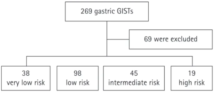
A total of 269 patients underwent surgery for gastric GIST, of whom 69 were excluded because they had simultaneous gastric adenocarcinoma, which would have confounded the analysis of recurrence rates. There were 200 patients in the fi- nal analysis. Based on their pathological results, 38 patients were classified in the very low risk group, 98 in the low risk group, 45 in the intermediate risk group, and 19 in the high risk group (Fig. 1). The patients’ mean age was 60.8 ± 11.7 years, and there were was a predominance of females (127 pa- tients). There were 6 patients with a tumor size <1 cm (3%), 37 with a tumor size of 1-2 cm (18.5%), 104 with a tumor size of 2-5 cm (52%), and 53 with a tumor size of >5cm (26.5%).
The mean follow-up duration was 67 months (0.2-200 months) (Table 1).
Risk of recurrence according to tumor size
The 6 patients with tumors <1 cm were all in the very low
Fig. 1. Recurrence risk classification. The very low, low, intermediate, and high risk groups were classified based on the 2007 JNCCN. Sixty nine patients who had simultaneous gastric adenocarcinoma were excluded.
269 gastric GISTs
69 were excluded
19 high risk 45
intermediate risk 98
low risk 38
very low risk
risk group. Of the 37 patients with a tumor size of 1-2 cm, 32 (86.5%) were in the very low risk group and 5 (13.5%) were in the low risk group. Of the 104 patients with a tumor size of 2-5 cm, 69 (66.3%) were in the low risk group and 35 (33.7%) were in the intermediate risk group. The 53 patients with a tumor size of >5 cm varied in risk, with 24 (45.3%) in the low risk group, 10 (18.9%) in the intermediate risk group, and 19 (35.8%) in the high risk group.
Tumor recurrence
During an average follow-up period of 67 months (0.2-200 months), recurrence was observed in 7 patients (3.5%). There was 1 case of recurrence in the very low risk group in a patient with a tumor size of 1-2 cm, and 2 cases in the very low risk group in patients with tumor sizes of 2-5 cm. In the high risk group, there were 3 cases of recurrences in patients with tu- mors >5 cm (5.7%). Table 2 shows the clinical characteristics of the 7 patients who developed a recurrence. The most com- mon sites of recurrence were the stomach and liver. All the pa- tients were subsequently treated with imatinib. There was 1 death during the follow-up period, of a patient in the high risk group with a tumor size of ≥10cm, and the cause of death was GI bleeding due to GIST. The 5-year recurrence-free survival rates were 100% in the very low risk group, 98.9% in the low risk group, 95.5% in the intermediate risk group, and 86.7% in the high risk group; these differences were statistically signifi- cant (log-rank test, p <0.05) (Fig. 2).
Discussion
This study was a retrospective analysis of recurrence in pa- tients who underwent surgery for gastric GIST. Compared to overseas studies, in which the mean age of gastric GIST pa- tients was around 63 years and there were more male patients than female patients [10,11], the mean age in our study was 60.8 years, and there was a predominance of female patients.
Other Korean studies have also reported younger patient ages (around 60 years on average) and more females than overseas studies [12,13].
The first aim of treatment for gastric GIST is complete sur- gical resection with no infiltration of the margin. The desired resection margin has been reported to be 1-2 cm [14]. How- ever, recent studies have reported that extensive resection to ensure a wide margin is not necessary, and is unrelated to good surgical outcomes [15,16]. Therefore, the objectives of gastric GIST surgery have shifted recently toward only com- plete resection with a clear margin, and since lymph node metastasis is extremely rare, lymph node dissection is not generally performed [7].
The postoperative recurrence rates of gastric GIST reported in Western studies is 17-24% [17,18]. However, studies from Korea and Japan report lower postoperative recurrence rates.
Similarly, we observed a postoperative recurrence rate of Table 1. Clinical characteristics of the 200 patients with gastric
GIST Characteristics
Age, years 60.8 ± 11.7
Sex, male : female 73 : 127
Tumor size, n (%)
< 1 cm 6 (3)
1-2 cm 37 (18.5)
> 2 cm ≤ 5 cm 104 (52)
> 5 cm 53 (26.5)
Risk of recurrence, n (%)
Very low 38 (19)
Low 98 (49)
Intermediate 45 (22.5)
High 19 (9.5)
Mean follow up period, months (range) 67 (0.2-200) GIST, gastrointestinal stromal tumor.
Fig. 2. The 5-year recurrence-free survival rates according to risk groups. The recurrence-free survival rates in the very low, low, intermediate, and high risk groups were 100%, 98.9%, 95.5%, and 86.7%, respectively. Statistical analysis showed a significant lower recurrence-free survival rate in the high risk group (log-rank test, p <0.05).
Time after surgery (months) 100
90 80 70 60 50 40 30 20 10 0
Very low: 100%
Low: 98.9%
Intermediate: 95.5%
High: 86.7%
0 50 100 150 200
Cumulative survival (%)
3.5%, albeit in a cohort with a large number of patients in the very low and low risk groups. Other Korean studies have also reported similar recurrence rates. Yang et al. [19] reported re- currence in 6 out of 105 patients (5.7%) after surgical resec- tion, and Kim et al. [7,20] reported postoperative recurrence rates of 2.7% and 4.8%. Japanese studies have shown even lower recurrence rates; for example, Honda et al. [21] report- ed recurrence in only 1 out of 78 patients (1.3%) after laparo- scopic surgery for gastric GIST. These low recurrence rates are thought to be due to recent improvements in surgical techniques and early diagnosis. In particular, Korea and Japan have high rates of early gastric cancer endoscopic examina- tions for early detection are performed more often than in Western countries. As a result, smaller GISTs are more likely to be found.
The 5-year recurrence-free survival rates in the very low and low risk groups in our study were 100% and 98.9%, re- spectively, which were similar to other studies [5]. In addi- tion, there were no deaths during the follow-up period in the low risk group or below, which is also consistent with other studies [22]. However, the high risk group showed a low 5-year recurrence-free survival rate of 86.7%, and there was also a case of death due to GIST-related bleeding in the high risk group. This is also the same as other studies, with high risk GIST patients showing high recurrence rates and poor prognoses; the 5-year survival rate after complete resection of high risk GIST has been reported in the range of 35-70%
[23-25].
Our study had several limitations. First, as a retrospective study, the numbers of patients in the very low, low, intermedi- ate, and high risk groups are all different, and the follow-up duration and timing of follow-up examinations also differ.
Second, there were not many patients who underwent endo- scopic ultrasound to determine the risk of gastric GIST. In
particular, 1 case of recurrence was observed even among pa- tients with a tumor size of <2cm, which is generally managed by monitoring without surgery. Had the internal appearance of the tumor been inspected by endoscopic ultrasound, it might have been possible to analyze the risk of recurrence in more detail.
In conclusion, although the recurrence rate after complete resection of gastric GIST is very low, the high risk group re- quires thorough postoperative follow-up to monitor for re- currence. In addition, even in the very low risk group, with tumor size <2cm and low mitotic index, cases of recurrence do occur, and so regular postoperative follow-up is essential.
Conflict of interest
All authors declare no conflicts-of-interest related to this article.
References
1. Blay JY, Bonvalot S, Casali P, Choi H, Debiec-Richter M, Dei Tos AP, et al. Consensus meeting for the management of gastrointes- tinal stromal tumors. Report of the GIST consensus conference of 20-21 March 2004, under the auspices of ESMO. Ann Oncol.
2005;16:566-78.
2. Fletcher CD, Berman JJ, Corless C, Gorstein F, Lasota J, Longley BJ, et al. Diagnosis of gastrointestinal stromal tumors: a consen- sus approach. Hum Pathol. 2002;33:459-65.
3. Heinrich MC, Corless CL, Demetri GD, Blanke CD, von Meh- ren M, Joensuu H, et al. Kinase mutations and imatinib response in patients with metastatic gastrointestinal stromal tumor. J Clin Oncol. 2003;21:4342-9.
4. Miettinen M, Lasota J. Gastrointestinal stromal tumors: review on morphology, molecular pathology, prognosis, and differential Table 2. Characteristics of the patients that developed a recurrence after surgical resection
Case
No. Age/Sex Method Maximum
diameter (cm) Mitotic index
(/50 HPF) Resection margin Time until
recurrence (Mo) Site of recurrence
1 58/F Wedge resection 1.8 1-2 clear 3 Stomach
2 50/F Total gastrectomy 18.5 7 clear 12 Liver
3 57/M Subtotal gastrectomy 24 50 clear 6 Liver
4 60/M Wedge resection 4.2 20 clear 24 Peritoneum
5 69/F Wedge resection 5.2 15-20 clear 14 Liver
6 45/F Total gastrectomy 3.5 5 clear 19 Liver
7 66/M Wedge resection 20 30 clear 60 Stomach
HPF, high power field.
diagnosis. Arch Pathol Lab Med. 2006;130:1466-78.
5. Kim MC, Yook JH, Yang HK, Lee HJ, Sohn TS, Hyung WJ, et al.
Long-term surgical outcome of 1057 gastric GISTs according to 7th UICC/AJCC TNM system: multicenter observational study from Korea and Japan. Medicine (Baltimore). 2015;94. DOI:
10.1097/md.0000000000001526.
6. DeMatteo RP, Lewis JJ, Leung D, Mudan SS, Woodruff JM, Brennan MF. Two hundred gastrointestinal stromal tumors: re- currence patterns and prognostic factors for survival. Ann Surg.
2000;231:51-8.
7. Hassan I, You YN, Shyyan R, Dozois EJ, Smyrk TC, Okuno SH, et al. Surgically managed gastrointestinal stromal tumors: a com- parative and prognostic analysis. Ann Surg Oncol. 2008;15:52-9.
8. Bennett JJ, Rubino MS. Gastrointestinal stromal tumors of the stomach. Surg Oncol Clin N Am. 2012;21:21-33.
9. Joensuu H, Vehtari A, Riihimaki J, Nishida T, Steigen SE, Brabec P, et al. Risk of recurrence of gastrointestinal stromal tumour af- ter surgery: an analysis of pooled population-based cohorts.
Lancet Oncol. 2012;13:265-74.
10. Miettinen M, Sobin LH, Lasota J. Gastrointestinal stromal tu- mors of the stomach: a clinicopathologic, immunohistochemi- cal, and molecular genetic study of 1765 cases with long-term follow-up. Am J Surg Pathol. 2005;29:52-68.
11. Tran T, Davila JA, El-Serag HB. The epidemiology of malignant gastrointestinal stromal tumors: an analysis of 1,458 cases from 1992 to 2000. Am J Gastroenterol. 2005;100:162-8.
12. Kim IH, Kim IH, Kwak SG, Kim SW, Chae HD. Gastrointestinal stromal tumors (GISTs) of the stomach: a multicenter, retrospec- tive study of curatively resected gastric GISTs. Ann Surg Treat Res. 2014;87:298-303.
13. Cho MY, Sohn JH, Kim JM, Kim KM, Park YS, Kim WH, et al.
Current trends in the epidemiological and pathological charac- teristics of gastrointestinal stromal tumors in Korea, 2003-2004.
J Korean Med Sci. 2010;25:853-62.
14. Fujimoto Y, Nakanishi Y, Yoshimura K, Shimoda T. Clinicopath- ologic study of primary malignant gastrointestinal stromal tu- mor of the stomach, with special reference to prognostic factors:
analysis of results in 140 surgically resected patients. Gastric Cancer. 2003;6:39-48.
15. Das A, Wilson R, Biankin AV, Merrett ND. Surgical therapy for gastrointestinal stromal tumours of the upper gastrointestinal tract. J Gastrointest Surg. 2009;13:1220-5.
16. Privette A, McCahill L, Borrazzo E, Single RM, Zubarik R. Lapa- roscopic approaches to resection of suspected gastric gastroin- testinal stromal tumors based on tumor location. Surg Endosc.
2008;22:487-94.
17. Demetri GD, von Mehren M, Blanke CD, Van den Abbeele AD, Eisenberg B, Roberts PJ, et al. Efficacy and safety of imatinib mesylate in advanced gastrointestinal stromal tumors. N Engl J Med. 2002;347:472-80.
18. Iesalnieks I, Rummele P, Dietmaier W, Jantsch T, Zülke C, Schlitt HJ, et al. Factors associated with disease progression in patients with gastrointestinal stromal tumors in the pre-imatinib era. Am J Clin Pathol. 2005;124:740-48.
19. Yang HK, Park DJ, Lee HJ, Kim HH, Kim WH, Lee KU. Clinico- pathologic characteristics of gastrointestinal stromal tumor of the stomach. Hepatogastroenterology. 2008;55:1925-30.
20. Kim KH, Kim MC, Jung GJ, Kim SJ, Jang JS, Kwon HC. Long term survival results for gastric GIST: is laparoscopic surgery for large gastric GIST feasible? World J Surg Oncol. 2012;10. DOI:
10.1186/1477-7819-10-230.
21. Honda M, Hiki N, Nunobe S, Ohashi M, Kiyokawa T, Sano T, et al. Long-term and surgical outcomes of laparoscopic surgery for gastric gastrointestinal stromal tumors. Surg Endosc. 2014;
28:2317-22.
22. Bucher P, Taylor S, Villiger P, Morel P, Brundler MA. Are there any prognostic factors for small intestinal stromal tumors? Am J Surg. 2004;187:761-6.
23. Benjamin RS, Blanke CD, Blay JY, Bonvalot S, Eisenberg B. Man- agement of gastrointestinal stromal tumors in the imatinib era:
selected case studies. Oncologist. 2006;11:9-20.
24. DeMatteo RP. The GIST of targeted cancer therapy: a tumor (gastrointestinal stromal tumor), a mutated gene (c-kit), and a molecular inhibitor (STI571). Ann Surg Oncol. 2002;9:831-9.
25. An JY, Choi MG, Noh JH, Sohn TS, Kang WK, Park CK, et al.
Gastric GIST: a single institutional retrospective experience with surgical treatment for primary disease. Eur J Surg Oncol. 2007;
33:1030-5.