• 검색 결과가 없습니다.

Snoring during Bronchoscopy with Moderate Sedation Is a Predictor of Obstructive Sleep Apnea

N/A
N/A
Protected

Academic year: 2021

Share "Snoring during Bronchoscopy with Moderate Sedation Is a Predictor of Obstructive Sleep Apnea"

Copied!
6
0
0

로드 중.... (전체 텍스트 보기)

전체 글

(1)

Introduction

Obstructive sleep apnea (OSA) is recognized as the occur- rence of repetitive episodes of partial airway collapse, manifest- ed as loud snoring and hypopnea, or complete airway collapse, manifested as apnea, which results in oxygen desaturation or arousal during sleep. In the 1990s, the prevalence of OSA was 4% in middle-aged men and 2% in middle-aged women

1

. However, the prevalence of OSA has increased substantially over the past two decades, ranging from 10% to 20%, with the increasing prevalence of obesity and advanced age

2,3

. OSA is frequently comorbid with cardiovascular, cerebrovascular, and metabolic diseases

4

, and is responsible for poor quality of life and increased health care use costs

5

. Furthermore, OSA is as- sociated with high morbidity and mortality rates

6,7

.

However, OSA remains underdiagnosed; up to 80% of pa-

Snoring during Bronchoscopy with Moderate Sedation Is a Predictor of Obstructive Sleep Apnea

Jaeyoung Cho, M.D.

1

, Sun Mi Choi, M.D.

1

, Young Sik Park, M.D.

1

, Chang-Hoon Lee, M.D., Ph.D.

1

, Sang-Min Lee, M.D., Ph.D.

1,2

and Jinwoo Lee, M.D.

1,2

1

Division of Pulmonary and Critical Care Medicine, Department of Internal Medicine, Seoul National University Hospital, Seoul,

2

Department of Internal Medicine, Seoul National University College of Medicine, Seoul, Korea

Background: Snoring is the cardinal symptom of obstructive sleep apnea (OSA). Snoring and upper airway obstruction associated with major oxygen desaturation may occur in populations undergoing flexible bronchoscopy.

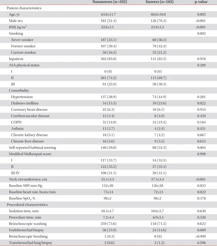
Methods: To evaluate the prevalence of patients at a high risk of having OSA among patients undergoing bronchoscopy with sedation and to investigate whether snoring during the procedure predicts patients who are at risk of OSA, we prospectively enrolled 517 consecutive patients who underwent the procedure with moderate sedation. Patients exhibiting audible snoring for any duration during the procedure were considered snorers. The STOP-Bang (Snoring, Tiredness, Observed apnea, high blood Pressure-Body mass index, Age, Neck circumference and Gender) questionnaire was used to identify patients at high (score ≥3 out of 8) or low risk (score <3) of OSA.

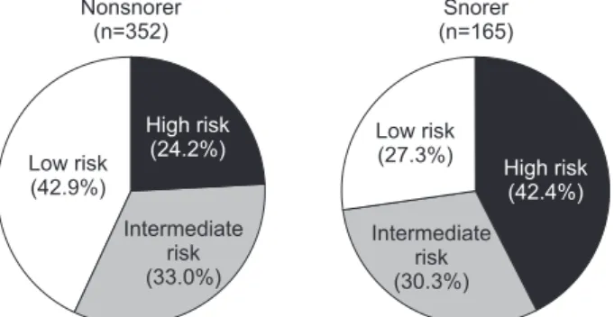
Results: Of the 517 patients, 165 (31.9%) snored during bronchoscopy under sedation. The prevalence of a STOP-Bang score ≥3 was 61.9% (320/517), whereas 200 of the 352 nonsnorers (56.8%) and 120 of the 165 snorers (72.7%) had a STOP-Bang score ≥3 (p=0.001). In multivariable analysis, snoring during bronchoscopy was significantly associated with a STOP-Bang score ≥3 after adjustment for the presence of diabetes mellitus, chronic obstructive pulmonary disease, chronic kidney disease, and stroke (adjusted odds ratio, 1.91; 95% confidence interval, 1.26–2.89; p=0.002).

Conclusion: Two-thirds of patients undergoing bronchoscopy with moderate sedation were at risk of OSA based on the screening questionnaire. Snoring during bronchoscopy was highly predictive of patients at high risk of OSA.

Keywords: Snoring; Bronchoscopy; Conscious Sedation; Sleep Apnea, Obstructive

Address for correspondence: Jinwoo Lee, M.D.

Department of Internal Medicine, Seoul National University College of Medicine, 103 Daehak-ro, Jongno-gu, Seoul 03080, Korea

Phone: 82-2-2072-7593, Fax: 82-2-762-9662 E-mail: [email protected]

Received: Jan. 28, 2019 Revised: Apr. 1, 2019 Accepted: Apr. 5, 2019 Published online: May. 31, 2019

cc

It is identical to the Creative Commons Attribution Non-Commercial License (http://creativecommons.org/licenses/by-nc/4.0/).

Copyright © 2019

The Korean Academy of Tuberculosis and Respiratory Diseases.

(2)

tients with moderate to severe OSA are estimated to remain undiagnosed in the general population

8

. Even in surgical patients, approximately 60% of those with moderate to severe OSA are not recognized preoperatively

9,10

. Thus, efforts have been made to devise simple screening instruments to identify patients who are undiagnosed but at high risk of OSA because overnight polysomnography is time-consuming, labor-inten- sive, and costly

11-14

.

Snoring is the cardinal symptom of OSA. Both male and female habitual snorers are more likely to have an apnea-hy- popnea index (AHI) of 15 or higher

1

. By contrast, the absence of snoring makes OSA less likely

4

.

Although flexible bronchoscopy is a generally safe and minimally invasive diagnostic procedure, patients with OSA may have an increased risk of obstructive episodes during procedures with moderate sedation

15

. Snoring and upper airway obstruction associated with oxygen desaturation have been reported to occur in patients undergoing bronchos- copy

16

. However, the association between snoring during bronchoscopy and OSA remains largely unexplored. Here, we aimed to evaluate the prevalence of patients at a high risk of having OSA in a population undergoing bronchoscopy with moderate sedation and to investigate whether snoring during the procedure predicts patients who are at risk of OSA. De- tecting additional clinical predictors of OSA may help identify patients for whom further evaluation of OSA is necessary.

Materials and Methods

1. Study design and patients

We prospectively enrolled consecutive patients who were 18 years or older and undergoing flexible bronchoscopy with moderate sedation in the bronchoscopy center at Seoul Na- tional University Hospital between 27 December 2016 and 1 October 2018. Patients were excluded from the study if they had a previous diagnosis of OSA, an American Society of An- esthesiologists physical status >3, or baseline pulse oximeter oxygen saturation (SpO

2

) <90%, or used a tracheostomy tube, needed supplemental oxygen before the procedure, or had been scheduled to undergo bronchoalveolar lavage, endo- bronchial ultrasound-guided transbronchial needle aspira- tion, or interventional bronchoscopy.

This study was approved by the Institutional Review Board of Seoul National University Hospital (H-1612-041-813). All participants provided written informed consent and the study was conducted in accordance with the tenets of the Declara- tion of Helsinki.

2. Screening for OSA

The STOP-Bang (Snoring, Tiredness, Observed apnea, high

blood Pressure-Body mass index, Age, Neck circumference and Gender) questionnaire was adopted as a screening tool for OSA in the present study

11

. The STOP-Bang questionnaire was developed and validated to identify patients with undi- agnosed OSA in preoperative clinics. The questionnaire com- prises eight yes-or-no questions (snoring, tiredness, observed apnea, high blood pressure, body mass index [BMI] >35 kg/

m

2

, age >50 years, neck circumference >40 cm, and male sex).

It has a high sensitivity at a cutoff score of ≥3: 84% in detecting any OSA (AHI >5/hr) and 93% in detecting moderate to severe OSA (AHI >30/hr)

11

. A recent study proposed an alternative model for scoring the STOP-Bang questionnaire to improve the specificity for detecting moderate to severe OSA

17,18

. Ac- cording to the alternative model, participants are classified into three groups based on the STOP-Bang score: low (0–2), intermediate (3–4), and high risk (5–8). Those with STOP- Bang scores of 3 or 4 can be further classified as having a high risk of moderate to severe OSA if they have both a STOP (snoring, tiredness, observed apnea, high blood pressure) score of ≥2 and meet one of the following criteria: (1) BMI >35 kg/m

2

, (2) neck circumference >40 cm, or (3) male sex

17,18

. The results of the questionnaire were not disclosed to the attend- ing nurses and bronchoscopists.

3. Sedation and monitoring

Our routine practice for flexible bronchoscopy is to use moderate sedation with intravenous midazolam. Midazolam was administered by an experienced nurse and titrated by the attending bronchoscopist. If required, 50 μg of fentanyl as an adjuvant was infused at the bronchoscopist’s discretion. From the onset of sedation, all patients were provided with 3 L/min of supplemental oxygen by nasal cannula with continuous pulse oximetry and electrocardiography, and intermittent blood pressure monitoring. Snoring during bronchoscopy was also monitored. Patients exhibiting audible snoring for any duration during the procedure were considered snorers in this study.

4. Statistical analysis

Clinical characteristics were compared between snorers and nonsnorers using the independent samples t test for con- tinuous variables. Categorical variables were compared using either the chi-square test or Fisher exact test. Multivariable logistic regression was performed to evaluate the association of snoring during bronchoscopy with a STOP-Bang score ≥3 adjusted for variables with p-values of <0.05 in univariable analysis considering collinearity.

A p-value of less than 0.05 was considered significant. Statis-

tical analyses were performed using Stata statistical software

(version 13.1, StataCorp LP, College Station, TX, USA).

(3)

Results

Among the 544 patients who were enrolled during the

study period, 27 were excluded from the analysis because of incomplete documentation. Of the 517 included patients, 165 (31.9%) snored during bronchoscopy under moderate

Table 1. Characteristics of study patients

Nonsnorers (n=352) Snorers (n=165) p-value Patient characteristics

Age, yr 63.0±11.7 66.0±10.0 0.003

Male sex 181 (51.4) 126 (76.4) <0.001

BMI, kg/m

2

22.6±3.1 23.9±3.3 <0.001

Smoking 0.002

Never-smoker 187 (53.1) 60 (36.4)

Former smoker 107 (30.4) 70 (42.4)

Current smoker 58 (16.5) 35 (21.2)

Inpatient 302 (85.8) 141 (85.5) 0.918

ASA physical status 0.289

I 0 (0) 0 (0)

II 261 (74.2) 115 (69.7)

III 91 (25.9) 50 (30.3)

Comorbidity

Hypertension 137 (38.9) 74 (44.9) 0.201

Diabetes mellitus 54 (15.3) 39 (23.6) 0.022

Coronary heart disease 22 (6.3) 10 (6.1) 0.934

Cerebrovascular disease 12 (3.4) 8 (4.9) 0.429

COPD 52 (14.8) 32 (19.4) 0.184

Asthma 13 (3.7) 4 (2.4) 0.451

Chronic kidney disease 18 (5.1) 7 (4.2) 0.667

Chronic liver disease 16 (4.6) 9 (5.5) 0.653

Self-reported habitual snoring 140 (39.8) 88 (53.3) 0.004

Modified Mallampati score 0.998

I 117 (33.7) 54 (33.5)

II 122 (35.2) 57 (35.4)

III/IV 108 (31.1) 50 (31.1)

Neck circumference, cm 35.1±3.3 37.3±3.4 <0.001

Baseline SBP, mm Hg 132±20 136±20 0.023

Baseline heart rate, beats/min 75±14 72±13 0.022

Baseline SpO

2

, % 98±2 98±2 0.378

Procedural characteristics

Sedation time, min 10.3±4.7 10.6±5.7 0.630

Procedure time, min 7.2±4.4 6.9±5.1 0.526

Bronchoscopic washing 259 (73.6) 118 (71.5) 0.622

Endobronchial biopsy 56 (15.9) 24 (14.6) 0.689

Bronchoscopic brushing 1 (0.3) 0 (0) >0.999

Transbronchial lung biopsy 2 (0.6) 2 (1.2) 0.596

(4)

sedation. Table 1 summarizes patient, procedural, and phar- macological characteristics. Snorers were significantly older and predominantly male compared with nonsnorers. Snorers had a higher BMI and neck circumference than nonsnorers.

Snorers were more likely to be ever-smokers and have comor- bid diabetes mellitus. Although patients who snored during the procedure were more likely to report habitual snoring, approximately half of snorers did not report habitual snoring, whereas nearly 40% of nonsnorers reported habitual snoring.

The modified Mallampati score was similar in both snorers and nonsnorers. The mean dose of midazolam did not differ significantly between the groups, but the use of fentanyl was more frequent in snorers.

The prevalence of a STOP-Bang score ≥3 was 61.9%

(320/517), whereas 200 of the 352 nonsnorers (56.8%) and 120 of the 165 snorers (72.7%) had a STOP-Bang score ≥3 (p=0.001). When the alternative scoring model of the STOP- Bang questionnaire was used, 42.4% of snorers and 24.2% of nonsnorers were classified as having a high risk of moderate to severe OSA (Figure 1).

In multivariable analysis, snoring during bronchoscopy was significantly associated with a STOP-Bang score ≥3 after adjustment for the presence of diabetes mellitus, chronic obstructive pulmonary disease, chronic kidney disease, and stroke (adjusted odds ratio, 1.91; 95% confidence interval, 1.26–2.89; p=0.002) (Table 2).

Discussion

In the present study, we found that two-thirds of patients undergoing bronchoscopy with moderate sedation were at risk of OSA based on the screening questionnaire. A third of patients snored during the procedure. Snoring during bron- choscopy predicted patients who were at risk of OSA after adjustment for comorbidities.

Snoring is a sleep-related sound caused by vibration of the soft tissue in the upper airway under conditions of inspiratory airflow limitation

4

. Snoring is generated by high-frequency opening and closing of the upper airway structures, including the tongue base and soft palate, aided by the adhesive proper- ties of mucosal secretions

4

. The associations of snoring with hypertension, myocardial infarction, and stroke were reported in the early 1990s

19-22

, and habitual snorers were found to be more likely to have moderate to severe OSA in the general Figure 1. Proportion of snorers and nonsnorers with low, interme-

diate, and high risk of obstructive STOP-Bang (Snoring, Tiredness, Observed apnea, high blood Pressure-Body mass index, Age, Neck circumference and Gender) questionnaire.

Table 1. Continued

Nonsnorers (n=352) Snorers (n=165) p-value Pharmacological characteristics

Total dose of midazolam, mg 4.2±1.4 4.0±1.2 0.055

Fentanyl 10 (2.8) 12 (7.3) 0.020

Values are presented as number (%) or mean±standard deviation.

BMI: body mass index; ASA: American Society of Anesthesiologists; COPD: chronic obstructive pulmonary disease; SBP: systolic blood pres- sure; SpO

2

: pulse oximeter oxygen saturation.

Table 2. Multivariable analysis of factors associated with the risk of obstructive sleep apnea

aOR 95% CI p-value Snoring during bronchoscopy

No 1

Yes 1.91 1.26–2.89 0.002

Diabetes mellitus

No 1

Yes 2.51 1.42–4.44 0.001

COPD

No 1

Yes 1.65 0.97–2.79 0.062

Chronic kidney disease

No 1

Yes 2.62 0.85–8.14 0.095

Stroke

No 1

Yes 2.89 0.80–10.4 0.104

aOR: adjusted odds ratio; CI: confidence interval; COPD: chronic

obstructive pulmonary disease.

(5)

population

1

. Recently, the amount of time spent snoring was demonstrated to be correlated with the extent of asymptom- atic carotid artery stenosis

23

.

Intravenous sedation should be provided to patients under- going flexible bronchoscopy, particularly when no contrain- dications to sedation are present

24

. Intravenous midazolam is the preferred sedative for bronchoscopy because of its rela- tively rapid onset of action and short half-life compared with those of other benzodiazepines. Midazolam induces central nervous system depression, partially by increasing the activity of γ-aminobutyric acid, the major inhibitory neurotransmitter in the brain

25

, while also increasing upper airway resistance leading to obstructive apnea

26

. Moreover, when patients un- dergo bronchoscopy in the supine position, they may have a higher risk of upper airway collapse than patients who un- dergo gastrointestinal endoscopy in the left lateral decubitus position.

Studies evaluating the risk of OSA in patients undergoing bronchoscopy with moderate sedation are scarce, but it has been reported that approximately 40% of patients undergoing routine and advanced gastrointestinal endoscopy are at risk of OSA

15,27,28

. In our study regarding flexible bronchoscopy, ap- proximately 60% of patients were at risk of OSA.

Sharara et al.

29

reported that persistent snoring under mod- erate sedation during colonoscopy was a strong predictor of OSA, which was confirmed by out-of-center sleep testing.

However, in their study, participants who had major comor- bidities and underwent the procedure on an inpatient basis were excluded. By contrast, we enrolled both inpatients and outpatients regardless of comorbidities. In this unselected cohort, snoring during bronchoscopy was significantly associ- ated with the risk of OSA after adjustment for the comorbidi- ties frequently associated with OSA.

Patients with OSA may experience adverse events dur- ing procedures with sedation

15

. However, adverse events in undiagnosed OSA patients undergoing bronchoscopy with moderate sedation remain poorly recognized. Previous stud- ies on the risk of adverse events have focused on populations undergoing gastrointestinal endoscopy. Patients at high risk of OSA had increased cardiopulmonary events when they underwent advanced endoscopic procedures such as endo- scopic retrograde cholangiopancreatography and endoscopic ultrasound

15

but not when they underwent routine esophago- gastroduodenoscopy and colonoscopy

27,28,30-32

. Differences in the duration and depth of sedation as well as in patient posi- tioning during procedures may account for the different rates of cardiopulmonary events. Although flexible bronchoscopy is a minimally invasive diagnostic procedure, it is generally considered to have a greater risk of cardiopulmonary events than gastrointestinal endoscopy. Consequently, further inves- tigation is warranted to evaluate cardiopulmonary events in patients undergoing bronchoscopy with moderate sedation.

To appreciate the results of our study appropriately, we have

to recognize its limitations. First, we were unable to confirm the diagnosis of OSA using polysomnography in patients with a STOP-Bang score ≥3. However, the STOP-Bang question- naire has been validated and its high sensitivity in detecting moderate to severe OSA has been documented repeatedly

18

. Second, capnography monitoring was not applied; although capnography detects episodes of apnea or hypoventilation, it is not widely used in routine practice.

In conclusion, we found that undiagnosed OSA, estimated using a screening questionnaire, was prevalent in patients undergoing bronchoscopy with moderate sedation. Snoring during bronchoscopy was highly predictive of patients at high risk of OSA. Snoring during the procedure may help uncover undiagnosed OSA and triage patients who need referral for polysomnography.

Authors’ Contributions

Conceptualization: Lee J, Cho J. Methodology: Lee J, Cho J. Formal analysis: Cho J. Investigation: Lee J, Cho J, Choi SM, Park YS, Lee CH, Lee SM. Writing - original draft preparation:

Lee J, Cho J. Writing - review and editing: Lee J, Cho J. Approval of final manuscript: all authors.

Conflicts of Interest

No potential conflict of interest relevant to this article was reported.

Funding

No funding to declare.

References

1. Young T, Palta M, Dempsey J, Skatrud J, Weber S, Badr S. The occurrence of sleep-disordered breathing among middle- aged adults. N Engl J Med 1993;328:1230-5.

2. Peppard PE, Young T, Barnet JH, Palta M, Hagen EW, Hla KM.

Increased prevalence of sleep-disordered breathing in adults.

Am J Epidemiol 2013;177:1006-14.

3. Jordan AS, McSharry DG, Malhotra A. Adult obstructive sleep apnoea. Lancet 2014;383:736-47.

4. Kryger MH, Roth T, Dement WC. Principles and practice of sleep medicine. Philadelphia: Elsevier; 2016.

5. Leger D, Bayon V, Laaban JP, Philip P. Impact of sleep apnea on economics. Sleep Med Rev 2012;16:455-62.

6. Marshall NS, Wong KK, Liu PY, Cullen SR, Knuiman MW,

Grunstein RR. Sleep apnea as an independent risk factor

(6)

for all-cause mortality: the Busselton Health Study. Sleep 2008;31:1079-85.

7. Young T, Finn L, Peppard PE, Szklo-Coxe M, Austin D, Nieto FJ, et al. Sleep disordered breathing and mortality:

eighteen-year follow-up of the Wisconsin sleep cohort. Sleep 2008;31:1071-8.

8. Young T, Evans L, Finn L, Palta M. Estimation of the clinically diagnosed proportion of sleep apnea syndrome in middle- aged men and women. Sleep 1997;20:705-6.

9. Finkel KJ, Searleman AC, Tymkew H, Tanaka CY, Saager L, Safer-Zadeh E, et al. Prevalence of undiagnosed obstructive sleep apnea among adult surgical patients in an academic medical center. Sleep Med 2009;10:753-8.

10. Singh M, Liao P, Kobah S, Wijeysundera DN, Shapiro C, Chung F. Proportion of surgical patients with undiagnosed obstructive sleep apnoea. Br J Anaesth 2013;110:629-36.

11. Chung F, Yegneswaran B, Liao P, Chung SA, Vairavanathan S, Islam S, et al. STOP questionnaire: a tool to screen patients for obstructive sleep apnea. Anesthesiology 2008;108:812-21.

12. Netzer NC, Stoohs RA, Netzer CM, Clark K, Strohl KP. Using the Berlin Questionnaire to identify patients at risk for the sleep apnea syndrome. Ann Intern Med 1999;131:485-91.

13. Ramachandran SK, Josephs LA. A meta-analysis of clinical screening tests for obstructive sleep apnea. Anesthesiology 2009;110:928-39.

14. Ramachandran SK, Kheterpal S, Consens F, Shanks A, Doherty TM, Morris M, et al. Derivation and validation of a simple perioperative sleep apnea prediction score. Anesth Analg 2010;110:1007-15.

15. Cote GA, Hovis CE, Hovis RM, Waldbaum L, Early DS, Edmundowicz SA, et al. A screening instrument for sleep apnea predicts airway maneuvers in patients undergoing ad- vanced endoscopic procedures. Clin Gastroenterol Hepatol 2010;8:660-5.e1.

16. Chhajed PN, Aboyoun C, Malouf MA, Hopkins PM, Plit M, Grunstein RR, et al. Management of acute hypoxemia during flexible bronchoscopy with insertion of a nasopharyngeal tube in lung transplant recipients. Chest 2002;121:1350-4.

17. Chung F, Yang Y, Brown R, Liao P. Alternative scoring models of STOP-Bang questionnaire improve specificity to detect undiagnosed obstructive sleep apnea. J Clin Sleep Med 2014;10:951-8.

18. Chung F, Abdullah HR, Liao P. STOP-Bang Questionnaire:

a practical approach to screen for obstructive sleep apnea.

Chest 2016;149:631-8.

19. Koskenvuo M, Kaprio J, Heikkila K, Sarna S, Telakivi T, Partin- en M. Snoring as a risk factor for ischaemic heart disease and

stroke in men. Br Med J (Clin Res Ed) 1987;294:643.

20. Schmidt-Nowara WW, Coultas DB, Wiggins C, Skipper BE, Samet JM. Snoring in a Hispanic-American population. Risk factors and association with hypertension and other morbid- ity. Arch Intern Med 1990;150:597-601.

21. D’Alessandro R, Magelli C, Gamberini G, Bacchelli S, Cris- tina E, Magnani B, et al. Snoring every night as a risk fac- tor for myocardial infarction: a case-control study. BMJ 1990;300:1557-8.

22. Seppala T, Partinen M, Penttila A, Aspholm R, Tiainen E, Kaukianen A. Sudden death and sleeping history among Finnish men. J Intern Med 1991;229:23-8.

23. Lee SA, Amis TC, Byth K, Larcos G, Kairaitis K, Robinson TD, et al. Heavy snoring as a cause of carotid artery atherosclero- sis. Sleep 2008;31:1207-13.

24. Du Rand IA, Blaikley J, Booton R, Chaudhuri N, Gupta V, Kha- lid S, et al. British Thoracic Society guideline for diagnostic flexible bronchoscopy in adults: accredited by NICE. Thorax 2013;68 Suppl 1:i1-44.

25. Reves JG, Fragen RJ, Vinik HR, Greenblatt DJ. Midazolam:

pharmacology and uses. Anesthesiology 1985;62:310-24.

26. Montravers P, Dureuil B, Desmonts JM. Effects of i.v. midazol- am on upper airway resistance. Br J Anaesth 1992;68:27-31.

27. Mehta PP, Kochhar G, Kalra S, Maurer W, Tetzlaff J, Singh G, et al. Can a validated sleep apnea scoring system predict cardio- pulmonary events using propofol sedation for routine EGD or colonoscopy? A prospective cohort study. Gastrointest Endosc 2014;79:436-44.

28. Boese ML, Ransom RK, Roadfuss RJ, Todd A, McGuire JM.

Utility of the Berlin Questionnaire to screen for obstructive sleep apnea among patients receiving intravenous sedation for colonoscopy. AANA J 2014;82:38-45.

29. Sharara AI, El Zahabi L, Maasri K, Hashash JG, Mansour N, Skoury A, et al. Persistent snoring under conscious sedation during colonoscopy is a predictor of obstructive sleep apnea.

Gastrointest Endosc 2010;71:1224-30.

30. Khiani VS, Salah W, Maimone S, Cummings L, Chak A. Seda- tion during endoscopy for patients at risk of obstructive sleep apnea. Gastrointest Endosc 2009;70:1116-20.

31. Cha JM, Jeun JW, Pack KM, Lee JI, Joo KR, Shin HP, et al. Risk of sedation for diagnostic esophagogastroduodenoscopy in obstructive sleep apnea patients. World J Gastroenterol 2013;19:4745-51.

32. Andrade CM, Patel B, Gill J, Amodeo D, Kulkarni P, Goldsmith

S, et al. Safety of gastrointestinal endoscopy with conscious

sedation in patients with and without obstructive sleep ap-

nea. J Clin Gastroenterol 2016;50:198-201.

수치

Table 1. Characteristics of study patients
Table 2. Multivariable analysis of factors associated with  the risk of obstructive sleep apnea

참조

관련 문서

The pur- pose of the present study was to identify patients at a rel- atively high risk for OSA by measuring the upper airway diameter (UAD) and mandibular-to-hyoid bone

Background and Purpose zzA population-based door-to-door study of cross-sectional methods for assessing the prevalence and factors related to a high risk of obstructive sleep