Ⅰ.
서 론점액섬유육종(myxofibrosarcoma)은 여러 다양한 방추형
(spindle) 세포로 이루어진 악성 종양으로 점액섬유모세포
(fibroblast)에서 기원하며, 일부 학자들은 이 종양을 악성 섬유 조직구종의 점액양 이형( ) (myxoid variant of malignant fibrous histiocytoma)으로 묘사하기도 한다
1. 이 종 양은 나이가 많은 성인층 환자의 사지에서 호발하며, 50%
이상의 경우에서 진피 또는 피하에 발생하고, 등, 후 복막 등에서도 발생하며, 두경부 영역에서의 발생은 드물어 지 금까지 보고된 증례는 10증례 미만으로, 병소의 크기는 1.5 cm에서 12 cm까지 다양하여, 평균 크기는 3.4 cm 정도로 알려져 있다
2,3.
점액섬유육종의 등급은 점액질 또는 고형질 영역의 비 율, 종양의 괴사, 세포분열 정도 등의 다양한 요소에 의해
나누어지는데, 한 병소에서도 여러 등급이 혼재되어 나타 날 수 있으며, 간혹 치료 및 경과관찰 중에 등급이 변하기 도 하며, 비활동성의 공격력이 낮은 저등급의 병소부터 높 은 공격성이 있는 중간-고등급까지 분류는 다양하다
4,5.
이 종양은 비교적 높은 재발률을 보이며, 국소적으로 재 발이 반복될수록 점점 조직 분화도는 나빠질 수 있으며, 일 부에서는 원발병소의 원격전이에 의해 환자가 사망하기도 한다
6. 또한 이 종양은 임상 및 병리조직학적으로 양성 종 양으로 오진하기 쉽고, 유사육종형태(pseudosarcomatous conditions)의 병소로 보이기도 하기 때문에 정확한 진단이 치료계획 수립에 무엇보다도 중요하다.
저자 등은 하악골에 매우 드물게 발생하는 저등급의 점 액섬유육종을 하악골절제술 및 장골이식과 수술 후 방사 선-항암화학요법의 병용치료를 통하여 양호한 결과를 얻 었기에 문헌고찰과 함께 보고하는 바이다.
Ⅱ.
증례보고59세 여성이 좌측 하악 후방부에 간헐적인 통증이 있는 종창성 병소에 대한 검사와 치료를 위해 내원하였다. 특기 할 만한 외과적 병력이나 외상력도 없었다. 구강 내 검사에 서 좌측 하악 및 병소 치은 부위에 직경 4 cm정도의 잘 경
김 진 수700-412 대구광역시 중구 삼덕동2가188-1번지 경북대학교 치의학전문대학원 구강악안면외과학교실 Chin-Soo Kim
Department of Oral and Maxillofacial Surgery, School of Dentistry, Kyungpook National University
188-1 Samduck-dong 2ga, Jung-gu, Daegu, 700-412, Korea TEL: +82-53-600-7551 FAX: +82-53-426-5365 E-mail: [email protected]
하악에 발생한 저등급의 점액섬유육종: 증례보고
박지훈∙최소영∙권대근∙김진수
경북대학교 치의학전문대학원 구강악안면외과학교실
Low-grade myxofibrosarcoma in the mandible: a case report
Ji-Hoon Park, So-Young Choi, Tae-Geon Kwon, Chin-Soo Kim
Department of Oral and Maxillofacial Surgery, School of Dentistry, Kyungpook National University, Daegu, Korea
Myxofibrosarcoma, also known as a myxoid variant of a malignant fibrous histiocytoma (MFH), is one of the most common sarcomas in the extremi- ties of elderly people. The lesion is characterized by a high frequency of local recurrence but is uncommon in the head and neck regions.
Low-grade myxofibrosarcoma, which is commonly misinterpreted as being benign, has a tendency for histological and biological progression in local recurrences, highlighting the importance of an accurate diagnosis and wide surgical excision of the primary lesion.
We report a rare case of low-grade myxofibrosarcoma of the mandible located in the left mandibular body and angle area. The tumor was first diag- nosed as a myxofibroma and was resected initially. After the final biopsy the patient underwent combined chemo-radiotherapy. The progress of the patent was uneventful until the one year follow up.
Key words:Myxofibrosarcoma, Mandible, Recurrence, Biologic grading
[paper submitted 2010. 9. 26 / revised 2010. 12. 8 / accepted 2011. 1. 19 ] Abstract (J Korean Assoc Oral Maxillofac Surg 2011;37:67-71)
계진 종창이 관찰되었으며(Figs. 1. A, B), 파노라마방사선 사진에서 좌측 하악 우각부를 포함한 광범위한 부위에 골 파괴상이 관찰되었고, 인접 피질골 외형이 소실되었 다.(Fig. 2. A) 컴퓨터단층촬영에서도 좌측 하악에 경계가 불분명한 다방성의 방사선투과성 병소가 협설측 및 하연 의 필질골을 팽윤시키고 파괴시켰으며, 이를 종합할 때 악 성 종양 병소로 의심되었다.(Fig. 2. B) 골스캔에서는 좌측 하악에 약간의 방사선동위원소 섭취증가로 보여 종양에 의한 골파괴 가능성을 시사하였으며, 기타 부위에 특기할 만한 방사선동위원소 섭취증가 및 골파괴 소견은 없었다.
초진 시 국소마취하에서 실시한 조직검사결과로 점액섬 유종(myxofibroma)으로 진단하였으며, 이에 전신마취하에 구내, 구외 접근을 통한 좌측 하악 소구치부터 후방 상행지 의 일부까지 포함한 하악골절제술을 계획하였다. 하악지
체부골절단술로 병소를 제거한 후, 결손 부위에 대하여 자 가장골과 재건용 금속판을 이용한 즉시 재건을 시행 하였 다.(Figs. 3. A-C)
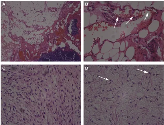
절제된 종물의 육안적 소견은 단단하고 평활한 소엽성의 덩어리로 구성되어 있었으며, 표면은 적색-회색빛을 띠었 다.(Fig. 3. B) 조직소견으로, 방추형 또는 성상형(stellate) 세포와 작은 혈관, 그리고 점액양 기질이 성기게 혼재되어 나타났으며, 늘어지고 각진(elongated and angular) 세포들 이 무정형의 기질 내에 산재되어있고, 굵고 가는 교원질 섬 유다발이 군데군데 있었으며, 병소 자체에 큰 혈관은 없으 나, 많은 작은 혈관들이 초자체 주위에 분포하였다. 일부에 서는 다형성 또는 과염색성 소견이 거의 보이지 않았으며 (Figs. 4. A, B), 또 다른 부위에서는 다양한 형태의 세포, 많 은 핵분열 등이 이상 형태로 나타났다.(Figs. 4. C, D) 최종
Fig. 1. A. Extraoral photograph. Left facial swelling. B. Intraoral photo- graph. A well-circumscribed bony and gingival swelling of the left mandible, which measured 4 cm at its greatest diameter, suggesting benign tumor.
A B
A
B
Fig. 2. A. Preoperative Panorama.
A destructive process with osteolytic changes near the angle, effacement of adjacent cortical outline on these lesion. B. Preoperative CT view. Ill- defined multilocular radiolucent lesion with buccolingual, inferior cortical thinning and buccolingual cortical dis- ruption.(CT: computed tomography)
적으로, 조직학적으로 경도의 악성도의 일부 침윤이 있는 점액섬유육종(myxofibrosarcoma)으로 진단하였다. 환자는 술후 각 220 Gy, 총 27회 5,940 Gy 방사선치료(intensity modulated radiation therapy)를, doxorubicin hydrochloride
(Adriamycin PFS, Pfizer, NY, USA) 20 mg/m
2항암치료를 2 회 intravenous (IV)를 시행하였고, 경과관찰 12개월 후, 현 재 치유상태는 양호하며, 현재까지 국소적 재발소견은 없 는 것으로 관찰되고 있다.(Fig. 5)
Fig. 3. A. An intra/extraoral resection of the left mandible from the premolar to the angle was carried out. B. The specimen consisted of a smooth, lobulated mass, the edge of which was well defined and covered with periosteum, there was no evi- dence of a capsule. C. After the patient underwent extirpation of the lesion, the defect area was reconstructed by iliac bone and reconstruction plate.
A B C
Fig. 4. A. The tumor is composed of spindle or stellate cells lying in an abundant amorphous myxoid stroma that also con- tains isolate coarse and fine collagen fiber bundles.(H&E staining, original magnification ×10) B. The tissue is not very vas- cular, and many of the small vessels are surrounded by a zone of hyalinization.(arrows, H&E staining, original magnification
×40) C. The tumor is composed of elongated and angular cells lying in an abundant amorphous stroma that also contains isolate coarse and fine collagen fiber bundles.(H&E staining, original magnification ×40) D. In sone parts, there is little pleomorphism or hyperchromatism. Mitotic figures are very infrequent, and the appearances are like those of a myxofibroma.
In other parts, however, there is greater variation in the cells, mitotic figures are frequent, and these include occasional abnormal forms.(arrows, H&E staining, original magnification ×40)
A B
C D
Ⅲ.
고 찰육종은 두경부 영영에서 드물게 발생하는 질환으로, osteosarcomas, angiosarcomas, rhabdomyosarcomas, 그리고 malignant fibrous histiocytomas (MFH) 등이 두경부 영역에 서 발생하는 모든 육종의 50% 이상을 차지 한다
7. 특히, 점 액섬유육종은 20세 이하에서는 드물며, 나이가 많은 환자 에서 호발하고, 약간 남성 우성의 발생률을 보이며, 주로 사지에 발생하는 경우가 많고, 두경부 영역에서는 매우 드
물어
8, 이 전에도 상악동, 접형골, 하악골 등에 발생한 몇몇
증례만이 보고되고 있으며
9-11특히 하악골에는 Kummoona
9만이 보고하였다.
점액섬유육종은 Angervall 등
5이1977년 처음으로 점액성 기질의 양이 다양한 악성 섬유모세포성 병변(malignant fibrous lesion)의 한 종류로, 임상병리학적으로 독립적인 독 특한 병으로 발표하였다. Weiss와 Enzinger
12는 조직학적으 로 점액질이 많고 세포의 양이 적을수록 점액질이 없는 형 태보다 좀 더 나은 예후를 나타냄을 보고하였다. 비록 이전 까지 보고된 증례가 매우 드물긴 하지만, 이 종양은 MFH 에서 갈라져 나온 것으로 보이며, 발생하는 각각의 장기마 다 다양한 생물학적 특징을 보인다. Weiss와 Enzinger
12는 이 종양이 깊은 조직에서 발생할수록 전이성이 커지고 좀 더 공격적인 성격을 나타내며, 단일 종괴로 나타나 침윤성 경계를 보인다고 하였다. 반면, 표층에 가깝게 발생하는 육 종일수록 다양한 젤라틴성 결절을 포함 다발성의 병소를 보인다고 하였다. 점액성 조직 내에 혈관성 조직이나 거대 세포가 얼마나 다양하게 있느냐에 따라서 조직학적인 유 형이 달라지며, 이러한 유형에 따라 전이성, 국소적 재발성 등 예후에 차이가 있다고 알려져 있다
7. 방사선학적 소견으 로, 특징은 좀먹은 모양(mottled appearance), 또는 벌집모양 (honeycomb appearance)의 팽윤된 골파괴성의 방사선투과 상을 보이며, 때때로 미맹출 매복치아를 포함하는 경우에
는 낭종과 감별이 어려울 수도 있다
9. 본 증례에서도 광범 위한 골파괴가 진행되고 있었으며, 선상의 골경화성 격벽 (septum)이 관찰되었다.
Mentzel 등
6은 점액섬유육종을 저, 중, 고등급의 병소로 나누었는데, 등급은 세포 포함성, 핵의 다형성, 세포분열성 등에 의해 결정된다. 본 증례에서와 같은 저등급의 점액섬 유육종은 세포가 많지 않고 주로 점액성 기질이 많이 나타 났으며, 세포형태는 방추상형, 원형, 성상형이며, 핵은 경 미하게 다형성의 불규칙한 모양으로 과염색성을 나타내 고, 세포분열은 좀처럼 보기 어렵다
1.
저등급의 점액섬유육종은 점액성 신경섬유종(myxoid neurofibroma)등과 같은 양성 점액성 종양과 임상적, 조직 학적으로 비슷한 소견을 보여 혼동이 될 때가 있으며, 전이 성이 높은 섬유성 점액육종(fibromyxoid sarcoma), 결절성 근막염(nodular fasciitis), 악성 주변성 신경종양(malignant peripheral nerve sheet tumor) 등도 임상적, 조직학적 특징이 비슷하여 감별진단에 포함해야 한다
1.
진단은 조직학적인 결과에 기초로 한다. 이 종양은 길고 굴곡진, 얇은 벽의 혈관을 풍부하게 분포하며, 혈관 주위로 곧은 방추형 종양세포가 밀집되어 있으며, 특별하게 호산 성 세포질과 불규칙한 모양의 핵이 있는 다핵성 거대세포, 섬유모세포 등을 발견할 수 도 있다
9. 면역조직학적 염색에 서 S-100 단백질과 평활근 항원에 음성을 나타내며, CD-34 와 증식성 인자인 Mib1 등에는 양성을 보인다
1,3. 본 증례의 조직학적 소견은 일반적인 점액섬유육종의 특징과 일치하 며, S-100단백질 음성반응, CD-68 부분 양성반응, smooth muscle actin (SMA) 양성반응을 보였다.
두경부 영역에서 점액섬유육종의 국소적 재발률은 그 등 급과 병소의 깊이에 따라 달라지는데, 재발률은 50-61% 정 도이고, 5년 생존율은 65%로 보고되고 있다
13. 원격 전이율 이 20-24%를 보인다는 발표도 있으나
6,13, 저 등급의 병소일 경우에는 전이성의 거의 없는 것으로 보이며, 예후는 좋은
Fig. 5.Postoperative panorama. Dental implant were installed on both canine a premolar areas.
것으로 보고된다
6.
치료방법으로는 단순 소파술이나 적출술과 같은 보존적 술식보다는 악골의 절제술 같은 근치적 술식이 요구된다
9. 이 종양에 대해서 방사선치료나 화학적 치료요법을 사용 한 몇몇의 증례발표가 있으나, 다양한 화학치료요법은 효 과가 없는 것으로 보고되며, 대부분은 방사선에 대한 저항 성을 보여, 두 치료요법 모두 효과가 크게 없는 것으로 나
타났다
14,15. 그러므로, 병소를 모두 포함하는, 깨끗한 경계
의 완벽한 절제술만이 점액섬유육종의 치료를 위한 효과 적인 방법이라 할 수 있다
1,9.
하지만 본 증례에서는 절제된 종물이 일부 침윤성 경계 (invasive margin)를 보였기에, 추가적으로 2차 수술이 필요 할 것으로 사료되었으나 환자분이 추가적 수술을 원치 않 으시고, 자가장골로 즉시 재건된 하악을 다시 제거하는 것 에 대한 부담이 있었기에 외과적 절제술 후 방사선치료와 항암치료를 병행하여 잔여 병소에 대한 처치를 계획하였 다. 상기 환자는 현재 면밀한 추적관찰 중이며, 수술 후 2년 까지는 6개월 마다, 수술 후 2년 이후는 2년에 1회씩 positron emission tomography-computed tomography (PET/CT)를 촬영하여 재발 및 전이 여부를 관찰하고자 한 다.
Ⅳ.
결 론저등급의 점액섬유육종은 악골에서 매우 드물게 발생하 며, 나이가 많은 환자에서 호발하고, 임상방사선학적으로 양성 종양으로 오진하기 쉬우나, 조직생물학적으로 공격 성과 국소적 재발의 가능성이 높기 때문에 조직생검을 통 한 정확한 진단이 매우 중요하다. 등급은 조직학적 소견에 의해 나눠지며, 저등급일수록 예후는 양호하고, 재발률을 낮추기 위해서는 보존적 술식보다는 원발병소의 광범위한 근치적 절제술이 필요하다. 본 증례에서는 초진 시 점액섬 유종으로 오인하어 절제술만 하였고, 이후 점액섬유육종 으로 확진하였으나 종물의 일부에서 침윤성 경계를 보여 추가적인 방사선-항암요법 병용치료를 시행하였으며, 술 후 1년이 경과한 지금까지 양호한 예후를 보이고 있다.
References
1.Gugatschka M, Beham A, Stammberger H, Schmid C, Friedrich G. First case of a myxofibrosarcoma of the vocal folds: case re- port and review of the literature. J Voice 2010;24:374-6.
2.Nishimura G, Sano D, Hanashi M, Yamanaka S, Tanigaki Y, Taguchi T, et al. Myxofibrosarcoma of the hypopharynx. Auris Nasus Larynx 2006;33:93-6.
3.Wada T, Hasegawa T, Nagoya S, Kawaguchi S, Kaya M, Ishii S.
Myxofibrosarcoma with an infiltrative growth pattern: a case re- port. Jpn J Clin Oncol 2000;30:458-62.
4.Huang HY, Lal P, Qin J, Brennan MF, Antonescu CR. Low- grade myxofibrosarcoma: a clinicopathologic analysis of 49 cas- es treated at a single institution with simultaneous assessment of the efficacy of 3-tier and 4-tier grading systems. Hum Pathol 2004;35:612-21.
5. Angervall L, Kindblom LG, Merck C. Myxofibrosarcoma. A study of 30 cases. Acta Pathol Microbiol Scand A 1977;85A:127- 40.
6.Mentzel T, Calonje E, Wadden C, Camplejohn RS, Beham A, Smith MA, et al. Myxofibrosarcoma. Clinicopathologic analysis of 75 cases with emphasis on the low-grade variant. Am J Surg Pathol 1996;20:391-405.
7.Sturgis EM, Potter BO. Sarcomas of the head and neck region.
Curr Opin Oncol 2003;15:239-52.
8.Nascimento AF, Bertoni F, Fletcher CD. Epithelioid variant of myxofibrosarcoma: expanding the clinicomorphologic spectrum of myxofibrosarcoma in a series of 17 cases. Am J Surg Pathol 2007;31:99-105.
9.Kummoona R. Central myxofibrosarcoma of the mandible treat- ed by radical resection. Oral Surg Oral Med Oral Pathol 1975;39:
713-7.
10.Lam PK, Trendell-Smith N, Li JH, Fan YW, Yuen AP.
Myxofibrosarcoma of the sphenoid sinus. J Laryngol Otol 2002;
116:464-6.
11.Pomerantz JM, Sanfacon DG, Dougherty TP, Hanson S.
Myxofibrosarcoma of the maxillary sinus. Del Med J 1982;54:
147-52.
12.Weiss SW, Enzinger FM. Myxoid variant of malignant fibrous histiocytoma. Cancer 1977;39:1672-85.
13.Soper CP, Andrews PA, Bending MR, Singh L, Fisher C.
Cervical myxofibrosarcoma in a renal allograft recipient treated with murine anti-CD3 monoclonal antibody therapy. Nephrol Dial Transplant 1998;13:1902-3.
14.Merck C, Angervall L, Kindblom LG, Ode′n A. Myxofibrosarcoma.
A malignant soft tissue tumor of fibroblastic-histiocytic origin. A clinicopathologic and prognostic study of 110 cases using multi- variate analysis. Acta Pathol Microbiol Immunol Scand Suppl 1983;282:1-40.
15.Kearney MM, Soule EH, Ivins JC. Malifnant fibrous histiocy- toma: a retrospective study of 167 cases. Cancer 1980;45:167-78.