391
Korean J Radiol 14(3), May/Jun 2013
kjronline.org
INTRODUCTION
Subcutaneous panniculitis-like T-cell lymphoma (SPTCL) is a rare subtype of cutaneous lymphoma, characterized by neoplastic T-cell infiltration of the subcutaneous tissue, mimicking panniculitis. Clinically, it appears as multiple palpable nodules on the lower extremities or trunk, and less commonly on the upper extremities or face. We report a woman with SPTCL, who was presented with a breast mass.
CASE REPORT
A 43-year-old woman came to our hospital due to a
Subcutaneous Panniculitis-Like T-Cell Lymphoma of the Breast
Seo In Jeong, MD
1, Hyo Soon Lim, MD
1, You Ri Choi, MD
2, Jin Woong Kim, MD
1, Min Ho Park, MD
3, Jin Seong Cho, MD
3, Ji Shin Lee, MD
4, Heoung Keun Kang, MD
1Departments of 1Radiology, 3Surgery and 4Pathology, Chonnam National University Medical School, Chonnam National University Hwasun Hospital, Hwasun 519-763, Korea; 2Department of Radiology, Chonnam National University Hospital, Gwangju 501-757, Korea
Subcutaneous panniculitis-like T-cell lymphoma (SPTCL) is a rare subtype of cutaneous lymphoma. There have been a few case reports describing the radiologic imaging findings of SPTCL. We report a case of SPTCL, rarely presented with a breast mass. Here, we review her clinical history and radiologic (mammography and ultrasound) findings.
Index terms: Subcutaneous panniculitis-like T-cell lymphoma; Breast; Mammography; Ultrasound
Received September 7, 2012; accepted after revision November 30, 2012.
Corresponding author: Hyo Soon Lim, MD, Department of Radiology, Chonnam National University Medical School, Chonnam National University Hwasun Hospital, 322 Seoyang-ro, Hwasun- eup, Hwasun 519-763, Korea.
• Tel: (8261) 379-7112 • Fax: (8261) 379-7133
• E-mail: nico1220@dreamwiz.com
This is an Open Access article distributed under the terms of the Creative Commons Attribution Non-Commercial License (http://creativecommons.org/licenses/by-nc/3.0) which permits unrestricted non-commercial use, distribution, and reproduction in any medium, provided the original work is properly cited.
Korean J Radiol 2013;14(3):391-394
palpable mass on the left breast that had been present for more than 3 months. On physical examination, a subcutaneous, poorly defined nodule was palpated in the upper inner portion of the left breast. No axillary lymphadenopathy was palpable.
Bilateral mammography performed with a Senographe 2000 D system (GE Healthcare, Milwaukee, WI, USA) showed a 5-cm, increased opacity with indistinct margin in the upper portion of the left breast (Fig. 1A). No calcifications or architectural distortion were seen. On sonographic examination (LOGIQ 9 with a 12-MHz transducer; GE Healthcare, Milwaukee, WI, USA) performed immediately after the mammography, a diffuse hyperechogenicity with a poorly defined margin was detected in a subcutaneous fat layer of the left upper inner breast (Fig. 1B). The multiple, linear hypoechoic areas were located within the diffuse hyperechogenicity. The sonographic findings suggested the possibility of edema or inflammation. However, a palpable mass had been present for more than 3 months and other symptoms suggesting the inflammation were not present, an ultrasound guided core biopsy was performed. On microscopic examination, the lesion disclosed no specific findings, except fibroadipose tissues with inflammation.
She also noticed nodules in both upper arms and she had http://dx.doi.org/10.3348/kjr.2013.14.3.391
pISSN 1229-6929 · eISSN 2005-8330
Case Report
| Breast Imaging392
Jeong et al.
Korean J Radiol 14(3), May/Jun 2013 kjronline.org symptoms of arthritis, photosensitivity, and severe fatigue.
She was clinically diagnosed as having lupus panniculitis and had a treatment with steroids. However, the symptoms were persistent and the number of the nodules was increased.
An excisional biopsy of the nodule in the left breast was performed. On microscopic examination, a dense lymphoid infiltrate located in the subcutaneous tissue.
The overlying dermis and epidermis were not involved (Fig. 1C). On high-power field, the lymphoid cells were atypical with irregular nuclei. There were numerous fat cells rimmed by atypical lymphoid cells. There were scattered and dispersed histiocytes, some with phagocytic cell debris (Fig. 1D). Immunophenotypically, atypical lymphocytes were positive for CD3, CD8, cytotoxic proteins granzyme B, and T-cell intracellular antigen-1, and negative for CD4, CD20, and CD56 (Fig. 1E). The rimming of individual
adipocytes by the CD8+ and CD56- neoplastic T-cell and strict subcutaneous localization were consistent with a diagnosis of SPTCL. Treatment with systemic chemotherapy (A-CHOP, alemtuzumab, cyclophosphamide, doxorubicin, vincristine, and prednisone) and local radiotherapy were performed. After a total of 6 cycles of chemotherapy and local radiotherapy, the skin nodules decreased in size over the several weeks. She remained clinically well with no lesions detected for a 5 years follow-up period.
DISCUSSION
Subcutaneous panniculitis-like T-cell lymphoma was first described by Gonzalez et al. (1) as a subtype of cutaneous T-cell lymphoma that preferentially infiltrates the subcutaneous tissue without overt lymph node involvement.
It was defined as a cytotoxic T-cell lymphoma characterized
C A
D E
B
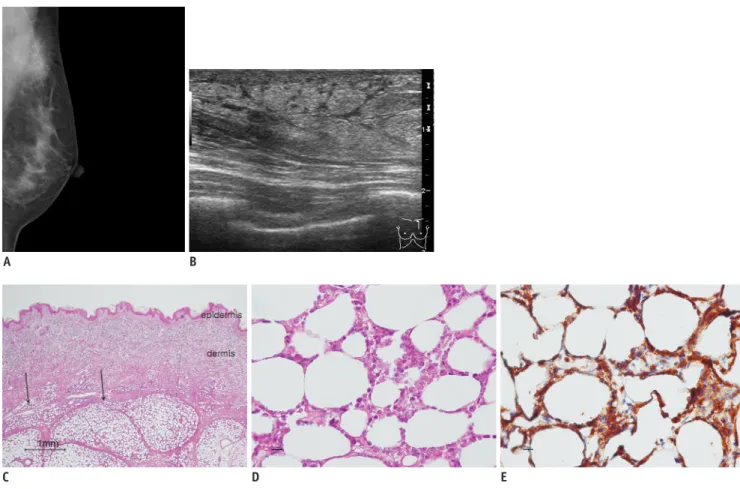
Fig. 1. Subcutaneous panniculitis-like T-cell lymphoma of breast in 43-year-old woman.
A. Mediolateral oblique mammographic view of left breast shows 5-cm, increased opacity with indistinct margin in upper portion of left breast.
There are no calcifications or architectural distortion. Craniocaudal mammographic view of left breast shows no abnormal finding (not shown).
B. Sonography of left breast shows diffuse hyperechogenicity with poorly defined margin in subcutaneous fat layer of left upper inner breast.
C, D. Microscopic examination shows infiltration of neoplastic lymphoid cells into subcutaneous tissue (arrows), but not into overlying dermis and epidermis in low-power view (C, H&E staining, x 20) and focal rimming of adipocytes by atypical lymphocytes and phagocytic macrophages in subcutaneous tissue in high-power view (D, H&E staining, x 400). E. Immunohistochemical stain shows rimming of individual fat spaces by tumor cells with staining for CD8 (x 400).
393 Subcutaneous Panniculitis-Like T-Cell Lymphoma
Korean J Radiol 14(3), May/Jun 2013
kjronline.org
by the presence of primarily subcutaneous infiltrates of pleomorphic T cells and many macrophages.
Clinically, SPTCL appears as multiple nodules and plaques, which mainly involve the legs, the arms, and/or the trunk, and less commonly the face. Ulceration is uncommon (2, 3). To our knowledge, there are only two case reports describing SPTCL manifesting as a breast mass (4, 5). Initial systemic symptoms, such as fever, fatigue, and weight loss, are frequent, and hemophagocystic syndrome may be present (1-3). SPTCL has been associated with autoimmune disorders, including (systemic) lupus erythematosus, juvenile rheumatoid arthritis, type 1 diabetes mellitus, idiopathic thrombocytopenia and multiple sclerosis. In addition, some patients were initially misdiagnosed as lupus erythematosus panniculitis, but lateral reclassified as SPTCL (3).
At first, our case was misdiagnosed as lupus panniculitis and had a treatment with steroids. This diagnosis was made because the patient had symptoms of lupus erythematosus, and inflammation in the fibroadipose tissue was shown on histopathologic examination. However, the symptoms were persistent and excisional biopsy of the nodule was carried out. Finally, she was diagnosed as SPTCL with characteristic histopathological and immunophenotypical features.
We reviewed the core biopsy specimen and microscopic examination revealed inflammation in the dermis and subcutaneous fat. Inadequate tissue sampling, which does not contain deeper subcutaneous tissue, may cause delayed diagnosis. However, in many reported cases, the lymphoma is preceded by an inflammatory phase with features of a lobular lymphocytic panniculitis (6).
Histopathologically, SPTCL reveals subcutaneous infiltrates simulating a panniculitis, showing a small and medium- sized pleomorphic T cell with hyperchromatic nuclei and often many macrophages. The overlying epidermis and dermis are typically uninvolved. Rimming of individual fat cells by neoplastic T cells is a helpful finding. Necrosis, karyorrhexia, and cytophagocytosis are common findings. In addition, immunophenotypically, SPTCL is positive for CD8 and negative for CD4 and CD56 (1-3).
There have been a few case reports describing the radiologic imaging findings associated with SPTCL (4, 5, 7, 8). Sy et al. (4) described late mammographic findings showing a shrunken breast with architecture distortion and extensive coarse flake-like calcifications, consistent with the areas of fat necrosis. There is no report about the initial mammographic finding of SPTCL. On our case, the
mammography showed an increased opacity with indistinct margin, and there was no calcification or architectural distortion. Previous reports on ultrasound finding of SPTCL in breast, abdomen and buttock showed poorly defined hyperechogenicity in the subcutaneous fat layer (4, 7, 8). These findings are similar to our case. Because of the difference in acoustic impedance between adipose cells and lymphocyte clusters, there were more reflected echoes, and greater echogenicity than in normal subcutaneous fat lobules. Therefore, the differential diagnosis on ultrasound examination should include panniculitis, subcutaneous edema, hemorrhage and cellulitis (7, 8). The CT and MRI findings of SPTCL, in previous case reports, were an indistinctly confined hyperdensity and diffuse marked enhancement of the subcutaneous tissue of the breast (4, 5). Unfortunately, there are no specific features that suggest SPTCL on image findings and accurate diagnosis of SPTCL is difficult because it is usually confused with benign panniculitis.
The lesions involving the subcutaneous fat layer include inflammatory conditions, such as fat necrosis or panniculitis or tumorous condition originating in the skin, the adipose tissue, or the superficial glandular tissue. The diagnosis of SPTCL, although rare, can be suspected in patients presenting breast mass, involving the subcutaneous adipose tissue with panniculitis features, especially when multiple sites are involved and the glandular tissue of the breasts are completely spared. Core needle biopsy can be diagnostic.
Because SPTCL is often confused with inflammatory panniculitis associated with connective disease, it is important to follow-up of inflammatory panniculitis and repeated biopsy should be performed if the symptoms are persistent in spite of the appropriate treatment.
In conclusion, although a rare condition, SPTCL should be included in the differential diagnosis of ill-defined subcutaneous lesion on the breast, especially when a patient presents with a long history of panniculitis-like lesions, and biopsy should be performed.
REFERENCES
1. Gonzalez CL, Medeiros LJ, Braziel RM, Jaffe ES. T-cell lymphoma involving subcutaneous tissue. A clinicopathologic entity commonly associated with hemophagocytic syndrome.
Am J Surg Pathol 1991;15:17-27
2. Willemze R, Jaffe ES, Burg G, Cerroni L, Berti E, Swerdlow SH, et al. WHO-EORTC classification for cutaneous lymphomas.
Blood 2005;105:3768-3785
394
Jeong et al.
Korean J Radiol 14(3), May/Jun 2013 kjronline.org 3. Willemze R, Jansen PM, Cerroni L, Berti E, Santucci M, Assaf
C, et al. Subcutaneous panniculitis-like T-cell lymphoma:
definition, classification, and prognostic factors: an EORTC Cutaneous Lymphoma Group Study of 83 cases. Blood 2008;111:838-845
4. Sy AN, Lam TP, Khoo US. Subcutaneous panniculitislike T-cell lymphoma appearing as a breast mass: a difficult and challenging case appearing at an unusual site. J Ultrasound Med 2005;24:1453-1460
5. Schramm N, Pfluger T, Reiser MF, Berger F. Subcutaneous panniculitis-like T-cell lymphoma with breast involvement:
functional and morphological imaging findings. Br J Radiol 2010;83:e90-e94
6. Guitart J. Subcutaneous lymphoma and related conditions.
Dermatol Ther 2010;23:350-355
7. Chiou HJ, Chou YH, Chiou SY, Chen WM, Chen W, Wang HK, et al. High-resolution ultrasonography of primary peripheral soft tissue lymphoma. J Ultrasound Med 2005;24:77-86
8. Kang BS, Choi SH, Cha HJ, Jung YK, Lee JH, Jeong AK, et al. Subcutaneous panniculitis-like T-cell lymphoma: US and CT findings in three patients. Skeletal Radiol 2007;36 Suppl 1:S67-S71