• 검색 결과가 없습니다.

Successful Management of a Rare Case of Stent Fracture and Subsequent Migration of the Fractured Stent Segment Into the Ascending Aorta in In-Stent Restenotic Lesions of a Saphenous Vein Graft

N/A
N/A
Protected

Academic year: 2022

Share "Successful Management of a Rare Case of Stent Fracture and Subsequent Migration of the Fractured Stent Segment Into the Ascending Aorta in In-Stent Restenotic Lesions of a Saphenous Vein Graft"

Copied!
4
0
0

로드 중.... (전체 텍스트 보기)

전체 글

(1)

58 Copyright © 2012 The Korean Society of Cardiology Korean Circulation Journal

Introduction

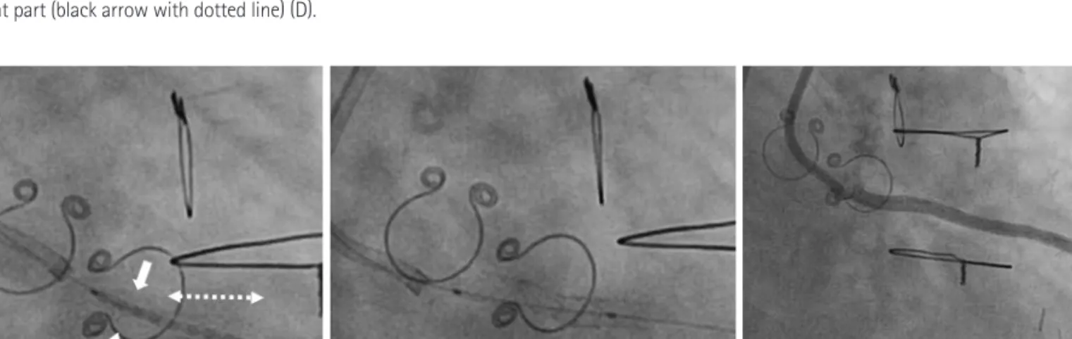
Although drug-eluting stents (DES) have significantly reduced the rate of restenosis by effectively suppressing neointimal growth compared with that of bare metal stents, complications such as in- stent restenosis and stent thrombosis still occur. Stent fracture is also a complication of DES. The incidence of stent fracture is 1-8%.

1)

Most stent fractures are found in patients who were treated with a sirolimus-eluting stent.

2)3)

We experienced an unusual case of stent fracture after implantation of a zotarolimus-eluting stent (ZES),

Case Report

http://dx.doi.org/10.4070/kcj.2012.42.1.58 Print ISSN 1738-5520 • On-line ISSN 1738-5555

Successful Management of a Rare Case of Stent Fracture and Subsequent Migration of the Fractured Stent Segment Into the Ascending Aorta in In-Stent Restenotic Lesions

of a Saphenous Vein Graft

Hoyoun Won, MD 1 , Jaewon Oh, MD 1 , Youngjun Yang, MD 1 , Mihyun Kim, MD 1 , Choongki Kim, MD 1 , Junbeom Park, MD 1 , Byeong-Keuk Kim, MD 1 , Donghoon Choi, MD 1 , and Myeong-Ki Hong, MD 1,2

1

Division of Cardiology, Severance Cardiovascular Hospital,

2

Severance Biomedical Science Institute, Yonsei University College of Medicine, Seoul, Korea

Stent fracture is a complication following implantation of drug eluting stents and is recognized as one of the risk factors for in-stent re- stenosis. We present the first case of successfully managing a stent fracture and subsequent migration of the fractured stent into the as- cending aorta that occurred during repeat revascularization for in-stent restenosis of an ostium of saphenous vein graft after implanta- tion of a zotarolimus-eluting stent. Although the fractured stent segment had migrated into the ascending aorta with a pulled balloon catheter, it was successfully repositioned in the saphenous vein graft using an inflated balloon catheter. Then, the fractured stent seg- ment was successfully connected to the residual segment of the zotarolimus-eluting stent by covering it with an additional sirolimus- eluting stent. (Korean Circ J 2012;42:58-61)

KEY WORDS: Coronary artery disease; Drug-Eluting stents; Complications.

Received: July 9, 2011 Accepted: August 30, 2011

Correspondence: Myeong-Ki Hong, MD, Division of Cardiology, Sever- ance Cardiovascular Hospital, Severance Biomedical Science Institute, Yonsei University College of Medicine, 50 Yonsei-ro, Seodaemun-gu, Seoul 120-752, Korea

Tel: 82-2-2228-8458, Fax: 82-2-393-2041 E-mail: [email protected]

• The authors have no financial conflicts of interest.

This is an Open Access article distributed under the terms of the Creative Commons Attribution Non-Commercial License (http://creativecommons.

org/licenses/by-nc/3.0) which permits unrestricted non-commercial use, distribution, and reproduction in any medium, provided the original work is properly cited.

which occurred during repeat revascularization for in-stent resteno- sis lesions of a saphenous vein graft.

Case

A 72-year-old male underwent coronary artery bypass surgery 10 years ago. He was free of symptoms for several years after the coro- nary artery bypass surgery. One year ago, he suffered from exer- tional chest pain and underwent a follow-up coronary angiogram.

Coronary angiography showed two newly developed significant ste- notic lesions with 80% luminal narrowing at the ostium and 90% lu- minal narrowing at the mid-portion of the saphenous vein graft from the ascending aorta to the left anterior descending artery (Fig. 1A).

The lesions were successfully treated with ZESs (3.5×15 and 3.0×

30 mm, respectively) (Endeavor Resolute®, Medtronic, Santa Rosa, CA, USA) (Fig. 1B). He was discharged without any complications.

Four months after stent implantation, he was admitted again due to exertional dyspnea. Coronary angiography revealed 90% in-stent restenosis at the ostium and a patent stent at the mid-portion of the saphenous vein graft (Fig. 1C). Fluoroscopic images showed an- gulation of the proximal portions of the ZES at the ostium (Fig. 1D).

After angioplasty with a cutting balloon catheter (3.25×15 mm) (Fig.

(2)

59 Hoyoun Won, et al.

http://dx.doi.org/10.4070/kcj.2012.42.1.58 www.e-kcj.org

2A), the inflated balloon catheter was suddenly pulled back outside of the saphenous vein graft during adjunct balloon dilatation with a noncompliance balloon catheter (3.5×15 mm) (Fig. 2B). At the same time, a stent fracture occurred and the fractured stent segment and migrated into the ascending aorta along the inflated balloon cath- eter (Fig. 2B). The migrated fractured struts were successfully pushed into the ostium of the saphenous vein graft without deflating the balloon catheter (Fig. 2C) and were inflated at higher inflation pres- sure with the same noncompliance balloon catheter. Although a distal part of the fractured stent segment was attached to the osti- um of the saphenous vein graft, a gap was evident between the frac- tured stent segment and the residual stent, and a proximal part of the fractured stent segment was located outside of the saphenous vein graft (Fig. 2D). There was a potential for systemic stent emboliz- ation of the fractured stent segment. Therefore, a sirolimus-eluting

stent (3.5×18 mm) (Cypher

TM

, Cordis, Miami, FL, USA) was deployed to connect the distal part of the fractured stent and the proximal part of the residual stent to prevent systemic stent embolization (Fig. 3A). Fluoroscopic images after implantation of the sirolimus- eluting stent showed that the separated ZES segments were well connected without a gap after using an additional stent (Fig. 3B).

The final angiogram with a noncompliance balloon catheter after ad- junct angioplasty showed no residual stenosis (Fig. 3C).

Discussion

Stent fracture is sometimes observed after DES implantation.

The most notable mechanisms of stent fracture are metal fatigue due to mechanical forces and shearing stress.

1)

The right coronary artery, tortuous vessels, and long stents are predictors of stent frac- A  

C  

B  

D  

Fig. 1. Two significantly stenotic lesions were detected in the saphenous vein graft from the ascending aorta to the left anterior descending artery; 80% lu-

minal narrowing at the ostium and 90% luminal narrowing at the mid-portion (A). These lesions were successfully treated with zotarolimus-eluting stent (ZES)

(B). The 4-month follow-up angiogram after stent implantation showed 90% in-stent restenosis at the ostium and a patent stent at the mid-portion of

the saphenous vein graft (C). Fluoroscopic images showed angulation (arrows with dotted line) of the proximal portion of the ZES at the ostium (D).

(3)

60 Management of Fractured Stent Segment Migrated Into Aorta

http://dx.doi.org/10.4070/kcj.2012.42.1.58 www.e-kcj.org

A  

C  

B  

D  

Fig. 2. Balloon angioplasty with a cutting balloon catheter was performed (A) A stent fracture occurred during adjunct balloon dilatation with a noncom- pliance balloon catheter and the fractured stent segment (two white thick arrows) had migrated into the ascending aorta along the inflated balloon cathe- ter (B, arrow with dotted line: residual stent part). The migrated fractured struts were successfully pushed into the ostium of the saphenous vein graft without deflating the balloon catheter (C). Fluoroscopic image shows a gap between the fractured stent segment (black arrow with solid line) and the re- sidual stent part (black arrow with dotted line) (D).

C   B  

A  

Fig. 3. A sirolimus-eluting stent was deployed connect the distal part of the fractured stent (two white thick arrows) with the proximal part of the residual

stent (arrow with dotted line) to prevent systemic stent embolization (A). Fluoroscopic images after implantation of the sirolimus-eluting stent showed

that the separated zotarolimus-eluting stent segments were well connected without a gap using an additional stent (B). Final angiogram revealed no re-

sidual stenosis (C).

(4)

61 Hoyoun Won, et al.

http://dx.doi.org/10.4070/kcj.2012.42.1.58 www.e-kcj.org

ture.

1)3)4)

Additionally, stents implanted in saphenous venous grafts seem to be more susceptible to fracture.

5)

The type of implanted DESs is also strongly related to stent fracture. Of those, the sirolim- us-eluting stent, which has a more rigid closed-cell stent design, has a tendency for more frequent fracture, compared with ZES, which has an open-cell stent design.

2)3)

In this case, we experienced a rare case of ZES fracture in an os- tial in-stent restenostic lesion of the saphenous vein graft. Stent an- gulation could be one of the contributing factors for stent fracture.

Furthermore, balloon dilatation for repeat revascularization of in- stent restenosis could be an additional aggravating mechanical force for stent fracture and separation of a DES implanted in an- gulated segments. Because the fractured stent segment migrated with the dilated balloon catheter along the guidewire system, we easily moved this fractured segment into the remnant distal seg- ments for re-union. This case shows how to manage the migration of a fractured ZES using balloon dilatation in a restenotic ostial le- sion of a saphenous vein graft followed by additional DES implan- tation.

REFERENCES

1. Chakravarty T, White AJ, Buch M, et al. Meta-analysis of incid- ence, clinical characteristics and implications of stent fracture. Am J Cardiol 2010;106:1075-80.

2. Nakazawa G, Finn AV, Vorpahl M, et al. Incidence and predictors of drug-eluting stent fracture in human coronary artery a patho- logic analysis. J Am Coll Cardiol 2009;54:1924-31.

3. Canan T, Lee MS. Drug-eluting stent fracture: incidence, contrib- uting factors, and clinical implications. Catheter Cardiovasc In- terv 2010;75:237-45.

4. Sianos G, Hofma S, Ligthart JM, et al. Stent fracture and restenosis in the drug-eluting stent era. Catheter Cardiovasc Interv 2004;61:

111-6.

5. Aoki J, Nakazawa G, Tanabe K, et al. Incidence and clinical im-

pact of coronary stent fracture after sirolimus-eluting stent im-

plantation. Catheter Cardiovasc Interv 2007;69:380-6.

수치

Fig. 1. Two significantly stenotic lesions were detected in the saphenous vein graft from the ascending aorta to the left anterior descending artery; 80% lu- lu-minal narrowing at the ostium and 90% lulu-minal narrowing at the mid-portion (A)
Fig. 3. A sirolimus-eluting stent was deployed connect the distal part of the fractured stent (two white thick arrows) with the proximal part of the residual  stent (arrow with dotted line) to prevent systemic stent embolization (A)

참조

관련 문서

• 이명의 치료에 대한 매커니즘과 디지털 음향 기술에 대한 상업적으로의 급속한 발전으로 인해 치료 옵션은 증가했 지만, 선택 가이드 라인은 거의 없음.. •

The proposal of the cell theory as the birth of contemporary cell biology Microscopic studies of plant tissues by Schleiden and of animal tissues by Microscopic studies of

Since every classical or virtual knot is equivalent to the unknot via a sequence of the extended Reidmeister moves together with the forbidden moves, illustrated in Section 2,

웰드라인의 위치를 위치를 옮기는 옮기는 방안 방안 - 게이트 위치 변경. - 제품 두께분포 변경 -

The meeting was attended by Assistant Foreign Minister for GCC Affairs, Ambassador, Nasser Al-Muzayyen, and Deputy Assistant Foreign Minister for the Office of the

“ Sheikh Nasser has a written message from HH the Amir, Sheikh Sabah Al-Ahmad Al-Jaber Al-Sabah to the Chinese President, Chi Gen Beng related to enhancing mutual

On his part, CEO of Express Roads Authority, Saud Al-Naqqi said that the heavy rains of the previous day led to clogging parts of the express

Kuwait will celebrate on Sunday the fourth anniversary of the UN honoring and proclamation of His Highness the Amir, Sheikh Sabah Al-Ahmad Al-Jaber Al-Sabah as