ISSN 0378-6471 (Print)⋅ISSN 2092-9374 (Online)
http://dx.doi.org/10.3341/jkos.2015.56.1.142
Case Report
편측성 급성 특발성 황반병증에서의 다양한 진단 이미지 검사
Multimodality Diagnostic Imaging in Unilateral Acute Idiopathic Maculopathy
김숙진⋅김기석
Suk Jin Kim, MD, Ki Seok Kim, MD, PhD
새빛안과병원
Saevit Eye Hospital, Goyang, Korea
Purpose: To report multimodality diagnostic imaging in a case of unilateral acute idiopathic maculopathy.
Case summary: A 32-year-old woman with reduced vision in the right eye had experienced fatigue and flu-like symptoms, includ- ing sore throat and fever a few days before. Her best corrected visual acuity (BCVA) was 1.0 in the right eye. There were no cells in the anterior chamber and vitreous. Fundus photographs of the right eye on presentation showed gray-white thickening of the fovea and retinal hemorrhage next to the fovea. Fluorescein angiography demonstrated ring-shaped mottled hyperfluorescence in the early phase and dye pooling in the late phase. Spectral-domain optical coherence tomography (OCT) showed abnormal hyper-reflective thickening at the level of the outer retina and retinal pigment epithelium (RPE) and detachment of the neuro- sensory retina in the foveal lesion. The inner segment/outer segment junction and photoreceptor elevation/disruption was noted.
Nineteen months after onset, the BCVA of the right eye was 1.0 and fundus photographs showed increased retinal pigment hy- perplasia, and residual RPE changes resembling a bull’s eye maculopathy. The OCT of the right macula showed that the inner segment/outer segment junction elevation/disruption almost completely regressed. The patient was diagnosed with unilateral acute idiopathic maculopathy.
Conclusions: We report a typical case with flu-like symptoms of unilateral acute idiopathic maculopathy. It is a very rare macular disease and a case such as ours with long-term follow-up using multimodality diagnostic imaging has not been previously reported.
J Korean Ophthalmol Soc 2015;56(1):142-147
Key Words: Bevacizumab, Choroidal neovascularization, Unilateral acute idiopathic maculopathy
■Received: 2014. 8. 8. ■ Revised: 2014. 12. 12.
■Accepted: 2014. 12. 18.
■Address reprint requests to Ki Seok Kim, MD, PhD
Saevit Eye Hospital, #1065 Jungang-ro, Ilsandong-gu, Goyang 410-817, Korea
Tel: 82-31-900-7700, Fax: 82-31-900-7777 E-mail: [email protected]
* Parts of this work have been presented at the fellow forum of The Korean Retina Society, Ramada Hotel, Gwangju, Korea, Feb.
14-15, 2014.
ⓒ2015 The Korean Ophthalmological Society
This is an Open Access article distributed under the terms of the Creative Commons Attribution Non-Commercial License (http://creativecommons.org/licenses/by-nc/3.0/) which permits unrestricted non-commercial use, distribution, and reproduction in any medium, provided the original work is properly cited.
편측성 급성 특발성 황반병증(unilateral acute idiopathic maculopathy)은 드문 질환으로 젊은 사람에게서 감기 증상
이후 급성의 단안의 중심시력 저하로 나타난다. 1991년 처 음 보고되었고 황반부 장액성 박리와 암슬러 검사에서 중 심 암점을 보인다.1 콕사키 바이러스 감염이 최근 이 질환 의 원인으로 추측되고 있으며, 몇 주 이내로 자연 회복되며 회복 이후 소 눈 모양(Bull´s eye pattern)의 망막색소상피의 과색소 침착이 특징이다.1,2
국내에서도 일부 증례가 보고된 바 있다.3-5 하지만, 감기 증상을 동반한 전형적인 형태의 편측성 급성 특발성 황반 병증에 대하여 여러 가지 진단 검사를 통한 망막 각층의 변 화를 장기간 경과 관찰한 보고가 없어 문헌 고찰과 함께 보 고하고자 한다.
Figure 1. (A) Fundus photographs of the right eye on presentation showed gray-white thickening of the fovea (white arrows)
and retinal hemorrhage next to the fovea (black arrowheads). (B) Fluorescein angiography demonstrated ring shape mottled hyperfluorescence in the early phase and dye pooling in the late phase at the initial visit. (C) Spectral-domain optical coher- ence tomography showed abnormal hyperreflective thickening at the level of the outer retina and retinal pigment epithelium and detachment of the neurosensory retina at the foveal lesion. The outer plexiform layer swelling was noted. The inner seg- ment/outer segment junction and photoreceptor elevation/disruption was noted with preservation of the external limiting membrane and inner retina (black arrows).증례보고
32세 여자 환자가 3일 전부터 시작된 우안의 시력 저하 와 갈색의 동그란 형태가 보이는 변시증을 주소로 내원하 였다. 환자는 내원 당시 1주 전부터 시작된 목 감기 및 피로 증상을 호소하였으나 전신적 특이소견은 발견하지 못하였 다. 최대 교정시력 양안 각각 1.0이었고, 안압은 정상 소견 이었다. 좌안은 특이소견이 없었지만, 우안 안저 검사에서 황반부 주변으로 회백색의 융기된 병변과 함께 점상 망막 출혈 소견이 관찰되었다(Fig. 1A). 우안의 전방 및 유리체 에 염증은 없었으나, 우안 형광 안저 혈관 조영술의 초기 소견에서 황반 주변부에 고리 모양의 얼룩덜룩한 염색이
나타나고 이는 후기로 갈수록 형광의 고임으로 관찰되었다 (Fig. 1B). 빛간섭 단층 촬영(SpectralisⓇ OCT; Heidelberg Engineering GmbH, Heidelberg, Germany)에서 동일한 황반 부 병변 부위의 외망막층 및 망막색소상피층에 걸쳐 국소 적 망막 두께 증가, 망막 박리 그리고 비정상적인 반사 증 가 소견을 보였고, 광수용체 내외절 경계 및 시세포층의 불 규칙한 배열을 보였다. 또한, 외망상층의 부종이 관찰되었 지만 상대적으로 외경계막과 내망막층은 정상 소견을 보였 다(Fig. 1C).
이에 저자는 편측성 급성 특발성 황반병증을 의심하였으 나 잠복성 특발성 맥락막 신생혈관을 배제할 수 없어 유리 체강내 베바시주맙 주입술을 진단 후 즉시 1회 시행하고
A B
C
A B
C D
E
Figure 2. (A) Nineteen months after onset, fundus photographs of the right eye showed increased retinal pigment
hyperplasia and these residual retinal pigment epithelium (RPE) changes resembling bull’s eye maculopathy (white arrows). (B) Fluorescein angiography demonstrated mild hyperfluorescence staining of the central macula in the early phase (upper left framed) and the late phase. (C) Indocyanine green angiography showed hypofluor- escence in the macular area. (D) Autofluorescence imaging showed hypoautofluorescence corresponding to the clinically significant lesion on funduscopic examination. (E) The spectral-domain optical coherence tomography of the right macula showed the inner segment/outer segment junction elevation/disruption almost completely re- gressed, but slight elevation of RPE and outer retina was remained (black arrows).A
B
C
D
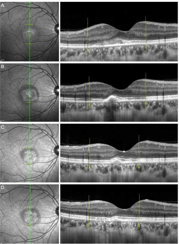
Figure 3. Optical coherence tomography (OCT) evaluation of the macular region in the vertical plane of the right
eye at initial presentation (A), and at one month (B), 19 months (C), 22 months (D) of follow-up. OCT showed abnormal hyperreflective thickening at the level of the outer retina and retinal pigment epithelium (RPE) and de- tachment of the neurosensory retina in the foveal lesion at presentation (A). One month follow-up presentation (B), it showed decrease in size of the lesion. 22 months after onset (D), RPE elevation was much decreased.Outer plexiform layer swelling was noted at presentation (A) and it decreased at one month after onset (B). The inner segment/outer segment junction and photoreceptor elevation/disruption was noted with preservation of the external limiting membrane and inner retina at presentation (A). Nineteen months after onset (C), the OCT showed the inner segment/outer segment junction elevation/disruption almost completely regressed, and 22 months after onset (D), the OCT showed the inner segment/outer segment junction elevation/disruption disappeared.
Outer photoreceptor segment irregularity was still remains.
경과 관찰하였다. 발병 1개월 뒤, 우안 최대 교정시력은 1.0 이었고, 증상이 호전되었으며 황반부 주변의 병변 부위에 색소가 침착되었다. 빛간섭 단층 촬영에서 병변의 형태적 변화는 초기와 비슷하였으나 병변의 크기가 줄어들고 외망 상층의 부종이 감소한 소견을 보였다. 발병 19개월 뒤 우안 최대교정시력은 1.0이었고 안저 소견에서 소 눈 모양 침착 (bull´s eye pigmentation)이 황반부에 형성되었다. 빛간섭 단층촬영에서 병변 부위의 광수용체 내외절경계의 분열이 감소되었으며 외망막층과 망막색소상피층의 두꺼워진 소 견이 일부 남아 있었다(Fig. 2A, 2E, 3). 우안 형광 안저 혈 관 조영술에서 초기와 후기 사진에서 형광 염색을 보이고, 인도사이아닌그린 형광 안저 혈관 조영술에서 초기와 후기 에서 저형광이 관찰되었다. 우안 자가 형광 촬영에서 저형 광이 관찰되었다(Fig. 2B, 2C, 2D). 발병 22개월 뒤 우안 최 대교정시력은 1.0이었고, 빛간섭 단층촬영에서 병변 부위의 광수용체 내외절경계의 분열이 소멸되고 망막색소상피층 의 두꺼워진 소견이 더 줄어든 소견을 보였다. 외광수용체 층의 불규칙한 모양은 남아있었다(Fig. 3).
고 찰
Yannuzzi et al1이 9안에서 보고한 편측성 급성 특발성 황 반병증에서의 병변의 양상은 황반부에 회백색의 병변과 함 께 감각 망막층의 박리가 관찰되고 점상의 망막 출혈과 유 리체 염증 소견을 나타낸다고 하였다. 보고한 증례들은 거 의 완전히 시력이 회복되었고 재발은 일어나지 않았다. 이 당시에는 병리 현상이 망막색소상피에 생긴 염증일 것이라 추측하였고 최근에는 면역학적 검사 등을 통해 콕사키 바 이러스 감염이 원인으로 추정되고 있다.2 이는 보통 수주 이내에 자연 회복되며 소 눈 모양의 망막색소상피의 변화 는 급성기가 지난 이후에 생긴다고 알려졌다.
기존에 보고된 국내의 증례는 1999년 Kim and Jang3에 의해 임상적으로 편측성 급성 특발성 황반병증으로 생각되 는 5안에 대한 보고가 있었으며, 또한 Choi and Kim,4 Lee et al5에 의해 양안을 침범하거나, 다수의 누출점을 갖는 비 전형적인 증례가 보고되기도 하였다. 하지만 이러한 국내 의 증례는 감기 증상 같은 전신 증상이 언급되지 않았고, 또한 빛간섭 단층 촬영 등의 정밀 검사를 통한 발병 후의 망막 각층 변화와 이의 시간 경과에 따른 회복의 양상에 대 한 내용도 알려진 것이 없다.
편측성 급성 특발성 황반병증은 급성기에는 형광 안저 혈관 조영술에서 망막색소상피의 병변 부위에서 불규칙한 과형광과 저형광이 초기에 나타나고, 후기에 누출에 의한 과형광 또는 마치 맥락막 신생혈관처럼 보이는 형광 염색
을 보이고 망막 출혈은 저반사로 나타나게 된다. 급성기 이 후에는 황반부 주변을 둘러싼 망막색소상피의 저반사와 고 반사 고리를 이루게 되어 소 눈 모양의 형상을 보이게 된 다. 인도사이아닌그린 형광 안저 혈관 조영술에서는 황반 부에 저형광을 보인다고 알려졌으며 이는 맥락막의 염증 소견과 망막색소상피층의 변화를 말해주고 있다고 알려졌
다.1,6,7 자가 형광 안저 촬영에서는 급성기에는 작은 점으로
이루어진 과형광과 저형광을 보이다가 병의 경과에 따라 점차 형광이 감소하여 결국 저형광으로 보이게 되며 이는 망막색소 상피층의 변화에 따른 것으로 생각한다.7
빛간섭 단층 촬영에서 급성기에 염증(inflammation)과 부 종이 외망막층과 망막색소 상피층에 영향을 주므로 외망막 층의 부종, 망막색소상피층 및 외망막층의 비정상적인 고반 사와 두꺼워짐이 관찰되며, 광수용체 내외절 경계의 분열을 보인다고 알려졌다. 급성기 이후에는 병변이 줄어들고, 광 수용체 내외절 경계의 분열이 줄어든다고 알려졌다. 이러한 광수용체층의 손상이 일시적이고 다시 가역적으로 돌아오 기 때문에 상대적으로 다른 질환에 비해 더 좋은 시력 예후 를 갖는다고 알려졌다.8-10 최근의 결과인 적응제어광학분야 (adaptive optics scanning laser opthalmoscopy)에서 밝혀진 결과는 급성기에 원추세포 밀도(cone density)가 감소하였다 가 회복된다고 알려졌다.11 다초점 망막전위도(multifocal electroretinogram)소견에서는 정상 잠복기이지만 N1-P1 진 폭 감소를 보였다가 급성기 이후에 정상 소견을 보인다.8
본 증례에서도 급성기의 형광 안저 혈관 조영술에서의 형광 고임, 빛간섭 단층 촬영에서의 외망막층 및 망막색소 상피층에 걸친 망막두께 증가, 비정상적인 반사 증가 및 광 수용체 내외절경계 및 시세포층의 불규칙한 배열을 보였다.
또한 회복 후, 안저 소견에서 소 눈 모양 침착과 더불어 빛 간섭 단층 촬영에서 광수용체 내외절경계의 분열이 소멸되 었고 초진 시부터 경과관찰까지 최대 교정 시력이 1.0으로 양호하여 문헌에 고찰된 증례와 비슷한 소견을 보였다.
이의 치료로 수주 또는 몇 달에 걸쳐 자연히 회복되기 때 문에 스테로이드 제제나, 항생제는 필요 없다고 하였으며, 일부 보고에서 스테로이드 복용이 후유증으로 남게 되는 과색소 침착을 줄일 수 있다고도 하였다. 콕사키 바이러스 감염에 의한 직접 또는 간접적인 망막색소상피와 외망막층 의 염증성 질환으로 생각되고 있으며 최근에는 맥락막이 병의 기전에 어느 정도 관여하고 있다고 생각되고 있다.12
임상적으로 유사한 몇 가지 질환은 중심성 장액맥락망막 병증, 하라다병, 다발성 소실성 백반 증후군(multiple evan- escent white dot syndrome:MEWDS) 또는 특발성 맥락막 신생 혈관으로 알려졌다. 특히 신경망막층의 박리와 망막 색소상피의 회백색의 병변 및 망막 출혈 소견이 마치 특발
= 국문초록 =
편측성 급성 특발성 황반병증에서의 다양한 진단 이미지 검사
목적: 편측성 급성 특발성 황반병증이 진단된 1예에서 다양한 진단 이미지 검사를 보고하고자 한다.
증례요약: 32세 여자 환자가 수일 전부터 열이 동반된 목 감기 및 피로 증상 이후, 우안 갈색의 동그란 형상이 보이며 시력이 저하됨을 주소로 내원하였다. 우안 최대 교정시력은 1.0이었다. 전방 및 유리체 염증은 없었으며, 황반부 주변으로 회백색의 융기된 병변 및 망막 출혈 소견이 있고 형광 안저 혈관 조영술에서 고리 모양의 얼룩덜룩한 염색과 후기 형광 고임이 관찰되었다. 빛간섭 단층 촬영에 서 외망막층 및 망막색소상피층에 걸쳐 국소적 망막 두께 증가, 망막 박리 그리고 비정상적인 반사 증가 소견과 광수용체 내외절 경계 및 시세포층의 불규칙한 배열을 보였다. 발병 19개월 뒤, 최대교정시력은 1.0이었고 소 눈 모양 침착이 관찰되고 빛간섭 단층촬영 에서 광수용체 내외절 경계의 분열이 감소된 소견을 보였다. 이에 편측성 급성 특발성 황반병증으로 진단하였다.
결론: 편측성 급성 특발성 황반병증은 매우 드문 망막 질환으로 감기 증상을 동반한 전형적인 증례에 대해 장기간의 경과 관찰을 통한 다양한 진단 이미지 검사에 대하여 보고하고자 한다.
<대한안과학회지 2015;56(1):142-147>
성 맥락막 혈관신생과 같이 보여 감별해야 할 진단으로 생 각한다. 본 증례에서도 역시 특발성 맥락막 혈관 신생을 의 심하여 유리체강내 베바시주맙 주입술을 시행하였으나, 형 광 안저 혈관 조영술에서의 병변의 활동성 즉 후기의 형광 고임이 아주 많은 것에 비해 빛간섭 단층 촬영 소견은 상대 적으로 정상에 비해 적은 변화를 보여 감별점이라 하겠다.
형광 안저 혈관 조영술, 인도사이아닌그린 형광 안저 혈 관 조영술, 자가 형광 안저 혈관 조영술 등 다양한 진단 이 미지 검사를 종합하여 볼 때, 편측성 급성 특발성 황반병증 은 맥락막, 망막색소상피, 외망막층의 염증 소견으로 생각 되며, 빛간섭 단층 촬영에서의 광수용체층의 회복과 연관 된 시력 회복 또한 중요한 점이라 하겠다.
본 증례는 처음에 여러 가지 진단을 같이 생각하지 못하 여, 추가적 다른 바이러스 감염에 대한 포도막염 검사, 콕사 키 바이러스 항체 검사 등이 이루어지지 못하였고, 병의 호 전을 보이기 전에 유리체강내 베바시주맙 주입술을 시행하 여 질병의 자연 경과에 대해 모호해진 측면이 있다. 하지만 감기 증상을 동반한 전형적인 편측성 급성 특발성 황반병 증을 빛간섭 단층 촬영 검사, 자가 형광 안저 촬영 등 다양 한 진단 이미지 검사와 같이 경험하여 보고하는 바이며, 또 한 장기간 경과 관찰하며 시간 경과 관찰에 따른 빛간섭 단 층촬영을 통한 망막층의 자세한 변화 및 광수용체층의 회 복 과정을 관찰한 것 또한 중요한 발견이라 생각한다.
REFERENCES
1) Yannuzzi LA, Jampol LM, Rabb MF, et al. Unilateral acute idio-
pathic maculopathy. Arch Ophthalmol 1991;109:1411-6.
2) Beck AP, Jampol LM, Glaser DA, Pollack JS. Is coxsackievirus the cause of unilateral acute idiopathic maculopathy? Arch Ophthalmol 2004;122:121-3.
3) Kim IT, Jang SD. Unilateral idiopathic maculopathy. J Korean Ophthalmol Soc 1999;40:1260-8.
4) Choi KS, Kim JS. Bilateral acute idiopathic maculopathy. J Korean Ophthalmol Soc 2000;41:1626-30.
5) Lee SS, Kim EC, Kim YY, Kim SD. Unilateral acute idiopathic maculopathy: A case of atypical presentation. J Korean Ophthalmol Soc 2000;41:1796-800.
6) de la Fuente MA, Cuadrado R. Unilateral acute idiopathic macul- opathy: angiography, optical coherence tomography and micro- perimetry findings. J Ophthalmic Inflamm Infect 2011;1:125-7.
7) Jung CS, Payne JF, Bergstrom CS, et al. Multimodality diagnostic imaging in unilateral acute idiopathic maculopathy. Arch Ophthalmol 2012;130:50-6.
8) Aggio FB, Farah ME, Meirelles RL, de Souza EC. STRATUSOCT and multifocal ERG in unilateral acute idiopathic maculopathy.
Graefes Arch Clin Exp Ophthalmol 2006;244:510-6.
9) Matsushita E, Fukuda K, Nakahira A, et al. Resolution of photo- receptor outer segment damage in a patient with unilateral acute idiopathic maculopathy observed using spectral-domain optical coherence tomography. Graefes Arch Clin Exp Ophthalmol 2012;
250:765-8.
10) Milani P, Cacioppo V, Raimondi G, Scialdone A. Spectral domain OCT and autofluorescence imaging of unilateral acute idiopathic maculopathy. Eur J Ophthalmol 2012;22:499-502.
11) Ooto S, Hangai M, Yoshimura N. Photoreceptor restoration in uni- lateral acute idiopathic maculopathy on adaptive optics scanning laser ophthalmoscopy. Arch Ophthalmol 2011;129:1633-5.
12) Srour M, Querques G, Rostaqui O, Souied EH. Early spectral-do- main optical coherence tomography findings in unilateral acute idi- opathic maculopathy. Retina 2013;33:2182-4.