INTRODUCTION
Skin defects caused by burns, trauma, infections, and chronic ulcers, such as venous ulcers and diabetic foot ulcers, are commonly presented to plastic and reconstructive surgeon in clinical practice. There are several therapeutic methods of the skin and soft tissue defect: inducing secondary healing from the wound dressing to the skin graft or flap surgery. Skin
grafting is the most frequently used procedure in the area of reconstructive surgery, defined as a technique for the transfer of cutaneous tissue from one site (donor site) of the body to another (recipient site). Each step of the skin grafting procedure is important, as is postoperative care. Many studies have been published about the three phases of skin graft success: serum imbibition, revascularization, and maturation. To improve graft survival, close contact between the graft and the wound bed is
Negative Pressure Wound Therapy Applied to a Meshed Split-Thickness Skin Graft
Dong-Hun Lee, Yu-Jin Kim*
Department of Plastic and Reconstructive Surgery, Gachon University Gil Medical Center, Incheon, Korea
CC This is an open-access article distributed under the terms of the Creative Commons Attribution Non-Commercial License (http://creativecommons.org/licenses/by-nc/4.0) which permits unrestricted noncommercial use, distribution, and reproduction in any medium, provided the original work is properly cited.
Copyright © 2016 by the Korean Society for Microsurgery. All Rights Reserved.
Received April 24, 2016 Revised July 22, 2016 Accepted July 28, 2016
*Correspondence to: Yu-Jin Kim Department of Plastic and Reconstructive Surgery, Gachon University Gil Medical Center, 21 Namdong-daero 774beon-gil, Namdong-gu, Incheon 21565, Korea Tel: +82-1577-2299
Fax: +82-32-461-2774
E-mail: pseugene@gilhospital.com ORCID: http://orcid.org/0000-0003-1333-4977
Financial support: None.
Conflict of interest: None.
Purpose: Skin grafting is used for the transfer of cutaneous tissue from one site of the body to another. To improve graft survival, close contact between the graft and the wound bed is essential for vessels to grow across the gap. Here, we introduce an easy and efficient dressing method to improve graft survival.
Materials and Methods: A retrospective chart review was performed to identify patients who underwent split thickness skin graft and negative pressure wound therapy (NPWT) or conventional treatment between January 2007 and April 2015. Overall, 25 consecutive patients were included in the NPWT group and 49 were included in the conventional dressing group to compare the outcome of the procedure. The data were obtained from medical records, including age, sex, cause of the skin defect, size of graft, time for healing, wound preparation time, and complications.
Results: Of the NPWT group, the average wound size was 147.04±146.74 cm2 (range, 9~900 cm2). With the exception of one patient, all wounds healed without the need for further procedure. The average duration of time required for the NPWT group, which was defined as removal of stitches (or staples) and no need for additional active dressing, was 6.4±1.97 days (range, 5~15 days). The average time for the conventional dressing group was 10.78±2.38 days (range, 5~15 days).
Conclusion: NPWT can be used to cover regions in which wound healing does not occur fully or when neither tie-over nor compressive dressings are applicable. This treatment also reduced wound healing time and allowed earlier patient mobilization and hospital discharge.
Key Words: Skin transplantation, Wound healing, Negative-pressure wound therapy
ARMS
Archieves of Reconstructive Microsurgery https://doi.org/10.15596/ARMS.2016.25.2.29critical for 1) vessels to grow across the gap, 2) elimination of graft movement during the immediate postoperative period, and 3) prevention of fluid accumulation. Conventional cotton bolster and tie-over dressings are useful for securing the graft site. However, when sealing off was done, recompression and fixation were difficult. Irregular and uneven wounds have a higher risk of fluid accumulation and graft tenting, resulting in partial or total graft loss. In contrast to traditional dressings, negative pressure wound therapy (NPWT) enhances wound healing via induction of growth factors, evacuation of fluid, bacterial clearance, and improved microcirculation.1
In the early 1990s, Argenta and Morykwas2 encountered many complex wounds. They designed several devices to draw the edges of the wound together to facilitate wound healing.
An open-pore polyurethane foam was placed in the wound, covered by a semi-occlusive dressing, and connected by a tube to a vacuum source. They called this method NPWT.3 The device that Argenta and Morykwas2 described has revolutionized the treatment of complex wounds, and it has become widely adopted over the last 10 years with over 1,000 peer-reviewed publications describing its use. We focused on its application to skin grafting and reported its outcomes.
Here, we introduce an easy and efficient dressing method with NPWT to improve outcomes of skin graft surgery to complicated cases observed in Gachon University Gil Medical Center compared to previously described procedures.
MATERIALS AND METHODS
Patient characteristics
A retrospective chart review was performed to identify patients who underwent split-thickness skin grafting and NPWT dressing for reconstruction of skin defect between January 2007 and April 2015. Twenty-five consecutive patients were included in this study. To compare the outcome of graft survival, we reviewed the 49 patients that were treated with skin graft and conventional bolster dressing in the same period. The data were obtained from the medical records, including age, sex, cause of the skin defect, graft size, removal timing of stitches or staples, wound preparing duration (if delayed reconstruction was performed), and complications. Wound preparing duration was defined period from the initial occurred wound time to the performing skin graft. Wound preparation was performed with
conservative treatment, such as saline soaking dressing, until healthy granulation tissue formation.
Outcome evaluation
The wound size measurement was performed by multiplying the perpendicular width and length of the wound with flexible ruler placed on it. Graft survival assessment was determined by the gross morphology of skin graft such as color, texture, capillary refill time and epithelization of wound margin and slit incision. These factors were performed by same study primary therapist.
Operative technique
All patients were treated with the same procedure. We harvested split-thickness skin graft at a thickness of 0.02794 cm from the patient’s own thigh using an air-powered dermatome (Zimmer®, Air Dermatome; Zimmer Surgical Inc., Warsaw, IN, USA) and harvested skin graft was meshed 1.0:1.5 with an EXPANDER (MeshgraftTM II; Zimmer Surgical Inc.). The potential for fluid collection beneath the graft is reduced by meshed graft. Recipient site was covered with meshed split- thickness skin graft and we sutured the laid split-thickness skin graft to the defect margin and to the wound bed for fixing the graft. Then, we applied antibiotic ointment (Teramycin®; Pfizer, New York, NY, USA) and non-adherent single layer of medicated paraffin gauze containing 0.5% chlorhexidine acetate (Bactigras®; Smith&Nephew, Mississauga, ON, Canada), as an antibacterial dressing, to laid skin grafts. Because a thin, porous barrier (medicated paraffin gauze) is then placed between the graft and open-cell polyurethane foam, we prevented the adherence of the foam to the transplanted skin graft. An Open-cell polyurethane foam (Curavac®; CGBio, Seongnam, Korea) that was trimmed to the size and geometric dimension of the uneven wound contour. Then trimmed open-cell polyurethane foam and poly vinyl chloride (PVC) suction tube was addedand wound was sealed with transparent film (IobanTM; 3MTM, Maplewood, MN, USA) to allow a negative pressure environment at the dressing site. PVC suction tube is brought out in an area where pressure will not be applied for preventing another injury, as pressure ulcer. PVC suction tube is then connected to a computerized suction pump that provides 80 mmHg of continuous negative pressure. The dressing was maintained for about 3 days, at which time most
grafts are well adhered to the recipient bed. On the first opening of the dressing, all grafts were opened and negative pressure therapy was stopped. After the negative suction dressing was carefully removed, the dressing was then changed every other day for the remainder of the hospitalization stay. Cleansing and applications of antibiotic ointment (Teramycin®; Pfizer), cotton ball molding, and mild compressive dressing were subsequently performed.
Conventional bolster dressing was an wet cotton ball applying instead of open-cell polyurethane foam. And mild compressive secondary dressing was following instead of applying the negative pressure therapy. Consecutively following dressing was same with NPWT group.
RESULTS
Patient demographics are shown in Table 1, 2. Of the NPWT applying group, the mean patient age was 48.15±25.49 years (range, 4~83 years). The average wound size was 147.04±146.74 cm2 (range, 9~450 cm2). Wound defects were located various areas of the upper/lower limbs and trunk.
Immediate skin graft reconstruction was performed to six patients and delayed reconstruction patients were nineteen.
Wound preparing average duration for delayed skin graft reconstruction is 40±2.19 days (range, 9~88 days). All patients were healed without having revision surgery except one patient (patient 1). The average duration of taken time was 6.4±1.97 days (range, 5~15 days).
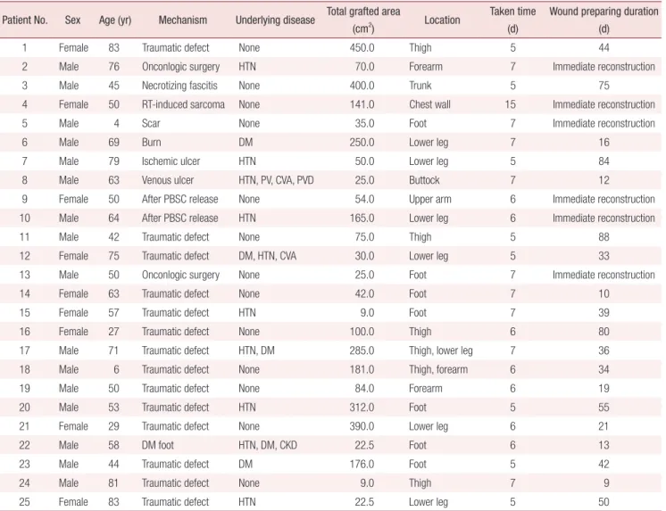
Table 1. Patient demographics and operative results (negative pressure wound therapy group) Patient No. Sex Age (yr) Mechanism Underlying disease Total grafted area
(cm2) Location Taken time (d)
Wound preparing duration (d)
1 Female 83 Traumatic defect None 450.0 Thigh 5 44
2 Male 76 Onconlogic surgery HTN 70.0 Forearm 7 Immediate reconstruction
3 Male 45 Necrotizing fascitis None 400.0 Trunk 5 75
4 Female 50 RT-induced sarcoma None 141.0 Chest wall 15 Immediate reconstruction
5 Male 4 Scar None 35.0 Foot 7 Immediate reconstruction
6 Male 69 Burn DM 250.0 Lower leg 7 16
7 Male 79 Ischemic ulcer HTN 50.0 Lower leg 5 84
8 Male 63 Venous ulcer HTN, PV, CVA, PVD 25.0 Buttock 7 12
9 Female 50 After PBSC release None 54.0 Upper arm 6 Immediate reconstruction
10 Male 64 After PBSC release HTN 165.0 Lower leg 6 Immediate reconstruction
11 Male 42 Traumatic defect None 75.0 Thigh 5 88
12 Female 75 Traumatic defect DM, HTN, CVA 30.0 Lower leg 5 33
13 Male 50 Onconlogic surgery None 25.0 Foot 7 Immediate reconstruction
14 Female 63 Traumatic defect None 42.0 Foot 7 10
15 Female 57 Traumatic defect HTN 9.0 Foot 7 39
16 Female 27 Traumatic defect None 100.0 Thigh 6 80
17 Male 71 Traumatic defect HTN, DM 285.0 Thigh, lower leg 7 36
18 Male 6 Traumatic defect None 181.0 Thigh, forearm 6 34
19 Male 50 Traumatic defect None 84.0 Forearm 6 19
20 Male 53 Traumatic defect HTN 312.0 Foot 5 55
21 Female 29 Traumatic defect None 390.0 Lower leg 6 21
22 Male 58 DM foot HTN, DM, CKD 22.5 Foot 6 13
23 Male 44 Traumatic defect DM 176.0 Foot 5 42
24 Male 81 Traumatic defect None 9.0 Thigh 7 9
25 Female 83 Traumatic defect HTN 22.5 Lower leg 5 50
RT: radiotherapy, PBSC: postburn scar contracture, DM: diabetes mellitus, HTN: hypertension, PV: polycythemia vera, CVA: cardiovascular accident, PVD: peripheral vessel disease, CKD: chronic kidney disease.
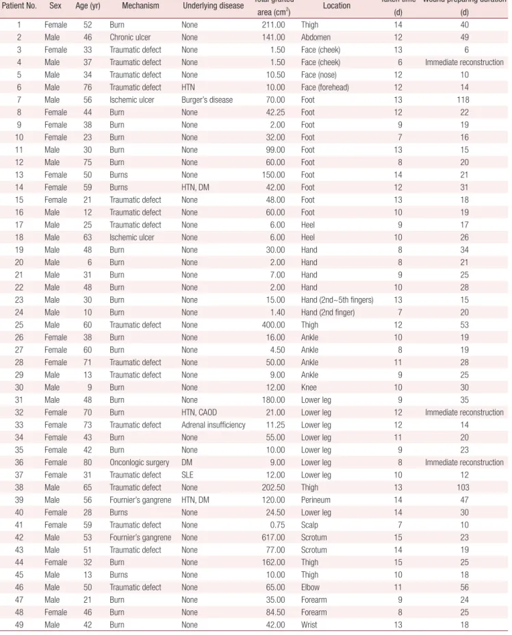
Table 2. Patient demographics and operative results (conventional dressing group) Patient No. Sex Age (yr) Mechanism Underlying disease Total grafted
area (cm2) Location Taken time (d)
Wound preparing duration (d)
1 Female 52 Burn None 211.00 Thigh 14 40
2 Male 46 Chronic ulcer None 141.00 Abdomen 12 49
3 Female 33 Traumatic defect None 1.50 Face (cheek) 13 6
4 Male 37 Traumatic defect None 1.50 Face (cheek) 6 Immediate reconstruction
5 Male 34 Traumatic defect None 10.50 Face (nose) 12 10
6 Male 76 Traumatic defect HTN 10.00 Face (forehead) 12 14
7 Male 56 Ischemic ulcer Burger’s disease 70.00 Foot 13 118
8 Female 44 Burn None 42.25 Foot 12 22
9 Female 38 Burn None 2.00 Foot 9 19
10 Female 23 Burn None 32.00 Foot 7 16
11 Male 30 Burn None 99.00 Foot 13 15
12 Male 75 Burn None 60.00 Foot 8 20
13 Female 50 Burns None 150.00 Foot 14 21
14 Female 59 Burns HTN, DM 42.00 Foot 12 31
15 Female 21 Traumatic defect None 48.00 Foot 13 18
16 Male 12 Traumatic defect None 60.00 Foot 10 19
17 Male 25 Traumatic defect None 6.00 Heel 9 17
18 Male 63 Ischemic ulcer None 6.00 Heel 10 26
19 Male 48 Burn None 30.00 Hand 8 34
20 Male 6 Burn None 2.00 Hand 8 21
21 Male 31 Burn None 7.00 Hand 9 25
22 Male 48 Burn None 2.00 Hand 10 28
23 Male 30 Burn None 15.00 Hand (2nd~5th fingers) 13 15
24 Male 10 Burn None 1.40 Hand (2nd finger) 7 20
25 Male 60 Traumatic defect None 400.00 Thigh 12 53
26 Female 38 Burn None 16.00 Ankle 10 19
27 Female 60 Burn None 4.50 Ankle 8 19
28 Female 71 Traumatic defect None 50.00 Ankle 11 28
29 Male 13 Traumatic defect None 9.00 Ankle 9 25
30 Male 9 Burn None 12.00 Knee 10 30
31 Male 48 Burn None 180.00 Lower leg 9 35
32 Female 70 Burn HTN, CAOD 21.00 Lower leg 12 Immediate reconstruction
33 Female 73 Traumatic defect Adrenal insufficiency 11.25 Lower leg 12 14
34 Female 43 Burn None 55.00 Lower leg 11 20
35 Female 42 Burn None 10.00 Lower leg 9 23
36 Female 80 Onconlogic surgery DM 9.00 Lower leg 8 Immediate reconstruction
37 Female 31 Traumatic defect SLE 12.00 Lower leg 10 12
38 Male 65 Traumatic defect None 202.50 Thigh 13 103
39 Male 56 Fournier’s gangrene HTN, DM 120.00 Perineum 14 47
40 Female 28 Burns None 24.50 Lower leg 14 30
41 Female 59 Traumatic defect None 0.75 Scalp 7 10
42 Male 53 Fournier’s gangrene None 617.00 Scrotum 15 23
43 Male 51 Traumatic defect None 77.00 Scrotum 14 19
44 Female 32 Burn None 162.00 Thigh 15 25
45 Male 13 Burns None 10.00 Thigh 10 18
46 Male 50 Traumatic defect None 65.00 Elbow 11 56
47 Male 21 Burn None 35.00 Forearm 9 24
48 Female 46 Burn None 84.50 Forearm 8 25
49 Male 42 Burn None 42.00 Wrist 13 18
HTN: hypertension, DM: diabetes mellitus, CAOD: coronary artery occlusive disease, SLE: systemic lupus erythematosus.
Of the conventional bolster dressing group, the mean patient age was 42.88±19.34 years (range, 6~80 years).
The average wound size was 66.94±109.94 cm2 (range, 0.75~617.00 cm2). Wound defects were located various areas same as NPWT group, except but facial wound. Delayed skin graft reconstruction patients were forty six and immediate reconstruction patients were three. Wound preparing average duration for delayed skin graft reconstruction is 27.56±20.92 days (range, 6~118 days). In fourteen patients (28.6%), fluid accumulation such as hematoma or seroma formation occurred and partial graft loss occurred in 8 patients (16.3%). The average duration of taken time was 10.78±2.38 days (range, 6~15 days).
Case 1
Patient 1 had an avulsion injury on the left thigh due to traffic accident with a very large and uneven wound bed surface.
An artificial dermis (Matriderm®; MedSkin Solutions Dr.
Suwelack, Billerbeck, Germany) was used as a skin graft to prevent joint stiffness because the location of the wound defect involved the knee joint (Fig. 1, 2). After 5 days of surgery, most of transplanted skin graft was well taken. But there still remain a few defects on grafted area. Cultured allogenic keratinocyte (Kaloderm®; Tego Science, Seoul, Korea) was onlaid over remnant defect. After 11 days of first skin graft surgery, we did revision surgery again (total re-grafted defect size was about 46 cm2).
Case 2
Patient 2 had a squamous cell carcinoma on his wrist, and we widely excised the skin cancer using a safety margin of 10 mm.
After that, we elevated the local flap (reverse flow distal forearm island flap) and treated the skin graft for coverage of the flap donor site. After excision of the cancer, the patient had a defect that had an irregular contour and poor vascular bed, including exposure of the paratenon. After 7 days of surgery, most of skin graft was well taken without any wound discharge (Fig. 3).
Case 3
Patient 3 had extensive skin loss in the abdominal region and right side that was not suitable for a compression dressing;
severe kyphosis made it difficult for the patient to move after surgery. NPWT was identified as a suitable dressing to maintain even pressure distribution across the large wound bed (Fig. 4).
All stitches (staples) were removed on postoperative day 5 and all transplanted skin graft was well epithelization and taken.
Case 4
Patient 4 had radiotherapy-induced sarcoma on the left chest wall and underwent wide excision and reconstruction with a free transverse rectus abdominis myocutaneous flap, but the flap had to be detached due to a thrombus at the anastomosed site. The subsequent defect on the chest wall was treated using a left gastroepiploic artery-pedicled omental flap and a mesh split thickness skin graft over the flap with NPWT application.
NPWT effectively allows drainage of exudates from the
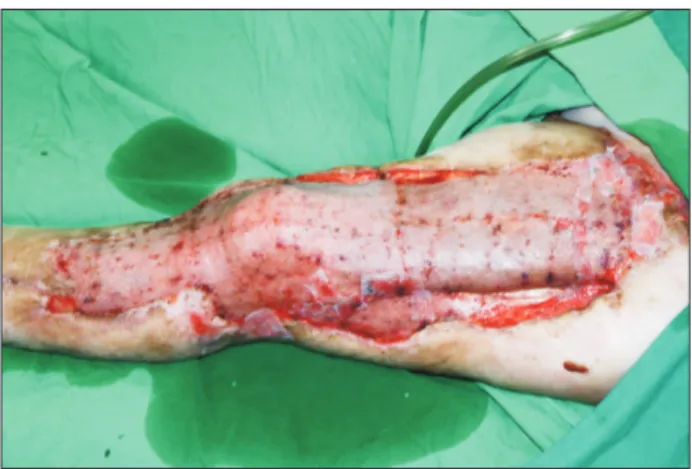
Fig. 1. Patient 1; Intraoperative clinical photo. An acellular dermal substitute (Matriderm®, MedSkin Solutions Dr. Suwelack, Germany) was used as a skin graft to prevent joint stiffness because the location of the wound defect involved the knee joint.
Fig. 2. Patient 1; Postoperative 5-day clinical photo. The graft was well taken and staples were completely removed, although focal raw surfaces requiring an additional surgery were seen.
omentum, reducing seroma or hematoma formation as well as lessening the chance of inflammation or infection (Fig. 5).
The graft site was complete healed on postoperative 15 days and there was not required any dressing except but applying ointment for keeping graft moisture.
Case 5
Patient 5 (5-year-old boy) had a hypertrophic scar on his ankle that was treated with scar excision; the resulting defect was covered by a mesh skin graft and application of NPWT over the grafted skin. He did not need a splint to immobilize the graft area on his ankle, which permitted full mobility of the
lower limb (Fig. 6).
As mentioned above, identical surgical techniques and postoperative management were done on all of the patients, yielding good aesthetic results without significant complications throughout the follow-up period.
DISCUSSION
A healthy wound bed, the prevention of fluid drainage between the wound bed and the graft, and the prevention of shearing forces from disrupting the graft are all critical for prompt graft success. Conventional tie-over or compressive
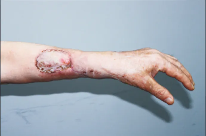
Fig. 3. Patient 2; Postoperative 11-day clinical photo. He did not need a splint for immobilization of the graft area on his wrist, which prevented stiffness in his hand and wrist.
Fig. 4. Patient 3; Postoperative 3-day clinical photo. Patient 3 had extensive skin loss in the abdominal region and right side that was not suitable for a compression dressing; severe kyphosis made it difficult for the patient to move after surgery. Negative pressure wound therapy was identified as a suitable dressing to maintain even pressure distribution across the large wound bed.
Fig. 5. Patient 4; Postoperative 2-day clinical photo. negative pressure wound therapy effectively allows drainage of exudates from the omental flap with onlaid skin graft, reducing seroma or hematoma formation as well as lessening the chance of inflammation or infection.
Fig. 6. Patient 5; Postoperative 6-day clinical photo. Non-compliant children was good candidate of applying negative pressure wound therapy for securing skin graft. He did not need a splint to immobilize the graft area on his ankle, which permitted full mobility of the lower limb.
molding dressings are sufficient for simple wounds, but for the complicated cases observed in our hospital, such methods do not suffice.
In our study, NPWT group shows more desirable clinical outcomes in aspects of graft taken or healing time and complication such as hematoma, seroma and partial graft loss.
Through many studies, expected split-thickness skin graft failure rate of 15% to 30% when using traditional dressing was reported.4-6 Comparing previous reports, our data suggest that conventional dressing group showed similar results of graft loss (16.3%) but a negative-pressure wound therapy over a split-thickness skin graft site may enhance overall graft survival (4%). Even graft taken or healing was required a shorter period at NPWT group (6.4±1.97 days) than conventional dressing group (10.78±2.38 days).
Several studies announce why NPWT may enhance graft survival.2,7 First, an important aspect to increase graft survival rate is maintaining study pressure to contact between the transplanted graft and the recipient wound surface.2,8 In our study, patient 2 and 3 were not suitable for a compression dressing to graft area because we were concerned about chocking the flap pedicle and compressing his trunk lead torespiration difficulty. In cases when compression dressing cannot be applied, compression of the graft was achieved without putting pressure on the surrounded structure using NPWT. After extensive three-dimensional skin grafting, it can be difficult to apply pressure effectively; however, NPWT evenly distributes pressure and secures transplanted skin firmly against the defect wound.
Second, accumulation of fluid (hematoma or seroma) beneath the graft has an adverse effect on graft survival. The use of NPWT contributes continuous evacuation immediately, which prevents the fluid accumulation and the graft site remains clean, thus limiting the chance of infection. Also the NPWT has been associated with lower bacterial counts at wound site,3 and this reduction in the local bacterial flora may enhance graft survival. In our study, patient 4 was treated using a left gastroepiploic artery-pedicled omental flap and a mesh split thickness skin graft over the flap with NPWT application.
NPWT effectively allows drainage of exudates from the omentum, reducing fluid collection as well as lessening the chance of inflammation or infection. Severe drainage from the wound can cause a hematoma under the graft or an infection at
the surgical site; however, fluid collection and infection can be prevented using negative pressure. It is critical to note that mesh or fenestrated grafts are likely to produce the best outcomes, as these maximize fluid removal and prevent accumulation of fluid.9 When applying NPWT, a slit incision is used to prevent fluid accumulation and better coaptation of the grafted skin. An incision using an expander without over-spreading the grafted skin is critical for both effectively removing fluid and producing better aesthetic outcomes at the same time. This will also reduce healing time.
As many studies suggest, NPWT yields better results for noncompliant children than do conventional methods. Major advantages of NPWT for pediatric patients are the superb success rates of the skin grafts and early mobilization. In age groups where compliance issue are highly expected, like in babies, toddlers and unteachable child, a firm and secure fixation of the skin grafts and high mobility of the patients are of utmost importance for the overall treatment outcome.10
We used a somewhat lower negative suction pressure (80 mmHg) than conventional pressure (125 mmHg). High pressure is not required for taken graft, but continuous firm contact between recipient bed and transplanted skin is important. As mentioned in previous studies, many surgeons are limited to a single vendor for NPWT. Therefore, the traditional approach of using 125 mmHg pressure and a polyurethane foam dressing was the only treatment described in the literature and taught to them for complex wounds. Pressure selection was based on classical swine studies conducted at Wake Forrest University. Argenta and Morykwas2 reported that local tissue perfusion was optimized at 125 mmHg. Recently, Borgquist et al.11 re-created the swine study and reported that pressures up to 80 mmHg increased local perfusion and higher pressures reduced perfusion.
Although no controlled studies have been performed, we have found this technique of dressing graft to be excellent for circumstances in which a skin graft must be placed over an uneven wound bed surface.
CONCLUSION
We suggest using NPWT under the following circumstances:
1. Presence of a very large and uneven wound bed surface (except a circumferential wound bed)
2. In an area that requires a non-compressible dressing (beside flap)
3. For use over the dermal substitute and skin graft 4. In non-compliant children
5. In cases of a poor vascular bed, paratenon, over the scar tissue.
NPWT can be used to cover wounds in which healing does not occur fully or when tie-over and compressive dressings are not applicable. The use of NPWT further contributes to reduced wound healing time and allows earlier patient mobilization and hospital discharge.
REFERENCES
1. Webb LX, Pape HC. Current thought regarding the mechanism of action of negative pressure wound therapy with reticulated open cell foam. J Orthop Trauma 2008;22:S135-7.
2. Argenta LC, Morykwas MJ. Vacuum-assisted closure: a new method for wound control and treatment: clinical experience.
Ann Plast Surg 1997;38:563-76; discussion 577.
3. Morykwas MJ, Argenta LC, Shelton-Brown EI, McGuirt W.
Vacuum-assisted closure: a new method for wound control and treatment: animal studies and basic foundation. Ann Plast Surg
1997;38:553-62.
4. Brandes SB. Urethral reconstructive surgery. New Jersey:
Humana Press, Springer Science & Business Media; 2008.
5. Thourani VH, Ingram WL, Feliciano DV. Factors affecting success of split-thickness skin grafts in the modern burn unit. J Trauma 2003;54:562-8.
6. Henderson NJ, Fancourt M, Gilkison W, Kyle S, Mosquera D.
Skin grafts: a rural general surgical perspective. ANZ J Surg 2009;79:362-6.
7. Deva AK, Buckland GH, Fisher E, Liew SC, Merten S, McGlynn M, et al. Topical negative pressure in wound management. Med J Aust 2000;173:128-31.
8. Molnar JA, DeFranzo AJ, Marks MW. Single-stage approach to skin grafting the exposed skull. Plast Reconstr Surg 2000;105:
174-7.
9. Avery CM, Pereira J, Brown AE. Suprafascial dissection of the radial forearm flap and donor site morbidity. Int J Oral Maxillofac Surg 2001;30:37-41.
10. Hoeller M, Schintler MV, Pfurtscheller K, Kamolz LP, Tripolt N, Trop M. A retrospective analysis of securing autologous split- thickness skin grafts with negative pressure wound therapy in paediatric burn patients. Burns 2014;40:1116-20.
11. Borgquist O, Ingemansson R, Malmsjö M. The influence of low and high pressure levels during negative-pressure wound therapy on wound contraction and fluid evacuation. Plast Reconstr Surg 2011;127:551-9.