Korean J Gastroenterol Vol. 77 No. 3, 132-135 https://doi.org/10.4166/kjg.2021.006 pISSN 1598-9992 eISSN 2233-6869
CASE REPORT
Korean J Gastroenterol, Vol. 77 No. 3, March 2021 www.kjg.or.kr
대량의 혈변을 동반한 원발성 대동맥-십이지장루 1예
김병연, 김기배
충북대학교 의과대학 충북대학교병원 내과
Primary Aortoduodenal Fistula Causes Massive Melena: A Case Report
Byung Yen Kim and Ki Bae Kim
Department of Internal Medicine, Chungbuk National University Hospital, Chungbuk National University College of Medicine, Cheongju, Korea A primary aortoenteric fistula is defined as the spontaneous development of communication between the gastrointestinal tract and the native aorta. This is unlike a secondary aortoenteric fistula that occurs after surgery, such as a vascular graft. A primary aortoenteric fistula is an extremely rare cause of upper gastrointestinal bleeding. The condition is often overlooked because of its extremely low incidence. This paper reports a case of a 75-year-old man who presented with massive melena. Esophagogastroduodenoscopy re- vealed an ulcer measuring approximately 1.3 cm with a huge pulsating vessel in the third portion of the duodenum. Later, the diagnosis of primary aortoduodenal fistula was confirmed. The patient died in the preoperative stage due to massive bleeding. The findings of this case suggest that the endoscopists should recognize aortoenteric fistula as a potential cause of gastrointestinal bleeding.
(Korean J Gastroenterol 2021;77:132-135)
Key Words: Gastrointestinal hemorrhage; Melena; Endoscopy; Fistula; Ulcer
Received January 6, 2021. Revised January 20, 2021. Accepted January 26, 2021.
CC This is an open access article distributed under the terms of the Creative Commons Attribution Non-Commercial License (http://creativecommons.org/licenses/
by-nc/4.0) which permits unrestricted non-commercial use, distribution, and reproduction in any medium, provided the original work is properly cited.
Copyright © 2021. Korean Society of Gastroenterology.
교신저자: 김기배, 28644, 청주시 서원구 1순환로 776, 충북대학교 의과대학 충북대학교병원 내과
Correspondence to: Ki Bae Kim, Department of Internal Medicine, Chungbuk National University Hospital, Chungbuk National University College of Medicine, 776 1Sunhwan-ro, Seowon-gu, Cheongju 28644, Korea. Tel: +82-43-269-7242, Fax: +82-43-269-6354, E-mail: [email protected], ORCID: https://orcid.org/
0000-0001-6372-432X
Financial support: None. Conflict of interest: None.
INTRODUCTION
Aortoenteric fistula is defined as an abnormal communica- tion between any portion of the gastrointestinal tract and the aorta.1 Primary aortoenteric fistula (PAEF) is the spontaneous development of direct communication between the gastro- intestinal tract and native aorta, unlike secondary aortoen- teric fistula that occurs after surgery, such as a vascular graft.2 PAEF is an extremely rare cause of upper gastro- intestinal bleeding.3,4 This paper reports the case of a 75-year-old man who presented with massive melena and was diagnosed with primary aortoduodenal fistula (PADF). This case is unique because the diagnosis was made by endoscopy
rather than CT or autopsy.
CASE REPORT
A 75-year-old man visited the emergency department due to a large amount of melena that started 4 hours earlier.
He had experienced abdominal pain after dinner 12 hours earlier. At the time of admission, there was no abdominal pain, and his blood pressure, heart rate, respiratory rate, and body temperature were 123/72 mmHg, 63 beats/min, 20 breaths/min, and 36.2℃, respectively. The hemoglobin level and hematocrit were 11.3 g/dL and 33.1%, respectively. He had hypertension and was being followed-up for an abdominal
Kim BY and Kim KB. Primary Aortoduodenal Fistula Leads to Melena 133
Vol. 77 No. 3, March 2021
AA BB
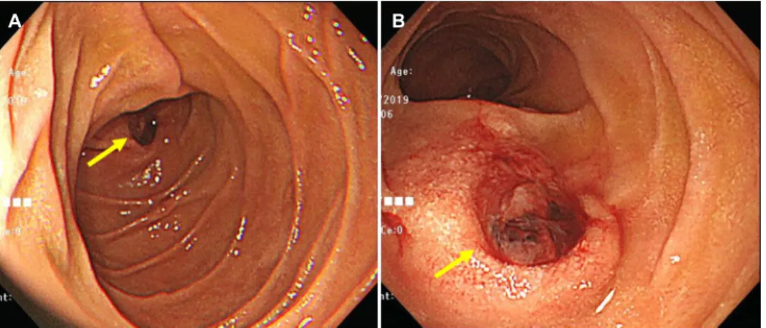
Fig. 1. Duodenal ulcer with a pulsating huge vessel at the inferior duodenal flexure. (A) Duodenal ulcer as viewed from the second portion of the duodenum. (B) Duodenal ulcer observed in the proximal third portion of the duodenum. Large blood vessels with pulsation are also observed. The yellow arrows indicate the duodenal ulcer suggesting the presence of an aortoenteric fistula.
A
A BB
Fig. 2. Abdominal computed tomography (CT) scan shows an infrarenal abdominal aortic aneurysm measuring 5 cm, along with an adjacent fat strand abutting the small bowel. (A) Coronal view of the abdominal CT scan. (B) Axial view of the abdominal CT scan. The yellow arrows indicate the suspected site of the aortoenteric fistula.
aortic aneurysm at another hospital. No abdominal tender- ness was observed during the physical examination at the time of admission. Esophagogastroduodenoscopy revealed an ulcer measuring approximately 1.3 cm with a huge pulsating vessel in the proximal third portion of the duodenum (Fig. 1).
An abdominal CT scan revealed an infrarenal abdominal aort- ic aneurysm measuring 5 cm with adjacent fat stranding abut- ting to the small bowel (Fig. 2). These observations confirmed the clinical diagnosis of PADF. The patient was prepared for emergency surgery, but the massive bleeding occurred again.
Unfortunately, he died in the preoperative stage.
DISCUSSION
PAEF is an extremely rare cause of upper gastrointestinal bleeding, with an incidence of 0.04-0.07% in autopsy
studies.3,4 The annual incidence rate of PAEF has been esti- mated to be 0.007 per million population.3-5 PADF is prevalent in the third and fourth portions of the duodenum because of its location close to the aorta.6,7 A report published in 2008 reviewed 366 cases of PAEF reported over the last 165 years since Salmon reported the first case of PAEF in 1843. Among 366 cases of PAEF, 267 cases (72.9%) were PADF.6 Approximately 80% of PAEFs are associated with athero- sclerotic aortic aneurysms, while the other 20% are asso- ciated with infections, foreign bodies, post-traumatic pseudoa- neurysms, tumors, and radiation therapy.8
The classic triad of PAEF is abdominal pain, upper gastro- intestinal bleeding, and a pulsating abdominal mass. On the other hand, these symptoms have been reported to appear in only 11% of cases. Other known symptoms include hema- temesis, pain in the back with or without the abdomen, mele-
134 김병연, 김기배. 원발성 대동맥-십이지장루 1예
The Korean Journal of Gastroenterology
na, shock, hematochezia, and syncope.4 PAEF usually pres- ents with spontaneous intermittent gastrointestinal bleeding before massive bleeding. Intermittent gastrointestinal bleed- ing is usually mild and self-limiting and is probably due to low blood pressure and the formation of thrombus.4 Thrombus may plug the fistula and temporarily stop bleeding. On the other hand, upper endoscopy and excessive volume supply can promote massive gastrointestinal bleeding.7 The time in- terval between intermittent herald gastrointestinal bleeding and massive bleeding can range from a few hours to months.9,10 In this case, a patient visits the hospital because of a small amount of gastrointestinal bleeding and eventually dies from massive bleeding; it is sometimes mistaken for a treatment error.
Upper endoscopy is the diagnostic modality of choice for acute upper gastrointestinal bleeding.11 On the other hand, stable patients often do not have current bleeding, and endos- copy rarely reveals confirmatory evidence of PAEF. In addition, endoscopic visualization of the lower third part of the duode- num, the most common area of PAEF, is complicated and is generally not observed by the endoscopist.4,7 Owing to the aforementioned reasons, the diagnosis of PAEF is achieved mainly by an abdominal CT scan or autopsy and is rarely diag- nosed with endoscopy.
Currently, the standard for a diagnosis is CT with intra- venous contrast.12 Compared to endoscopy or angiography, CT is less invasive, easier to use, and has no risk of promoting massive gastrointestinal bleeding. The presence of air in the retroperitoneum and thrombus, along with the loss of the fat plane, is probably an indicator of PADF.6 Arterial angiography is useful only when there is active bleeding. If there is no active bleeding or a small amount of bleeding, the condition is impossible to diagnose, even with an examination. As a result, a re-examination may be required to confirm the diag- nosis of PADF.6
The treatment for PAEF is the same as that for a secondary aortoenteric fistula and involves an aortic reconstruction and duodenal repair with an exploratory laparotomy.1 Multivariate analysis of 791 patients with primary and secondary aorto- duodenal fistula suggested that an aortic reconstruction is an independent predictor of survival.13 Recently, successful endovascular treatment of PADF was reported. Endovascular repair is becoming an attractive treatment option. On the oth- er hand, endovascular stent removal is required in patients
with PADF months after successful initial endovascular treat- ment because of the high risk of infection with endovascular stent grafts.14 To the best of the authors’ knowledge, there are no reports of success with PAEF treatment through endoscopy. A case of attempting endoscopic treatment of PAEF was reported. In that case, the patient had a large amount of bleeding several hours after the endoscopy and died while being prepared for emergency surgery.15 In the authors’ opinion, endoscopic devices, such as clips used for endoscopic hemostasis, can damage the aneurysm wall. A damaged aneurysm causes massive bleeding and is asso- ciated with a poor prognosis.
Early diagnosis of PADF is difficult because of its non-spe- cificity and subtle clinical features.15,16 Moreover, it is often overlooked as the cause of upper gastrointestinal bleeding owing to its rarity. The mortality rate of untreated PAEF is almost 100%.2,4,16 The key to an early diagnosis of PAEF is the suspicion of the endoscopist. The doctors should recog- nize PAEF as a potential cause of gastrointestinal bleeding.
This is particularly true for patients with a history of abdominal aortic aneurysms. Finally, if a duodenal ulcer has a pulsating nature at a location other than a common peptic ulcer in the endoscope, endoscopic hemostasis should not be at- tempted, and an abdominal CT scan should be performed for confirmation.
REFERENCES
1. Ishimine T, Tengan T, Yasumoto H, et al. Primary aortoduodenal fistula: a case report and review of literature. Int J Surg Case Rep 2018;50:80-83.
2. Ranasinghe W, Loa J, Allaf N, Lewis K, Sebastian MG. Primary aortoenteric fistulae: the challenges in diagnosis and review of treatment. Ann Vasc Surg 2011;25:386.e1-386.e3865.
3. Voorhoeve R, Moll FL, de Letter JA, Bast TJ, Wester JP, Slee PH.
Primary aortoenteric fistula: report of eight new cases and review of the literature. Ann Vasc Surg 1996;10:40-48.
4. Saers SJ, Scheltinga MR. Primary aortoenteric fistula. Br J Surg 2005;92:143-152.
5. Hughes FM, Kavanagh D, Barry M, Owens A, MacErlaine DP, Malone DE. Aortoenteric fistula: a diagnostic dilemma. Abdom Imaging 2007;32:398-402.
6. Lozano FS, Muñoz-Bellvis L, San Norberto E, Garcia-Plaza A, Gonzalez-Porras JR. Primary aortoduodenal fistula: new case re- ports and a review of the literature. J Gastrointest Surg 2008;12:1561-1565.
7. Ihama Y, Miyazaki T, Fuke C, et al. An autopsy case of a primary aortoenteric fistula: a pitfall of the endoscopic diagnosis. World
Kim BY and Kim KB. Primary Aortoduodenal Fistula Leads to Melena 135
Vol. 77 No. 3, March 2021 J Gastroenterol 2008;14:4701-4704.
8. Vilas-Boas F, Azevedo F, Marques M, et al. Primary aortoenteric fistula. Clin J Gastroenterol 2013;6:299-302.
9. Sweeney MS, Gadacz TR. Primary aortoduodenal fistula: mani- festation, diagnosis, and treatment. Surgery 1984;96:492-497.
10. Finch L, Heathcock RB, Quigley T, Jiranek G, Robinson D.
Emergent treatment of a primary aortoenteric fistula with N-butyl 2-cyanoacrylate and endovascular stent. J Vasc Interv Radiol 2002;13:841-843.
11. Barkun AN, Almadi M, Kuipers EJ, et al. Management of non- variceal upper gastrointestinal bleeding: guideline recom- mendations from the international consensus group. Ann Intern Med 2019;171:805-822.
12. Korkut AK, Arpinar E, Yasar T, Guney D. Primary aortoduodenal fistula complicated by abdominal aortic aneurysm. J Cardiovasc
Surg (Torino) 2000;41:113-115.
13. Rodrigues dos Santos C, Casaca R, Mendes de Almeida JC, Mendes-Pedro L. Enteric repair in aortoduodenal fistulas: a for- gotten but often lethal player. Ann Vasc Surg 2014;28:756-762.
14. Keunen B, Houthoofd S, Daenens K, Hendriks J, Fourneau I. A case of primary aortoenteric fistula: review of therapeutic challenges. Ann Vasc Surg 2016;33:230.e5-230.e13.
15. Bae HJ, Kim BW, Kim JS. a rare case of primary aortoduodenal fistula. Korean J Helicobacter Up Gastrointest Res 2020;20:
164-166.
16. Šumskienė J, Šveikauskaitė E, Kondrackienė J, Kupčinskas L.
Aorto-duodenal fistula: a rare but serious complication of gastro- intestinal hemorrhage. A case report. Acta Med Litu 2016;
23:165-168.