대흉외지 2006;39:802-804 □ 증례보고 □
- 802 - 증 례
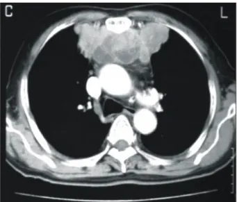
71세의 남자 환자가 2004년 8월 외부병원에서 시행한 건강 검진에서 발견된 전종격동의 종양에 대한 검사를 위 해 2004년 9월 본원에 입원하였다. 환자는 혈액 검사상 백 혈구 증가 소견과 입원 8일째부터 발생한 원인 불명의 발 열 외에 특별한 소견은 관찰되지 않았다. 입원하여 검사 한 흉부 X-ray와 외부병원에서 시행한 흉부 전산화 단층 촬영상 다엽성 전종격동 종양이 관찰되었고(Fig. 1) 전산 화 단층 촬영 하 세침 흡입술이 시행되었다. 이때 얻은 조 직으로 시행한 조직검사상 악성 섬유조직구종, 염증성 근 섬유모세포성 종양 등을 포함하는 육종이 의심되어 2004 년 9월 13일 정중 흉골 절제술을 통한 종양 제거술을 시
행하였다. 종양의 크기는 10×8×4 cm 정도로 측정되었으 며 양측 폐상엽으로의 유착이 관찰되어 종양 제거술과 함 께 양측 폐상엽의 부분 절제술, 흉선 절제술을 동시에 시 행하였다. 육안적으로 종양은 괴사와 출혈을 동반한 회백 색의 연부조직으로 관찰되었다. 조직학적으로는 소용돌이 모양의 다형성 방추 세포들과 거대 세포들로 구성되어 있 었으며 다형성의 핵과 유사분열, 그리고 산재되어 있는 림프구들이 관찰되었다(Fig. 2). 양측 폐와 흉선으로의 침 습 소견은 관찰되지 않았다. 면역조직화학검사에서는 vi- mentin, CD68, HLA-DR에 양성으로 염색되었으며 CD34, S-100, CD31, MSA 등에는 음성으로 나타났다. 이상과 같 은 결과에서 소용돌이-다형성형(storiform-pleomorphic) 악 성 섬유조직구종에 합당한 것으로 진단되었다. 수술 후
전종격동에 발생한 악성 섬유 조직구종
-1예 보고-
김 혁*․노선균*․강정호*․김영학*․정원상*․박문향**
Malignant Fibrous Histiocytoma of the Anterior Mediastinum
-A case report-
Hyuck Kim, M.D.*, Sun Kyun Ro, M.D.*, Jeong Ho Kang, M.D.*, Young Hak Kim, M.D.*, Won Sang Chung, M.D.*, Moon Hyang Park, M.D.**
Malignant fibrous histiocytoma (MFH) is a tumor which most often develops in the soft tissues of the extremities and retroperitoneum, but very rarely originates in the mediastinum. We report a 71-year-old man who admitted with anterior mediastinal tumor and underwent surgical resection of tumor in our hospital. The mass was histologically confirmed as MFH.
(Korean J Thorac Cardiovasc Surg 2006;39:802-804) ꠏꠏꠏꠏꠏꠏꠏꠏꠏꠏꠏꠏꠏꠏꠏꠏꠏꠏꠏꠏꠏꠏꠏꠏꠏꠏꠏꠏꠏꠏꠏꠏꠏꠏꠏꠏꠏꠏꠏꠏꠏꠏꠏꠏꠏꠏꠏꠏꠏꠏꠏꠏꠏꠏꠏꠏꠏꠏꠏꠏꠏꠏꠏꠏꠏꠏꠏꠏꠏꠏꠏꠏꠏꠏꠏꠏꠏꠏꠏꠏꠏꠏꠏꠏꠏꠏꠏꠏꠏꠏꠏꠏ Key words: 1. Tumor, malignant
2. Mediastinum
3. Mediastinal neoplasms
*한양대학교 의과대학 흉부외과학교실
Department of Thoracic and Cardiovascular Surgery, College of Medicine, Hanyang University
**한양대학교 의과대학 병리학교실
Department of Pathology, College of Medicine, Hanyang University 논문접수일:2006년 5월 25일, 심사통과일:2006년 7월 18일
책임저자:김영학 (133-792) 서울시 성동구 행당동 산 17번지, 한양대학교 의과대학 흉부외과학교실 (Tel) 02-2290-8465, (Fax) 02-2290-8462, E-mail: [email protected]
본 논문의 저작권 및 전자매체의 지적소유권은 대한흉부외과학회에 있다.
김 혁 외 Malignant Fibrous Histiocytoma
- 803 -
특별한 문제는 없었으며 방사선 치료 계획 하에 21일째 퇴원하였다.이후 타병원에서 방사선 치료를 시행하였으며 본원 외 래 추적관찰 중 2005년 10월 기침, 가래 등의 증상이 발현 하여 흉부 전산화 단층 촬영을 시행하였고 우상엽에 새로 이 나타난 종괴를 관찰할 수 있었다(Fig. 3). 입원 후 세침 흡입술 검사를 시행하였고, 조직검사상 악성 섬유조직구종 의 전이로 진단되어 2005년 10월 4일 우측 개흉을 통한 폐 우상엽 절제술을 시행하였다. 전이된 종양은 5×4×3 cm의 크기로 흉막으로의 침습 소견은 관찰되지 않았고 조직학적 으로 2004년 9월 전종격동에 발생했던 악성 섬유조직구종
의 전이로 확진되었다.
이후 추가 항암 치료를 시행하였으며(cytoxan, adriamycin, cisplatin) 2차 항암치료(ifosfamide, vincristin, adriamycin)까지 시행 후 추적 관찰 중이다.
고 찰
악성 섬유조직구종은 O'Brien과 Stout에 의해 1964년 처 음으로 기술된 질환으로[1] 성인기에 가장 흔한 연부조직 육종이다[2]. 주로 사지(68%) 또는 몸통(16%)의 심부 근막 과 골격근에 발생하며 종격동에 발생하는 경우는 매우 드
Fig. 1. Initial chest CT showed a multilobulated mass in the anterior
mediastinum.
Fig. 3. Chest CT 13 months after
complete surgical resection (B) show- ed a newly developed large mass in the right upper lobe, which had not been seen on the chest CT 10 months after operation (A).A B
Fig. 2. The cellular tumor is composed mainly of highly pleomorphic
spindle-shaped cells and mononuclear or multinucleated giant cells.The spindle tumor cells show focal storiform pattern. Multinuleated giant cells show a foamy or vacuolated cytoplasm (H&E stain, ×400).
대흉외지 2006;39:802-804
- 804 -
물다[2]. 현재까지 보고된 바에 따르면 종격동에 발생하는 악성 섬유조직구종 중에서 후종격동에 발생하는 경우가 가장 많으며 전종격동에 발생하는 경우는 그 다음으로 나 타난다[3].악성 섬유조직구종은 조직학적으로 (1) storiform-pleomor- phic형, (2) myxoid형, (3) giant cell형, (4) inflammatory형, (5) angiomatoid형의 5가지로 나뉘며 다엽성 회백색의 종괴, 세 포학적 다형성증, 소용돌이 모양이나 다형성 또는 섬유성 세포 배열 등의 특징을 통해 진단할 수 있다[2].
종격동에 발생하는 악성 섬유조직구종의 경우 임상적 증상이 나타나는 경우 주로 흉통으로 나타나며 발열이나 체중감소, 호흡 곤란, 식욕 감소 등으로 나타나는 경우도 있다[3]. 본 증례의 경우 특별한 임상 증상 없이 건강 검 진상 발견되었으나 입원 후 발열과 백혈구 증가 소견이 관찰되었으며 술 후 발열과 백혈구 증가 소견은 사라졌 다. 이렇게 발열과 백혈구 증가 등이 나타나는 원인에 대 해서는 명확히 밝혀지지 않았으나 종양에서 나오는 lym- phokine의 분비 때문이라는 보고도 있다[4].
악성 섬유조직구종의 진단에 도움이 되는 검사로는 α1- antichymotrypsin과 α1-antitrypsin 등이 종양 표지자로 사용 될 수 있으며[5] 종양의 침범 부위나 혈액 공급과 같은 주 변 조직과의 관계를 파악하기 위해 술 전 자기 공명 영상 이 도움이 되는 경우도 있다[6].
종격동에 발생한 악성 섬유조직구종의 치료는 수술적 절제가 우선이며 가장 효과적인 방법으로 현재 알려져 있 다[3]. 술 후 추가적인 방사선 치료가 국소 재발의 위험성 을 낮추는데 도움이 된다는 보고도 있으나[7] 추가 방사선 치료에도 불구하고 국소 재발을 하는 경우가 종종 있어 이에 대한 추가적인 연구가 필요할 것으로 보인다. 또한 항암 약물치료는 대부분의 경우에서 효과적이지 않은 것 으로 보고하고 있으며[3] Muneta 등은 후종격동의 악성 섬유조직구종의 interferon α-2a와 OK-432를 이용한 면역
치료 결과 장기적으로 부분적 관해가 나타났다고 보고하 고 있다[8]. 그러나 대부분 경우에서 수술적 절제에도 불 구하고 그 예후가 좋지 않은 것으로 보고하고 있어[3] 보 다 많은 연구를 통한 치료 방법의 확립이 필요할 것으로 보인다.
저자들은 국내에서는 처음으로 전종격동에 발생한 악 성 섬유조직구종을 경험하였기에 문헌 고찰과 함께 보고 하는 바이다.
참 고 문 헌
1. O'Brein JE, Stout AP. Malignant fibrous xanthomas. Cancer 1964;17:1445-55.
2. Enzinger FM, Weiss SW. Soft tissue tumors. 2nd ed. St Louis:
CV Mosby. 1988.
3. Murakawa T, Nakajima J, Fukami T, Tanaka M, Takeuchi E, Takamoto S. Malignant fibrous histiocytoma in the anterior
mediastinum. Jpn J Thorac Cardiovasc Surg 2001;49:722-7.
4. Aggarwal P, Sharma SK, Dey AB, Chattopadhyay TK, Mathur M. Malignant fibrous histiocytoma of the mediastinum. Postgrad Med J 1989;65:929-31.
5. Chowdhury LN, Swerdlow MA, Jao W, Kathpalia S, Desser RK. Postirradiation malignant fibrous histiocytoma of the lung:
demonstration of alpha1-antitrypsin-like material in neoplastic cells. AJCP 1980;74:820-6.
6. Morshuis WJ, Cox AL, Lacquet LK, Mravunac M, Barentsz JO. Primary malignant fibrous histiocytoma of the mediastinum.
Throax 1990;45:154-5.
7. Le Doussal V, Coindre JM, Leroux A, Hacene K, Terrier P, Bui NB, et al. Prognostic factors for patients with localized
primary fibrous histiocytoma: a multicenter study of 216 pa- tients with multivariate analysis. Cancer 1996;77:1823-30.
8. Muneta S, Kohno N, Matsuoka H, Hiwada K, Ueda N. Pro-
longed partial remission of malignant fibrous histiocytoma in posterior mediastinum by immunotherapy. Chest 1992;101:
1163-5.
=국문 초록=
악성 섬유 조직구종은 사지나 후복막의 연부조직에 주로 발생하는 종양으로 종격동에 발생하는 경우 는 극히 드물다. 환자는 71세 남자 환자로 전종격동에 발생한 원발성 종양에 대해 종양 제거술을 시 행하였고 조직학적으로 악성 섬유 조직구종으로 진단되었다.
중심 단어: 1. 악성 종양 2. 종격동 3. 종격동종양