The Antifibrotic and Antioxidant Activities of Hot Water Extract of Adventitious Root Culture of Panax ginseng (ARCP)
Hee Kyoung Lim
1, Youn Woo Kim
2, Dae Ho Lee
3, Somi Kim Cho
4,5and Moonjae Cho
1,*
1Departments of Biochemistry, 2Department of Pediatrics, 3Department of Internal Medicine, College of Medicine,
4Faculty of Biotechnology, College of Applied Life Sciences, and 5TheResearch Institute for Subtropical Agriculture and Biotechnology, Cheju National University, Jeju 690-756, Korea
Received April 11, 2007; Accepted June 15, 2007
The anti-fibrotic effects of hot water extract of adventitious root culture of Panax ginseng (ARCP) and the possible mechanisms were investigated on CCl4-induced hepatotoxicity model mice.
Fibrosis was induced by a mild treatment of CCl4. Then silymarin as a positive control drug and ARCP or carrier alone as a negative control were treated. Serum GPT, GOT and ALP activity levels were lowered by 25, 21 and 11% for silymarin treated group and by 48, 39 and 14% for ARCP treated group compared to carriertreated alone. Hepatic collagen for ARCP treatment group was reduced by 18% and MDA contents decreased a little more. Pro-fibrotic gene (TGF-β1, TIMP-1 and α-SMA) expression increased following the CCl4 treatment, but both the silymarin and the ARCP treatments decreased the expressions of these genes by 20% to 50%. The antioxidant effect of ARCP was studied by DPPH free radical scavenging activity. In addition, a generation of reactive oxygen species (ROS) was also reduced in H2O2-treated HepG2 cells upon the ARCP treatment. In summary, ARCP has antioxidant property, and can have some protection against oxidative stress; more importantly, ARCP can efficiently protect mice against CCl4- induced fibrosis.
Key words: adventitious root culture, anti-fibrotic effect, antioxidant effect, Panax ginseng, tetrachloromethane
Recently, improvements of the quality of life worldwide has spurred great interests in human health and the longevity of life. Wild ginseng has been considered as a remedy for a broad spectrum of diseases from the past.
Panax ginseng C.A. Meyer (Araliaceae) is a valuable herb in East Asia, and because of its pharmacological properties, it has also gained popularity in the West [Mahady et al., 2000]. Saponins of wild ginseng have shown to improve immunity function. The immuno- modulating effects of wild and cultured ginseng were compared recently [Mizuno et al., 1994]. There are reports of decrease in cholesterol [Lee et al., 2003], and
whitening effects [Shin, 2001] of wild ginseng roots culture. Especially, ginseng has been found to have liver- protection effect via a cytochrom P450 inhibition to reduce CCl4-induced hepatic lipid peroxidation [Kim et al., 1997].
Traditionally, P. ginseng is categorized as an either cultivated or wild according to the different nurturing methods. Recently, adventitious root culture technique of wild P. ginseng has been developed to make mass production possible in a large culture system. However, only a few studies have been reported concerning the pharmacological activities of ARCP [Yoo et al., 2003].
Fibrosis reflects increased the deposition of the physiological components of extracellular matrix, which is the hallmark of aging of various organs, including heart and kidneys. Aging is also associated with variable degrees of fibrosis in the liver [Sun et al., 1998]. OS might represent a direct or an indirect profibrogenic stimulus for HSC, suggested by in vivo experimental studies of antioxidants preventing OS, lipid peroxidation and liver fibrosis [Chao et al., 2002]. Among the various cytokines, TGF-β1 plays an important role as a
*Corresponding author
Phone: +82-64-754-3837; Fax: +82-64-725-2593 E-mail: [email protected]
Abbreviations: ALP, alkaline phosphatase; α-SMA, α-smooth muscle actin; ALT, alanine transaminase; ARCP, adventitious root culture of Panax ginseng; AST, aspartate aminotransferase;
HSC, hepatic stellate cells; MDA, malondialdehyde; OS, oxida- tive stress; ROS, reactive oxygen species; TGF-β1, transforming growth factor β-1; TIMP-1, tissue inhibitors of metalloprotease
profibrogenic factor in chronic liver disease, triggering the expression of procollagen-I and TIMP-1, the key effectors of fibrogenesis [Jeong et al., 2005].
Tetrachloromethane (CCl4) induced liver damage has been extensively studied and widely used as a model for screening hepato-protectors. CCl4 is capable of causing liver necrosis, changing the activities of metabolic enzymes in liver [Noguchi et al., 1982], as well as increasing the hepatic lipid peroxidation [Comporti, 1985]. Because the damaging effects of CCl4 are generated by oxidative stress, many antioxidants [Ilavarasan et al., 2003]have been tried as hepato-protective agents.
In this study, we created a liver damage by pre- treatment of mice with CCl4 for a month and then treated them with water extracts of ARCP to test the protective or healing effects.This paper reports the mechanism of the anti-fibrotic effect of water extracts of ARCP on CCl4- induced mouse liver.
Materials and Methods
Chemicals. All the plastic materials were purchased from Falcon Labware (Becton-Dickinson, Franklin Lakes, NJ, USA). Silymarin, trichloroacetic acid and 2- thiobarbituric acid were obtained from Sigma (St Louis, MO, USA), and CCl4 was obtained from Aldrich (Milwaukee, WI, USA). Dulbecco’s Modified Eagle’s Medium and fetal bovine serum were purchased from Gibco BRL (Grand Island, NY, USA). Other chemicals and reagents were of the highest quality available.
Plants materials and extraction procedure. ARCP was supplied by Biovalue Co. (Jeju, Korea). Hot water extract of ARCPwas prepared as follow: Five kilograms of wet weight ARCP was placed in the extractor (Cosmos, Inchon, Korea) with 2 L distilled water and extracted for 9 h at 120oC under reduced pressure. The remaining liquid was collected, lyophilized and stored for further use.
Animals. Six-week-old male Balb/c mice about 22 g were obtained from Daehan Lab. (Animal Research Center Co. Ltd, Daejeon, Korea). They were acclimated for 1 week before dosage under controlled environmental conditions at 23oC and 65% relative humidity with a 12-h light/dark cycle. Feed and water were available ad libitum.
Experimental design. Mice were divided into five groups with five mice per group (control, CCl4, silymarin and two different dose ARCP treatment groups). Liver fibrosis was induced in mice by the intraperitoneal injection of CCl4 (0.15 mL per mouse (diluted 1 : 1 in mineral oil)) once a week for up to 4 weeks. After liver fibrosis was induced, the groups of mice were orally
given silymarin (50µg/g body weight) or ARCP (100 or 600µg/g body weight) dissolved in 0.5% sodium carboxymethylcellulose (CMC) every day during a week.
Measurement of AST, ALT and ALP. After treatment, blood was obtained by heart puncture and centrifuged at 2500×g for 20 min at 10oC to separate the sera. Activities of AST, ALT and ALP in the serum were measured according to manufacturer’s manual using Randox TM kit and Screenmaster colorimeter set.
Collagen contents and staining. After development of fibrosis, mice were anaesthetized and laparotomized. The liver was perfused with phosphate-buffered saline and a part of the liver was removed and quickly frozen in liquid nitrogen. The remaining part of the liver was perfusion- fixed with 4% formaldehyde, dehydrated and embedded in Polybed. Sections were cut at a thickness of 5 mm and stained for 1 h in 0.1% (w/v) Sirius red (Direct Red 80, Aldrich, Milwaukee, WI).
A portion of each liver was homogenized in 33 volumes (mL/g) of 0.5 M acetic acid at 4oC using a Polytron PCU- 2 homogenizer (Kinematica, Luzern, Switzerland), and the homogenate was disrupted by freeze-thawing and sonication for 2 min for collagen determination [Shimizu et al., 1999]. The results are expressed as micrograms of collagen per milligram of liver tissue.
Polymerase chain reaction for TGF-β1, TIMP-1, and α-SMA. Total RNA was extracted using Trizol (Biostar Biologic technology Co. Ltd. USA.) according to the manufacturer’s directions. Then total RNA was reverse-transcribed into cDNA. PCR system contained 2
µL cDNA, 5µL 10 × buffer, 5µL 25 mmol/L MgCl2, 1µL 10 mmol/L dNTP, 1µL 20 pmol/µL target gene sense and anti-sense primer, 1µL 20 pmol/µL β-actin primer pair, 3 Unit of Taq DNA polymerase. PCR was carried out as followed: initial denaturation at 95oC for 5 min, 30 amplification cycles (denaturation at 94oC for 45 s, annealing at 55oC for 30 s, extension at 72oC for 1 min) and final extension at 72oC for 7 min. The primers used were listed in Table 1.
Electrophoresis and semi-quantitative analysis. The PCR products were run on 2% agarose gel electrophoresis and visualized with ethidium bromide staining. The expected product sizes were 170 bp of TGF-β1, 170 bp of TIMP-1, 74 bp of α-SMA and 188 bp of β-actin. GS-710 calibrated imaging densitometer system (Bio-RAD) was employed to detect the density of bands of PCR products.
The values of TGF-β1, TIMP-1 and α-SMA expression were semi-quantified by scanning densitometry using the ratios of TGF-β1/β-actin , TIMP-1/β-actin and α-SMA to assess the relative level.
Determination of antioxidant activity using DPPH.
The free radical scavenging activity of the ARCP extracts
was measured by the 2,2-diphenyl-1-picrylhydrazyl (DPPH) radical assay described by Blois. ARCP at final concentrations of 2.5, 1, 0.5, and 0.25 mg/mL were tested individually by addition to 150 mM solution of DPPH in ethanol. The mixtures were vigorously mixed and allowed to stand in the dark at RT for 4hrs. The absorbance of the remaining DPPH was measured at 520 nm using a spectrophotometer.
These experiments were run in triplicate. The radical scavenging activity was calculated according to following equation:
Radical scavenging activity (%) = [(Ao-A)/Ao]*100%, where Ao is the A520 of DPPH without sample (control), A is the A520 of DPPH with sample.
Cell culture. The human hepatoma HepG2 (KCLB No.58065) cells were purchased from the Korea Cell Line Bank (Seoul, Korea). They were grown in Dulbecco’s Modified Eagle’s Medium supplemented with 10% (v/v) heat-inactivated fetal bovine serum, 100 units/mL of penicillin, and 100µg/mL of streptomycin. Cells were maintained in a humidified incubator at 37oC in a 5% CO2 atmosphere. The culture medium was changed twice a week, and the cells were subcultured at 1 : 4 ratio once a week.
Determination of reactive oxygen species. Cellular ROS were quantified by dichlorofluorescein (DCFH) assay, in which the esterified form of 2'’,7'-dichoro- fluoresceindiacetate (DCFH-DA) diffuses through the cell membrane and is enzymatically deacetylated by intracellular esterase. The resulting compound, dichloro- fluorescein (DCFH), is reactive with ROS to give an oxidized fluorescent compound, dichlorofluorescein (DCF). HepG2 cells were pretreated with ARCP for 30 min, then washed twice and incubated with 1 mM H2O2 for 30 min. DCFH-diacetate was added to the culture plates at a final concentration of 12 mM and incubated for 30 min at 37oC. DCF fluorescence was detected at an excitation wavelength of 485nm and an emission wavelength of 530 nm using Genios multiwell fluorescence plate reader (GENios, Tecan, Salzburg, Austria).
Effect of wild ginseng on lipid peroxidation inhibitory activity in vivo. Lipid peroxidation was assayed by measuring malondialdehyde (MDA) according to the method of Ohkawa et al. In order to evaluate the inhibitory activity of the ARCP on a lipid peroxidation
generated assay system in vivo. Liver tissue were sliced and homogenized (13,000 rpm, 3 min) with 25 mM Tris- HCl buffer (pH 7.2) (10% w/v). In a glass test tube, 0.1 mL of the liver homogenate was incubated with shaking for 1 h at 37°C in Tris-HCl buffer (pH 7.2), and then 1.5 mL of 1.0% TBA and 1.5 mL of 20% acetic acid were added and incubated continuously for 1 h at 95oC. The upper lipid solution was then extracted for spectro- photometer analysis at 532 nm absorption.
Statistical analysis. Experimental data were analyzed by Student’s t-test and expressed as mean ± SE. A probability value of p< 0.05 was considered statistically significant.
Results and Discussion
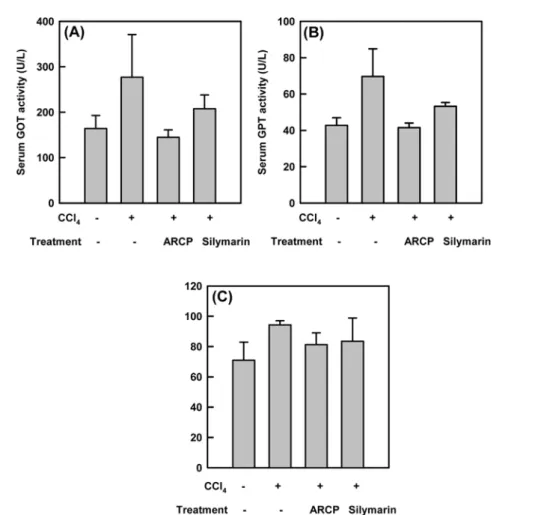
Serum GPT and serum GOT and alkaline phosphatase activities. After liver fibrosis was induced by the intraperitoneal injection of CCl4 once a week for 4 weeks, silymarin or ARCP was introduced orally to mice every day for a week. Then, blood was obtained to measure the activities of aspartate aminotransferase (AST/GOT), alanine transaminase (ALT/GPT) and alkaline phosphatase (ALP). Serum GPT, GOT and ALP activities for CCl4- treated groups were found to be 169%, 160% and 131%, respectively, compared to the non-treated groups (Fig. 1).
However, with the addition of silymarin to these CCl4- treated groups, serum GPT, GOT and ALP activity levels showed 25, 21 and 11%, decrease respectively, compared to the groups with CCl4 treatment alone. ARCP treated group was lowered by 48, 39 and 14%, respectively. It has been generally reported and accepted that the CCl4- induced hepatotoxicity results from its hepatotoxic metabolites and trichloromethyl free radical (•CCl3) [Rechnagel and Glende, 1973]. This free radical can react with sulfhydryl groups, such as glutathione (GSH) and thiol-groups in the protein side chain. Also, it covalently binds with cell proteins, and initiates the lipid peroxidation process in the cellular membrane, which eventually leads to various liver pathological processes [Connor et al., 1990].
Collagen accumulation. Serum levels of GPT, GOT, and ALP indicated fast recovery of hepatocytes. To estimate the progress of fibrosis, we measured collagen Table 1. Primer pairs used for RT-PCR in this study
Forward Reverse
TGF-β1 TIMP-1
α-SMA
β-actin
5'-tgacgtcactggagttgtacgg-3' 5'-attcaaggctgtgggaaatg-3' 5'-cctggcttcgctgtctacct-3' 5'-aggtgacagcattgcttctg-3'
5'-gttcatgtcatggatggtgc-3' 5'-aagaagctgcaggcattgat-3' 5'-ttgcggtggacgatgga-3' 5'-gctgcctcaacacctcaac-3'
contents. For CCl4-treated groups, hepatic collagen contents had 11.6µg/mg liver weight (Fig. 2). However, for ARCP group, the hepatic collagen contents was 9.48
µg/mg liver weight. These hepatic collagen contents of ARCP-treated group were reduced by 18 % compared to that of CCl4-treated groups. The state of fibrosis is Fig. 1. Effect of ARCP administration on SGOT, SGPT and ALP levels in animals recovering from CCl4 -induced fibrosis. After liver fibrosis was induced by the intraperitoneal injection of CCl4 once a week for 4 weeks, silymarin or ARCP was introduced orally to mice every day for a week. Then, blood was obtained to measure the activities of (A) aspartate aminotransferase (AST/GOT), (B) alanine transaminase (ALT/GPT) and (C) alkaline phosphatase (ALP). Enzyme activities as the average ± SE from 5 animals.
Fig. 2. Effects of ARCP treatment on spontaneous resolution of fibrosis. After liver fibrosis was induced by the intrap- eritoneal injection of CCl4 once a week for 4 weeks, vehicle or ARCP was introduced orally to mice every day for a week.
Livers were assessed for fibrosis by determination of (A) collagen content and examination of (B) sirus red-stained sec- tions. Collagen contents as the average ± SE from 5 animals.
controlled by both the rate of development and the reversion of fibrosis. Activation of HSC is the primary event that triggers the process of fibrogenesis and is characterized by the conversion of quiescent vitamin A- storing cells into proliferative, collagen-producing myofibroblastic cells. It is believed that the reversion of fibrosis starts with the apoptosis of activated HSC.
Effect of ARCP on TGF-β1, TIMP-1 and α-SMA expression. Because the fibrosis seems to be delayed based on the collagen contents, the genes related with fibrosis were checked. The major profibrotic cytokine, TGF-β1 expression level was measured by semi-quantitative PCR (Fig. 4-A). The TGF-β1 expression in the CCl4- treated group revealed 6.4 fold increase compared to CCl4-non-treated control group. The silymarin-treated group and the ARCP-treated group had increased TGF-
β1 expression by 3.9 fold and 4.6 fold, respectively.
Since the expression of tissue inhibitors of metalloprotease (TIMPs) is reported to decrease in fibrotic tissue, the expression of TIMP-1 was semi-quantified by RT-PCR.
Figure 4-B shows that the treatment of ARCP or silymarin after the CCl4 treatment decreased the expression of TIMP-1 to about 50% and 70%, respectively, compared to the CCl4 treatment alone.
The elevated expressions of α-SMA in the activated hepatic stellate cells also characterize chronic hepatic fibrosis. In order to conform the effects of ARCP on liver fibrosis, the expressions of α-SMA in the CCl4 induced fibrosis model were measured by RT-PCR. As expected, expressions of α-SMA increased in the CCl4 induced fibrosis model animal to about 6 fold. However, silymarin treatment after induction of fibrosis reduced expression of
α-SMA to about 70%. The ARCP treatment also reduced the expression of α-SMA to about 30% (Fig. 4-C).
Therefore, the results stated above clearly demonstrate that ARCP has anti-fibrosis effects in the CCl4 induced fibrosis model animal.
Effect of ARCP on lipid peroxidation inhibitory activity. Lipid peroxidation is a freely radical-mediated propagation of oxidative insult to the polyunsaturated
Fig. 3. Reduced expression of TGF-β1, TIMP-1 and α-SMA transcripts in ARCP treated livers. Semi-quentitative revers-transcription polymerase chain reaction for expression of (A) TGF-β1, (B) TIMP-1 and (C) α-SMA transcripts in livers of mice injured for 4 weeks with CCl4 followed by recovery for a week of treatment with ARCP and silymarin.
Actin was used as an internal control. The relative level of transcription as the average ± SE from the three independent experiments.
fatty acids, involving several types of free radicals.
Termination occurs through enzymatic means or free radical scavenging activities of antioxidants.
The control’s hepatic MDA contents was 11.6µg/mg liver weight (Table 2). However, for silymarin and ARCP group, the hepatic MDA contents were 0.125 and 0.145 nmol/mg protein, which showed a decrease of 32% and 21% respectively compared to those of the CCl4 group.
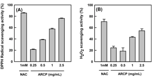
Antioxidant activity. The DPPH radical scavenging activity of ARCP was compared to that of the standard, 1 mM N-acetyl cysteine (NAC), at a concentration of 2.5 mg/mL (Fig. 4A). The radical scavenging activity of ARCP was similar to that of 1 mM NAC, which is equivalent to 0.16 mg/mL.
To investigate ROS scavenging effects of ARCP upon exposure to H2O2, the level of intracellular ROS was measured by quantification of DCF fluorescence in HepG2 cells. Cells were treated for 30 min with ARCP then it was removed from the culture medium. 1 mM of H2O2 was added to the 30 min of ROS assay. H2O2 scavenging activity increased by 24, 19, 43, and 55%
with the pretreatment of ARCP at a concentration of 0.25, 0.5, 1 and 2.5 mg/mL, respectively. The results suggest that water extractof wild ginseng is capable of amelioration of hepatocyte oxidation stress caused by H2O2.
Anti-oxidant activity of ARCP may relate to loosening of the CCl4-induced hepatotoxicity because ARCP showed strong radical and H2O2 scavenging activities (Fig. 3, 4). A Decrease of MDA contents in ARCP- treated group (Table 2) might be resulted from anti- oxidant activity of ARCP. Wild P. ginseng leaves have reported to decrease lipid peroxidation in streptozotocin diabetic rats [Jung et al., 2005]. Hot water extract of P.
notoginseng was reported to contain protective effects on ethanol-induced hepatotoxicity via the reduction of
oxygen-free radical production [Lin et al., 2003]. Hepatocyte apoptotic bodies can be phagocytosed by Kupffer and HSCs, and it significantly increased superoxide production via activation of NADPH oxidase [Canbay et al., 2003].
It is well known that an exposure of HSC to a soluble factor such as a reactive oxygen radical produced from Kupffer cells leads to morphological transition to myofibroblast-like cells [Nieto et al., 2002]. Therefore, it is not an unreasonable conclusion that the radical scavenging activity of ARCP will play anti-fibrotic role by reducing ROS, thus reducing apoptosis.
However, in the experimental design of ours, it opens other possibilities of reducing fibrosis. Because the intraperitoneal injection of CCl4 at a low dose for four weeks was induced, the treatment of ARCP or the control drug was started without a co-injection of CCl4. The effects of ARCP on reducing fibrosis may partially be depended on the reversion of fibrosis. Additionally, untreated control mice undergo reversion process upon stopping injection of CCl4, but the ARCP-or drug-treated mice may reverse fibrosis faster.
In this study, we employed the methods to induce liver fibrosis first by using CCl4, and then by treating ARCP to Fig. 4. DPPH radical scavenging activity (A) and H2O2 scavenging activity (B) of ARCP and 1 mM NAC. Each experiment was performed at least 4 times and data are expressed as the average percent change from control ± SE.
Table 2. Inhibitory effect of ARCP on CCl4 induced lipid peroxidation in the mouse liver homogenate in vivo
Group MDA
(nmol/mg protein) Inhibition rate Vehicle (%)
CCl4
0.128 ± 0.0015
0.182 ± 0.0014 -
ARCP (600µg/g) -
ARCP (100µg/g) 0.145 ± 0.0013
0.150 ± 0.0018 21%
Silymarin (50µg/g) 0.125 ± 0.0016 17%32%
evaluate anti-fibrotic effects. We found the decrease of serum GPT, GOT and ALT after the treatment of ARCP.
Collagen contents also decreased significantly in ARCP treated group than the control groups. Other factors related to fibrosis such as expressions of TGF-β1, TIMP- 1 and α-SMA also decreased in ARCP treated group. Our future work will focus on the fractionation of active compounds and more detailed study on the mechanism of reducing fibrosis. Especially, we will investigate the possibility of ARCP that induces apoptosis of activated HSC.
Acknowledgments. This work was supported by Korean’s Small and Medium Business Administration (SMBA) grant (S050422-M1640375-101000000), the research grant of the Cheju National University in 2006 and by the Brain Korea 21 Project in 2006.
References
Canbay A, Feldstein AE, Higuchi H, Werneburg N, Gram- bihler A, Bronk SF, and Gores GJ (2003) Kupffer cell engulfment of apoptotic bodies stimulates death ligand and cytokine expression. Hepatology38,1188-1198.
Chao C, Youssef J, Rezaiekhaleigh M, Birnbaum LS, and Badr M (2002) Senescence-associated decline in hepatic peroxisomal enzyme activities corresponds with dimin- ished levels of retinoid X receptor alpha, but not peroxi- some proliferator-activated receptor alpha. Mech Ageing Dev 123, 1469-1476.
Comporti M (1985) Lipid peroxidation and cellular damage in toxic liver injury. Lab Invest53, 599-623.
Connor HD, Lacagnin LB, Knecht KT, Thurman RG, and Mason RP (1990) Reaction of glutathione with a free radical metabolite of carbon tetrachloride. Mol Pharma- col37, 443-451.
Ilavarasan R, Vasudevan M, Anbazhagan S, and Venkatara- man S (2003) Antioxidant activity of Thespesia popul- nea bark extracts against carbon tetrachloride-induced liver injury in rats. J Ethnopharmacol87, 227-230.
Jeong DH, Lee GP, Jeong WI, Do SH, Yang HJ, Yuan DW, Park HY, Kim KJ, and Jeong KS (2005) Alterations of mast cells and TGF-β1 on the silymarin treatment for CCl4-induced hepatic fibrosis. World J Gastroenterol 11, 1141-1148.
Jung CH, Seog HM, Choi IW, Choi HD, and Cho HY (2005) Effects of wild ginseng (Panax ginseng C.A.
Meyer) leaves on lipid peroxidation levels and antioxi- dant enzyme activities in streptozotocin diabetic rats. J Ethnopharmacol98, 245-250.
Kim HJ, Chun YJ, Park JD, Kim SI, Roh JK, and Jeong TC (1997) Protection of rat liver microsomes against carbon tetrachloride-induced lipid peroxidation by red ginseng saponin through cytochrome P450 inhibition.
Planta Med63, 415-418.
Lee EJ, Jhao HL, Li DW, Jung CS, Kim JH, and Kim YS (2003) Effect of MeOH extract of adventitious root cul- ture of Panax ginseng on hyperlipidemic rat induced by high fat-rich diet. Kor J Pharmacogn34, 179-184.
Lee KJ and, Jeong HG (2002) Protective effects of Platy- codi radix on carbon tetrachloride-induced hepatotoxic- ity. Food Chem Toxicol40, 517-525.
Lin CF, Wong KL, Wu RS, Huang TC, and Liu CF (2003) Protection by hot water extract of Panax notoginseng on chronic ethanol-induced hepatotoxicity. Phytother Res17, 1119-1122
Mahady GB, Gyllenhall C, Fong HH, and Farnsworth NR (2000) Ginsengs: a review of safety and efficacy. Nutr Clin Care3, 90-101.
Mizuno M, Yamada J, Terai H, Kozukue N, Lee YS, and Tsuchida H (1994) Differences in immunomodualating effects between wild and cultured Panax ginseng. Bio- chem Biophys Res Commun200, 1672-1678.
Nieto N, Friedman SL, and Cederbaum AI (2002) Cyto- chrome P450 2E1-derived reactive oxygen species medi- ate paracrine stimulation of collagen I protein synthesis by hepatic stellate cells. J Biol Chem 277,9853-9864.
Noguchi T, Fong KL, Lai EK, Alexander SS, King MM, Olson L, Poyer JL, and . McCay PB (1982) Specificity of a phenobarbital-induced cytochrome P-450 for metab- olism of carbon tetrachloride to the trichloromethyl radi- cal. Biochem Pharmacol31, 615-624.
Rechnagel RO and Glende EA Jr (1973) Carbon tetrachlo- ride hepatotoxicity: an example of lethal cleavage. CRC Crit Rev Toxicol2, 263-297.
Shimizu I, Ma YR, Mizobuchi Y, Liu F, Miura T, Nakai Y, Yasuda M, Shiba M, Horie T, Amagaya S, Kawada N, Hori H, Ito S (1999a) Effects of Sho-saiko-to, a Japa- nese herbal medicine, on hepatic fibrosis in rats. Hepa- tology29, 149-160.
Shin MH (2001) Study of mountain ginseng adventitious culture and application. J Soc Cosmet Scientists Korea
27, 45-56.
Sun WB, Han BL, Peng ZM, Li K, Ji Q, Chen J, Wang HZ, and Ma RL (1998) Effect of aging on cytoskeleton system of Kupffer cell and its phagocytic capacity. World J Gastroenterol4, 77-79.
Yoo BS, Chang MS, and Byun SY (2003) Characterization of cell cultures and ginsenoside production by cultured ginseng and wild mountain ginseng. Korean J Biotech- nol Bioeng 18, 133-139.