351
Copyrights © 2013 The Korean Society of Radiology
INTRODUCTION
Chronic expanding hematoma is a clinicopathologic entity that is characterized by its increasing size over one month after the initial event of hemorrhage (1). It may resemble malignant neoplasm for its large size, and slow but progressive enlarge- ment (1). Chronic expanding hematoma may occur in various locations, and several studies have sporadically reported that it occurred in the soft tissue, sinonasal cavity, brain and lung, as well as rarely in the adrenal gland (2-9).
Here, we present a case of chronic expanding hematoma, which occurs in the adrenal gland mimicking a hemangioma on multi- phase CT, and review the relevant literature on this rare lesion.
CASE REPORT
A 57-year-old man was admitted to our hospital complaining of epigastric fullness and abdominal distension in the left upper
quadrant area for two months. He performed coronary artery bypass graft nine years ago for unstable angina and he had been taking aspirin after this procedure. He had also been treated with medication for hypertension and diabetes mellitus. Physi- cal examination showed a more than palm-sized, hard mass without tenderness in the left upper quadrant of the abdomen.
Laboratory findings were all in normal range, including the 24- hour urine catecholamine and serum aldosterone level, platelet number, prothrombin time and activated partial thromboplas- tin time. Multiphase CT was performed 40 and 70 seconds after the contrast injection to obtain the hepatic arterial and portal venous phase images. Unenhanced CT scans showed a well-de- fined, ellipsoid mass with heterogeneous attenuation between the spleen and left kidney in the left side of the retroperitoneum, measuring 20 cm in its maximal diameter (Fig. 1A). Calcifica- tion or macroscopic fat was not present within the mass. Con- trast-enhanced CT scans showed several enhancing foci of ir- regular and frond-like shape in the peripheral portion of the
Case Report
pISSN 1738-2637
J Korean Soc Radiol 2013;68(4):351-354 http://dx.doi.org/10.3348/jksr.2013.68.4.351
Received August 18, 2012; Accepted January 10, 2013 Corresponding author: Min-Jeong Kim, MD Department of Radiology, Hallym University Sacred Heart Hospital, Hallym University College of Medicine, 22 Gwanpyeong-ro 170beon-gil, Dongan-gu, Anyang 431-070, Korea.
Tel. 82-31-380-3885 Fax. 82-31-380-3878 E-mail: drkmj@hallym.or.kr
This is an Open Access article distributed under the terms of the Creative Commons Attribution Non-Commercial License (http://creativecommons.org/licenses/by-nc/3.0) which permits unrestricted non-commercial use, distri- bution, and reproduction in any medium, provided the original work is properly cited.
We report a rare case of unilateral chronic expanding hematoma in the left adrenal gland, mimicking a hemangioma on multiphase computed tomography (CT). On CT, the mass showed several enhancing foci of irregular and frond-like shape in the pe- riphery at the hepatic arterial phase and gradual fill-in pattern at the portal venous phase, which was similar with the enhancement pattern of hemangioma.
Index terms Adrenal Gland Hematoma Hemangioma
Chronic Expanding Hematoma of the Adrenal Gland Mimicking a Hemangioma: A Case Report
1혈관종으로 오인된 부신에 생긴 만성 확장성 혈종: 증례 보고1
Hyun Jung Lee, MD
1, Min-Jeong Kim, MD
1, Hongil Ha, MD
1, In Jae Lee, MD
1, Kwanseop Lee, MD
1, Jin Won Seo, MD
2, Seung-Gu Yeo, MD
3Departments of 1Radiology, 2Pathology, Hallym University Sacred Heart Hospital, Hallym University College of Medicine, Anyang, Korea
3Department of Radiation Oncology, Soonchunhyang University College of Medicine, Cheonan Hospital, Cheonan, Korea
Chronic Expanding Hematoma of the Adrenal Gland Mimicking a Hemangioma
submit.radiology.or.kr
J Korean Soc Radiol 2013;68(4):351-354
352
showed a well-demarcated, round mass with yellow-tan, mea- suring 22 cm in dimension. The cut surface of the mass con- tained blood and friable hemorrhagic blood clots, and showed white-tan fibrotic wall peripherally (Fig. 1E). Microscopically, the central area of the mass was full of organized blood clots.
The fibrotic wall of the mass consisted of granulation tissue and residual adrenal cortical tissue (Fig. 1F). It did not contain any neoplastic cells. Considering these pathologic findings and its clinical expanding nature over one month, the final diagnosis was determined as chronic expanding hematoma.
DISCUSSION
Adrenal hemorrhage is uncommon in adults, and it is mainly caused by trauma, stress, septicemia, anticoagulant therapy, and systemic illness, or associated with tumors (2-6). Most adrenal he- mass at the hepatic arterial phase (Fig. 1B). These enhancing
foci increased in size progressively at the portal venous phase (Fig. 1C), but most part of the mass was not enhanced. Coronal reformatted image (Fig. 1D) demonstrated the same huge mass that displaced the left kidney downward, as well as the spleen and splenic flexure of the colon upward. The left kidney showed delayed renal excretion in contrast to the right kidney, because the left renal artery and vein were stretched and compressed downward. However, the left adrenal gland was not recognized throughout all these images. Considering these CT findings, we diagnosed this lesion as a huge cavernous hemangioma in the left adrenal gland. In addition, the possibility of adrenocortical carcinoma, retroperitoneal gastrointestinal stromal tumor, and angiosarcoma could not be excluded.
The patient underwent surgical excision of the retroperitoneal mass without postoperative complication. The resected mass
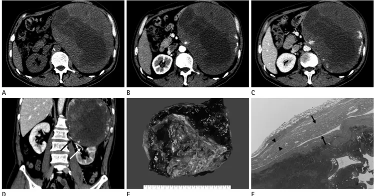
Fig. 1. A 57-year-old man with chronic expanding hematoma of the left adrenal gland.
A. Unenhanced CT scan shows a well-circumscribed ellipsoid mass with heterogeneous attenuation in the left retroperitoneal area.
B. Axial CT scan obtained during the hepatic arterial phase reveals that several strong enhancing foci of irregular shape are seen in the periphery of the mass.
C. Axial CT scan obtained during the portal venous phase demonstrates that strong enhancing foci increase in extent progressively, and some of them seem to arise slightly inside from the periphery of the mass.
D. On the coronal reformatted image, the huge mass shows the same enhancement pattern as that of Fig. 1C more clearly. Note that the mass is compressing the left kidney downward, and the left renal artery (black arrow) and vein (white arrow) are stretched, resulting in delayed excretion of the left kidney in contrast to the right kidney.
E. Gross specimen of the mass reveals smooth fibrotic wall and large hemorrhagic blood clots.
F. Microscopic examination (Hematoxylin-Eosin, × 40) demonstrates thin, peripheral fibrous tissue (arrowheads), residual adrenal cortical tissues (arrows), and central organized blood clots.
D A
E B
F C
Hyun Jung Lee, et al
submit.radiology.or.kr J Korean Soc Radiol 2013;68(4):351-354
353
As is known, this pattern of enhancement was similar to those of adrenal hemangioma (10). Besides, adrenal neoplasms ac- companying a hemorrhage, such as pheochromocytoma, ade- noma, adrenocortical carcinoma, and hemorrhagic metastases can be included in the differential diagnoses, because these neo- plasms contain both hemorrhagic components, centrally and variously, enhancing tumor component peripherally. In addi- tion, retroperitoneal gastrointestinal stromal tumor and angio- sarcoma are also needed to be included in the differential diag- noses. We also misdiagnosed our case as adrenal hemangioma before surgery. As shown in our case, differentiation between hemangioma and chronic expanding hematoma is most diffi- cult, and their enhancement pattern seemed to be somewhat different. Hemangioma usually shows globular or nodular pe- ripheral enhancement, because peripheral vascular spaces are filled with contrast material first during the hepatic arterial phase, whereas chronic expanding hematoma may demonstrate both central and peripheral enhancement of irregular shape depend- ing upon the distribution and shape of the granulation tissue (4, 10). Our case also showed peripheral enhancement of irregular and frond-like shape, some of which arose slightly inside from the periphery of the mass. Based on these CT findings, our case seems more likely to be chronic expanding hematoma rather than hemangioma.
In conclusion, we describe a rare case of chronic expanding hematoma in the adrenal gland, which is difficult to diagnose preoperatively, unless it is acknowledged and suspected. Despite its rarity, chronic expanding hematoma needs to be included in the differential diagnoses of unilateral adrenal mass, which shows various patterns of peripheral enhancement and contains internal hemorrhagic component. Moreover, understanding of their different enhancement patterns is helpful in diagnosing chronic expanding hematoma correctly.
REFERENCES
1. Reid JD, Kommareddi S, Lankerani M, Park MC. Chronic expanding hematomas. A clinicopathologic entity. JAMA 1980;244:2441-2442
2. Kawashima A, Sandler CM, Ernst RD, Takahashi N, Roubi- doux MA, Goldman SM, et al. Imaging of nontraumatic hemorrhage of the adrenal gland. Radiographics 1999;19:
matomas decrease in size over time, and resolve completely with- out significant clinical problems. However, some hematomas may persist for a long period and appear incidentally as adrenal mass simulating neoplasm. These chronic adrenal hematomas have sporadically been reported in the literature, and diversely called as idiopathic or spontaneous adrenal hematoma, organizing or or- ganized hematoma, or chronic expanding hematoma (2-6).
Chronic expanding hematoma is a term proposed by Reid et al. (1). It is characterized by a persistent hematoma that increas- es in size for more than one month after initial bleeding, no neoplastic cell on pathologic examination, and no bleeding ten- dency. The expanding nature of the hematoma is speculated to be caused by the irritation effects of blood and its breakdown products, which induces exudation and bleeding from capillar- ies in the granulation tissues (1).
Adrenal hematomas show variable CT findings according to the age and clinical condition of patients, presence of underlying adrenal lesions, and age of hematomas (2). According to the age of hematoma, acute to subacute hematomas (less than 7 weeks after onset) usually appear as a homogeneously high-attenua- tion mass with round or oval shape on the unenhanced CT scans, and show no definite enhancement on the contrast-enhanced CT scans (2). Chronic hematoma appears as a mass with homo- geneous or heterogeneous attenuation on the unenhanced CT scans and various enhancement patterns on the contrast-en- hanced CT scans (2-6).
In addition, Hoeffel et al. (3) reported that spontaneous adre- nal hematomas showed no contrast enhancement in four of the five patients, and thin, peripheral rim-enhancement in the re- maining one due to neovascularization of fibrous capsule. These hematomas were old and organized on pathological findings.
Yamada et al. (4) reported that chronic expanding hematoma in the adrenal gland showed heterogeneous enhancement, both pe- ripherally and centrally, at the hepatic arterial phase and gradual spread of contrast material at the portal venous phase of dynamic CT. These CT findings were similar with those of our case in that our case showed enhancing foci of irregular and frond-like shape peripherally at the hepatic arterial phase and gradual centripetal enhancement at the portal venous phase. In retrospective review, some of the peripheral enhancing foci seemed to arise slightly in- side from the periphery of the mass, particularly on the portal venous phase and coronal reformatted images (Fig. 1D).
Chronic Expanding Hematoma of the Adrenal Gland Mimicking a Hemangioma
submit.radiology.or.kr
J Korean Soc Radiol 2013;68(4):351-354
354
7. Akata S, Ohkubo Y, Jinho P, Saito K, Yamagishi T, Yoshimu- ra M, et al. MR features of a case of chronic expanding hematoma. Clin Imaging 2000;24:44-46
8. Asayama Y, Fukuya T, Honda H, Kaneko K, Kuroiwa T, Yo- shimitsu K, et al. Chronic expanding hematoma of the spleen caused by angiomyolipoma in a patient with tuber- ous sclerosis. Abdom Imaging 1998;23:527-530
9. Aoki T, Nakata H, Watanabe H, Maeda H, Toyonaga T, Hashimoto H, et al. The radiological findings in chronic expanding hematoma. Skeletal Radiol 1999;28:396-401 10. Xu HX, Liu GJ. Huge cavernous hemangioma of the adre-
nal gland: sonographic, computed tomographic, and mag- netic resonance imaging findings. J Ultrasound Med 2003;
22:523-526
949-963
3. Hoeffel C, Legmann P, Luton JP, Chapuis Y, Fayet-Bonnin P.
Spontaneous unilateral adrenal hemorrhage: computer- ized tomography and magnetic resonance imaging find- ings in 8 cases. J Urol 1995;154:1647-1651
4. Yamada T, Ishibashi T, Saito H, Sato A, Matsuhashi T, Taka- hashi S, et al. Case report: chronic expanding hematoma in the adrenal gland with pathologic correlations. J Com- put Assist Tomogr 2003;27:354-356
5. Kobayashi T, Uenoyama S, Miura K, Takehara Y. Idiopathic unilateral adrenal hematoma: report of a case. Surg Today 2004;34:279-282
6. Kim EY, Kim HJ, Chung SK, Dhong HJ, Kim HY, Yim YJ, et al. Sinonasal organized hematoma: CT and MR imaging findings. AJNR Am J Neuroradiol 2008;29:1204-1208
혈관종으로 오인된 부신에 생긴 만성 확장성 혈종: 증례 보고1
이현정
1· 김민정
1· 하홍일
1· 이인재
1· 이관섭
1· 서진원
2· 여승구
3만성 확장성 혈종은 출혈이 자연적으로 소실되지 않고 1개월 이상 지속되며 서서히 커지는 질환으로 종양으로 오인될 수 있 다. 체내 어느 부위에나 생길 수 있으나, 연부조직에서 많이 보고되어 왔으며, 부신에 생긴 경우는 매우 드물다. 이에 저자들 은 혈관종으로 오인된 좌측 부신에 생긴 만성 확장성 혈종 1예를 영상의학적 소견 및 병리 소견과 함께 보고하고자 한다.
한림대학교 의과대학 성심병원 1영상의학과, 2병리과, 3순천향대학교 의과대학 천안병원 방사선종양학과