• 검색 결과가 없습니다.

Successful Management of Delayed Esophageal Rupture with T-Tube Drainage Using Video-Assisted Thoracoscopic Surgery

N/A
N/A
Protected

Academic year: 2021

Share "Successful Management of Delayed Esophageal Rupture with T-Tube Drainage Using Video-Assisted Thoracoscopic Surgery"

Copied!
3
0
0

로드 중.... (전체 텍스트 보기)

전체 글

(1)

ISSN: 2233-601X (Print) ISSN: 2093-6516 (Online)

− 478 −

Received: February 3, 2016, Revised: April 26, 2016, Accepted: April 28, 2016, Published online: December 5, 2016

Corresponding author: Jin Gu Lee, Department of Thoracic and Cardiovascular Surgery, Severance Hospital, Yonsei University College of Medicine, 50-1 Yonsei-ro, Seodaemun-gu, Seoul 03722, Korea

(Tel) 82-2-2228-2140 (Fax) 82-2-393-6012 (E-mail) [email protected]

© The Korean Society for Thoracic and Cardiovascular Surgery. 2016. All right reserved.

This is an open access article distributed under the terms of the Creative Commons Attribution Non-Commercial License (http://creativecommons.org/

licenses/by-nc/4.0) which permits unrestricted non-commercial use, distribution, and reproduction in any medium, provided the original work is properly cited.

Successful Management of Delayed Esophageal Rupture with T-Tube Drainage Using Video-Assisted Thoracoscopic Surgery

Young Woo Do, M.D. 1 , Chang Young Lee, M.D. 1 , Sungsoo Lee, M.D. 2 , Ha Eun Kim, M.D. 1 , Bong Jun Kim, M.D. 1 , Jin Gu Lee, M.D. 1

1

Department of Thoracic and Cardiovascular Surgery, Severance Hospital, Yonsei University College of Medicine,

2

Department of Thoracic and Cardiovascular Surgery, Gangnam Severance Hospital, Yonsei University College of Medicine

Spontaneous perforation of the esophagus after forceful vomiting is known as Boerhaave syndrome, a rare and life-threatening condition associated with a high rate of mortality. The management of Boerhaave syn- drome is challenging, especially when diagnosed late. Herein, we report the successful management of late-di- agnosed Boerhaave syndrome with T-tube drainage in a 55-year-old man. The patient was transferred to our institution 8 days after the onset of symptoms, successfully managed by placing a T-tube, and was discha- rged on postoperative day 46 without complications.

Key words: 1. Esophagus, perforation 2. T-tube

3. Boerhaave syndrome 4. Delayed diagnosis

Case report

A 55-year-old man with hypertension was admit- ted to a local hospital with a 2-day history of persis- tent nausea, myalgia, and epigastric pain following vomiting after drinking alcohol. He had been receiv- ing anticoagulant medication due to thrombosis of the portal vein, superior mesenteric vein, and splenic vein and had a history of admission 3 months prior due to acute pancreatitis. A chest computed tomo- graphic (CT) scan performed 5 days later revealed a suspicious wall defect on the left side of the lower esophagus with an adjacent extraluminal air bubble and a large left pleural effusion.

The patient was transferred to Severance Hospital 8 days after the onset of symptoms. Upon arrival, his blood pressure was 148/81 mm Hg and his heart rate

was 137 bpm, and he had a fever of 38
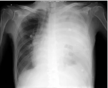
o

C. Laborato- ry tests revealed a low hemoglobin level of 9.6 g/dL, a leukocyte count of 18,250/μL, an elevated proth- rombin time of 19. 7 seconds, and an elevated inter- national normalized ratio of 1.72. Chest radiography revealed total opacification of the left chest (Fig. 1).

He underwent an emergency operation under the sus- picion of esophageal rupture.

On the operating table, upper gastrointestinal en- doscopy revealed an ulcerative fistula lesion measur- ing approximately 2.5 cm at the gastroesophageal ju- nction. The left side was approached by video-assisted thoracoscopic surgery (VATS). The pleural cavity was filled with food material and a foul-smelling fluid.

After decontamination with saline irrigation, a 2.5-cm transmural perforation of the esophagus was identi- fied 2 cm above the diaphragm (Fig. 2A). After the

Korean J Thorac Cardiovasc Surg 2016;49:478-480 □ CASE REPORT □

https://doi.org/10.5090/kjtcs.2016.49.6.478

(2)

Management of Boerhaave Syndrome with T-Tube Drainage

− 479 −

Fig. 2. (A) Ruptured esophagus. Levin tube placed inside the ruptured esophagus (white arrow). (B) T-tube, removed from the patient.

Fig. 1. Chest radiography on admission. Total opacification of the

left chest. Fig. 3. (A, B) Esophagogram on postoperative day 94 showing no

leakage.

defect was debrided, a rubber T-tube (16 Fr), gene- rally used for biliary tree disease, was positioned in- to the rupture and the esophageal wall was closed with polypropylene sutures (Prolene; Ethicon, Somerville,

NJ, USA). The T-tube was then brought out through the lateral chest wall and connected to a drain. Two drainage tubes were placed in the left pleural cavity and mediastinum. In addition, a Jackson-Pratt drain was placed next to the lower esophagus for irrigation. A feeding jejunostomy was also performed through a mini-laparotomy. After surgery, the patient was tra- nsferred from the operating room to the intensive care unit. The total operation time was 220 minutes, and the total anesthesia time was 290 minutes.

Postoperatively, the patient received broad-spectrum

antimicrobials and supportive care. On postoperative

day (POD) 1, the patient was weaned from a mechan-

ical ventilator and transferred to the general ward

on POD 8. He started feeding through a feeding jenu-

nostomy on POD 2. He was discharged on POD 41

with a T-tube and a Jackson-Pratt drainage in good

general condition. After confirming the absence of

discharge through the Jackson-Pratt drain on POD 68,

the T-tube was gradually withdrawn every week and

finally removed on POD 98 in an outpatient clinic

(Fig. 2B). An esophagogram on POD 94 showed no

leakage but a slightly stenotic appearance (Fig. 3). On

POD 94, he restarted an oral diet, and the feeding je-

junostomy was removed on POD 129 at an outpatient

clinic.

(3)

Young Woo Do, et al

− 480 −

Discussion

Prompt decision-making in the management of Bo- erhaave syndrome is crucial because it is a life-thre- atening condition characterized by the disruption of the distal esophagus due to a barotrauma, resulting in the contamination of the mediastinum and pleural cavity with gastric contents. Left untreated, the mor- tality rate exceeds 90%, and has been reported to be as high as 40% even after appropriate surgical inter- vention [1,2].

Patients with Boerhaave syndrome complain of a tearing substernal or epigastric pain, which may radi- ate to the left chest, shoulder, or back. However, some patients present with atypical symptoms such as shock or respiratory distress, and physical exam findings are often non-specific [3]. Unfortunately, the condition is often misdiagnosed as a perforated peptic ulcer or pancreatitis, which can lead to a delay in receiving appropriate treatment [4]. Further evaluation with a chest X-ray, contrast-enhanced CT scan, or gastro- intestinal endoscopy should be performed in any pa- tient suspected to have Boerhaave syndrome.

It is clear that primary surgical repair within 24 hours of spontaneous esophageal perforation reduces mortality. Some authors have reported that good out- comes can be obtained with surgical repair, even af- ter 24 hours [5]. However, delayed management of Boerhaave syndrome is clearly associated with an in- creased risk of mortality.

Sulpice et al. [6] compared T-tube repair and pri- mary repair for the management of Boerhaave syn- drome and found no difference between the 2 treat- ment strategies, even though the time from symptom onset to surgery was longer for T-tube repair. In our case, surgical management was performed 8 days af- ter symptom onset and the mucosa appeared too fri- able to hold sutures. For that reason, we inserted a T-tube and closed the esophagus wall around it for the purpose of controlling salivary drainage, with the hope that the patient could be managed successfully.

In addition to surgical drainage and debridement, adequate postoperative nutritional support is impo-

rtant for the management of Boerhaave syndrome. In this case, placement of a feeding jejunostomy was es- sential to provide nutritional support.

Previously, open thoracotomy was considered the only option for the surgical management of Boerh- aave syndrome. However, recent advances and expe- riences in VATS has led to VATS being used to treat Boerhaave syndrome. Specifically, Haveman et al. [2]

showed that VATS can be used as the first choice for Boerhaave syndrome. Indeed, VATS is a safe proce- dure associated with a lower rate of complications and similar results in comparison with open surgery.

In the present case, we performed VATS to minimize additional surgical trauma for an already critically ill patient.

In conclusion, by inserting a T-tube into the rup- tured esophagus, we successfully managed a late-di- agnosed case of Boerhaave syndrome.

Conflict of interest

No potential conflict of interest relevant to this article was reported.

References

1. Duehring GL. Boerhaave syndrome. Radiol Technol 2000;

72:51-5.

2. Haveman JW, Nieuwenhuijs VB, Kobold JP, van Dam GM, Plukker JT, Hofker HS. Adequate debridement and drain- age of the mediastinum using open thoracotomy or vid- eo-assisted thoracoscopic surgery for Boerhaave’s syndrome.

Surg Endosc 2011;25:2492-7.

3. Van der Weg G, Wikkeling M, van Leeuwen M, Ter Avest E.

A rare case of oesophageal rupture: Boerhaave’s synd- rome. Int J Emerg Med 2014;7:27.

4. Connelly CL, Lamb PJ, Paterson-Brown S. Outcomes follow- ing Boerhaave’s syndrome. Ann R Coll Surg Engl 2013;

95:557-60.

5. Kim KD, Chung KY, Kim CS, Park HG. Delayed primary re- pair of esophageal rupture. Korean J Thorac Cardiovasc Surg 1998;31:46-51.

6. Sulpice L, Dileon S, Rayar M, et al. Conservative surgical

management of Boerhaave’s syndrome: experience of two

tertiary referral centers. Int J Surg 2013;11:64-7.

수치

Fig. 2. (A) Ruptured esophagus. Levin tube placed inside the ruptured esophagus (white arrow)

참조

관련 문서

Clicker를 이용한 학습과 교육 그리고 학습관리 시스템과 clicker의 결합 Using Clicker for learning and teaching clicker with a Learning..

It considers the energy use of the different components that are involved in the distribution and viewing of video content: data centres and content delivery networks

This Presentation contains financial forecasts with respect to Bakkt’s projected financial results, including Total Revenue, Total Revenue less Transaction-Based Expenses

Natural history of OA: Progressive cartilage loss, subchondral

¡ Risk and issue management: It needs to come up with measures to prevent the project from being delayed between planning and implementation stage. The

Table 4.1 Efficiency parameters in the flat plate solar collector using MWCNT nanofluid and water ··· 40.. Table 4.2 Efficiency parameters in the evacuated tube solar

• Observed time difference of arrival with idle period downlink (OTDoA-IPDL). •

and Simmons, J.S., 2002, Long-term performance of passive acid mine drainage treatment systems, Proceedings of the 23rd AnnualWest Virginia Surface Mine Drainage Task