• 검색 결과가 없습니다.

Continuous “Over and Over” Suture for Tricuspid Ring Annuloplasty

N/A
N/A
Protected

Academic year: 2021

Share "Continuous “Over and Over” Suture for Tricuspid Ring Annuloplasty"

Copied!
5
0
0

로드 중.... (전체 텍스트 보기)

전체 글

(1)

http://dx.doi.org/10.5090/kjtcs.2012.45.1.19 ISSN: 2233-601X (Print) ISSN: 2093-6516 (Online)

Department of Thoracic and Cardiovascular Surgery, Dong-A University Medical Center, Dong-A University College of Medicine

†This article was presented at the 42nd Autumn Scientific Meeting of The Korean Society for Thoracic and Cardiovascular Surgery.

Received: March 8, 2011, Revised: October 22, 2011, Accepted: November 11, 2011

Corresponding author: Jong Soo Woo, Department of Thoracic and Cardiovascular Surgery, Dong-A University Medical Center, Dong-A University College of Medicine, 1 Dongdaesin-dong 3(sam)-ga, Seo-gu, Busan 602-715, Korea

(Tel) 82-51-240-5195 (Fax) 82-51-231-5195 (E-mail) jswoo@dau.ac.kr

C

The Korean Society for Thoracic and Cardiovascular Surgery. 2012. All right reserved.

CC
This is an open access article distributed under the terms of the Creative Commons Attribution Non-Commercial License (http://creative- commons.org/licenses/by-nc/3.0) which permits unrestricted non-commercial use, distribution, and reproduction in any medium, provided the original work is properly cited.

Continuous “Over and Over” Suture for Tricuspid Ring Annuloplasty

Kwon-Jae Park, M.D., Jong Soo Woo, M.D., Sang Seok Jeong, M.D., Jung Hoon Yi, M.D.

Background: A ring implantation in the tricuspid annulus requires many interrupted mattress sutures for correction of tricuspid regurgitation (TR). In this study, tricuspid ring annuloplasty was performed by 2-0 polypropylene con- tinuous suture instead of multiple interrupted 2-0 polyester mattress sutures, and the efficacy of the method was evaluated. Materials and Methods: This study included 20 patients who underwent tricuspid ring annuloplasty by continuous suture between May 2009 and July 2010. Four of the patients had an isolated TR, and the rest had a left-sided cardiac lesion. The concomitant tricuspid annuloplasty was performed after the left-sided heart surgery was completed and a Duran flexible ring prosthesis was used. Results: There was no perioperative mortality or conduction problem. More than a moderate degree of TR was improved to less than a mild degree after the procedure. After the ring annuloplasty, the right atrial volume decreased from 123.7±69.2 mL to 74.5±37.4 mL, and the mean right atrial pressure was lowered from 18.7±12.2 mmHg to 8.9±5.5 mmHg. Conclusion: The continuous

“over and over” suture may be a useful procedure for fixing the ring to the annulus and making an intentional an- nular placation in performing tricuspid ring annuloplasty.

Key words: 1. Tricuspid valve

2. Tricuspid valve insufficiency 3. Tricuspid valve repair

INTRODUCTION

Functional tricuspid valve regurgitation (TR) associated with mitral valve disease has not been considered to be seri- ous because it can be improved after the mitral valve lesion is surgically managed. In patients undergoing mitral valve surgery, an isolated tricuspid valve surgery for remnant or ag- gravated severe TR usually indicates a poor outcome with high perioperative mortality, poor survival, and no significant improvement in functional capacity [1,2].

Tricuspid annuloplasty (TA) without a ring prosthesis has shown a relatively high recurrence rate of TR [3]. Compared with it, ring annuloplasty for functional tricuspid valve regur- gitation improved the long-term clinical outcome and de- creased the recurrence rate of TR [4,5].

Tricuspid ring annuloplasty is usually performed with many interrupted mattress sutures to affix the ring to the annulus.

However, incorrect suturing in the annulus may poorly fix the

ring to the annulus and result in uneven plication of the di-

lated annulus, resulting in remnant TR. With this concern, we

(2)

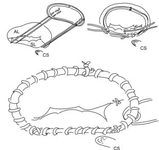
Fig. 1. Tricuspid ring annuloplasty using continuous suture technique. CS=coronary sinus; SL=septal leaflet; AL=anterior leaflet.

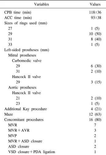
Table 1. Preoperative characteristics of patients undergoing tricus- pid ring annuloplasty (n=20)

Characteristics Preoperative

values Age (yr)

Sex (male:female) Follow-up interval (mo) NYHA III, IV Tricuspid valve lesion

Functional:traumatic (chorda rupture) Mitral valve lesion

Rheumatic:degenerative:prosthesis dysfunction Isolated TR

Previous operations (DVR, ASD closure, MVR, LA myxoma excision)

Atrial fibrillation Renal failure Previous CVA

60.2±11.6 8:12 8.4±5.4 10 (50)

19:1

9:3:1 4 (20) 4 (20)

13 (65) 3 (15) 3 (15) Values are presented as mean±standard deviation or number (%).

NYHA=New York Heart Association; TR=tricuspid regurgita- tion; DVR=double valve replacement; ASD=atrial septal defect;

MVR=mitral valve replacement; LA=left atrium; CVA=cere- brovascular accident.

performed a ring annuloplasty using the continuous suture technique and assessed the clinical outcome of the ring annu- loplasty for functional TR.

MATERIALS AND METHODS 1) Patients

From May 2009 to July 2010, 20 patients (male/female, 8/12; mean age, 60 years) with functional TR underwent tri- cuspid ring annuloplasty using a continuous suture technique.

The indications for tricuspid ring annuloplasty were mild to severe TR, marked annular dilatation (tricuspid annulus diam- eter 4 cm or more by echocardiography) irrespective of any TR grade. Nineteen of them had functional TR without an or- ganic change in the valve leaflet and one patient had TR with chordal rupture secondary to blunt chest trauma. Four patients had isolated TR and 16 had functional TR secondary to left heart valve disease or valve surgery. Ten patients were in more than New York Heart Association (NYHA) Class III (Table 1). Perioperative changes in the degree of TR were

evaluated by transthoracic echocardiography. The last post- operative echocardiographic results, which were available in 16 patients, were obtained more than 6 months postope- ratively.

2) Surgical procedures

After a median sternotomy, cardiopulmonary bypass was initiated with a standard aortic cannula and two vena cava cannulas. The tricuspid valve repair was performed after the left cardiac procedures were completed. The ring annuloplasty was performed on an arrested heart in 17 patients and on a beating heart in three. For the ring annuloplasty, a Duran ring was used in all patients. Two septal sutures at the ante- roseptal and posteroseptal commissures with 2-0 polypro- pylene were placed and then the suture was passed through the two markers of the ring. The ring was lowered down to the annulus and one arm of each suture was passed along the septal annulus in an over-and-over fashion and they were tied when they met. The other arm of each suture was passed along the anterior and posterior annulus and then tied. In this way, the annulus size was reduced equidistantly (Fig. 1).

3) Statistical analysis

Continuous values are expressed as the mean±standard de-

viation and were compared by paired and unpaired t-tests.

(3)

Table 2. Patient surgical characteristics

Variables Values

CPB time (min) ACC time (min) Sizes of rings used (mm) 27

29 31 33

Left-sided prostheses (mm) Mitral prostheses Carbomedic valve 29

31

Hancock II valve 29

Aortic prostheses Hancock II valve 21

23

Additional Kay procedure Maze

Concomitant procedures MVR

MVR+AVR MVP

MVR+ASD closure ASD closure

VSD closure+PDA ligation

118±36 93±38

1 (5) 10 (50) 8 (40) 1 (5)

6 (30) 2 (10)

3 (15)

2 (10) 1 (5) 4 (21) 12 (63) 16 (80)

7 3 2 1 2 1 Values are presented as mean±standard deviation or number (%).

CPB=cardiopulmonary bypass; ACC=aortic cross clamp; MVR=

mitral valve replacement; AVR=aortic valve replacement; MVP=

mitral valvuloplasty; ASD=atrial septal defect; VSD=ventricular septal defect; PDA=patent ductus arteriosus.

Categorical variables were analyzed by Fisher's exact test us- ing cross tabulation, and categorical ordinal variables were compared by the Wilcoxon signed rank test. The changes and differences of values were considered statistically significant at a p<0.05.

RESULTS

The surgical data are described in Table 2. There was no perioperative mortality or any conduction problems. One pa- tient required placement of a permanent pacemaker due to si- nus dysfunction. The NYHA class of the patients improved

and the grade of TR was reduced to less than mild after the operation. After the ring annuloplasty using the continuous suture technique, perioperative echocardiographic values were significantly improved (Table 3). At the mean follow-up du- ration, none has aggravation of the degree of TR or the an- nular dimension (Table 4).

DISCUSSION

In an early study, Braunwald et al. [6] suggested that sec- ondary TR resolves after mitral valve replacement (MVR) and a concomitant tricuspid procedure is not needed.

However, nowadays, it is accepted that TR might not im- prove after mitral valve surgery and even regurgitation might aggravate as much as 20 years later [7-10]. Although there is no complete consensus on the guidelines for tricuspid valve (TV) repair, TV repair is usually indicated for severe TR in patients undergoing mitral valve surgery, concomitant TV re- pair with a TA diameter >40 mm or moderate TR, sympto- matic functional TR despite medical management, or sympto- matic isolated TR late after left-sided valve surgery. Howev- er, the American College of Cardiology/American Heart Association guidelines do not clearly recommend TV repair when TR is less than severe in patients undergoing mitral valve surgery [11-13]. In general, a more aggressive con- comitant tricuspid annuloplasty is recommended at the initial major procedure because the delayed secondary TR and reop- eration for residual TR show a poor outcome [14-17].

The most common finding of functional TR is an annular

dilatation, which subsequently disturbs coaptation of tricuspid

leaflets, secondary to the right ventricular dilatation. Several

methods such as suture annuloplasty, ring annuloplasty, annu-

loplasty using tissue materials, and even tricuspid valve re-

placement have been used for elimination of functional

regurgitation. Recent studies have demonstrated that ring an-

nuloplasty is the most effective of the techniques, but 14% of

patients with ring annuloplasty had remnant or recurrent TR

of 3 to 4+ [18]. We believe that the early residual regur-

gitation was related to the suture technique for ring

implantation. The usual suture technique for ring implantation

is interrupted mattress sutures that are placed along the tricus-

pid annulus and enable gathering up the annulus, resulting in

(4)

Table 3. Preoperative and postoperative echocardiographic findings

Variables Preoperative value Postoperative value p-value

NYHA class

Tricuspid annulus size (mm) Pulmonary arterial pressure (mmHg) Right ventricular function

Size grade of right ventricle Right atrial pressure (mmHg) Right atrial volume (mL) Inferior vena cava diameter

Interventricular septal shifting to left ventricle (no.) Tricuspid regurgitation grade

2.7±0.8 31.7±6.3 67.6±22.9

1.1±0.9 1.3±1.2 17.9±10.9 120.2±68.1 2.35±0.44

7 2.47±0.49

1.2±0.4 19.8±6.2 48.1±15.3

0.6±0.9 0.2±0.6 8.1±3.9 69.7±30.9 2.07±0.39

4 0.82±0.51

0.00 0.00 0.006 0.013 0.002 0.002 0.001 0.024 0.026 0.00

Values are presented as mean±standard deviation. Ordinal values are expressed as follows: normal=0, mild=1, moderate=1, severe=3, none=0, trace=0.5, mild=1, moderate=2, severe=3.

NYHA=New York Heart Association.

Table 4. Echocardiographic findings at more than 6 months after operation (n=16)

Variables Postoperative

values Mean follow-up interval (mo)

Tricuspid annular dimension (mm) Pulmonary arterial pressure (mmHg) Right ventricular function

Right ventricular size Right atrial pressure (mmHg) Right atrial volume (mL) Inferior vena cava diameter

Interventricular septal shifting to left ventricle Tricuspid regurgitation grade

12.4±6.6 19.0±1.6 35.9±14.4 0.35±0.49 0.29±0.47 7.2±3.8 70.2±33.0

1.9±0.4 0 0.79±0.25 Values are presented as mean±standard deviation. Ordinal val- ues are expressed as follows: normal=0, trace=0.5, mild=1, moderate=2, severe=3.

a reduction in annular size [19,20]. The interrupted sutures cannot always make intentional even plication of the tricuspid annulus. However, when the annular plication is not made with the continuous suture technique, the suture can be re- leased before tying and the annular plication length be redistributed. We concluded that interrupted sutures make it difficult to adjust the annulus size by the areas of the anterior and posterior leaflets but a continuous suture can be easily performed to allow the adjustment of the size to fit the leaf- let area. For these reasons, we changed the suture technique.

TR is strongly related to the annulus dimension: TR was

seen when the dimension was larger than 34 mm [21]. In our study, the preoperative mean annulus dimension measured by echocardiography was 31.7 mm and even a tricuspid annulus measured 22 mm in diameter showed moderate regurgitation.

Postoperatively, the mean annular dimension was 19.8 mm and all the patients revealed less than mild regurgitation, which even 6 months later was still not aggravated in any of the patients. In addition, the ring annuloplasty improved the postoperative hemodynamic values and did not result in peri- operative mortality or conduction problems. These results suggest that the continuous suture technique for tricuspid ring implantation is safe and effective.

This study is limited by the small number of patients and short study duration; a report on longer-term surgical out- comes is needed.

CONCLUSION

Various methods of tricuspid annuloplasty have been used to manage TR. To obtain better outcomes, a ring prosthesis should be used when tricuspid annuloplasty is performed.

Instead of the interrupted mattress suture technique using

many sutures, a continuous suture technique for the ring im-

plantation in tricuspid annulus is associated with reasonable

postoperative clinical outcomes. Therefore, this method is

thought to be a useful way of performing tricuspid ring

annuloplasty.

(5)

REFERENCES

1. Mangoni AA, DiSalvo TG, Vlahakes GJ, Polanczyk CA, Fifer MA. Outcome following isolated tricuspid valve repla- cement. Eur J Cardiothorac Surg 2001;19:68-73.

2. Kwon DA, Park JS, Chang HJ, et al. Prediction of outcome in patients undergoing surgery for severe tricuspid regur- gitation following mitral valve surgery and role of tricuspid annular systolic velocity. Am J Cardiol 2006;98:659-61.

3. Rivera R, Duran E, Ajuria M. Carpentier's flexible ring ver- sus De Vega's annuloplasty: a prospective randomized stu- dy. J Thorac Cardiovasc Surg 1985;89:196-203.

4. Matsuyama K, Matsumoto M, Sugita T, et al. De Vega an- nuloplasty and Carpentier-Edwards ring annuloplasty for secondary tricuspid regurgitation. J Heart Valve Dis 2001;

10:520-4.

5. Tang GH, David TE, Singh SK, Maganti MD, Armstrong S, Borger MA. Tricuspid valve repair with an annuloplasty ring results in improved long-term outcomes. Circulation 2006;114(1 Suppl):I577-81.

6. Braunwald NS, Ross J Jr, Morrow AG. Conservative man- agement of tricuspid regurgitation in patients undergoing mitral valve replacement. Circulation 1967;35(4 Suppl):I63-9.

7. Porter A, Shapira Y, Wurzel M, et al. Tricuspid regurgita- tion late after mitral valve replacement: clinical and echo- cardiographic evaluation. J Heart Valve Dis 1999;8:57-62.

8. Simon R, Oelert H, Borst HG, Lichtlen PR. Influence of mi- tral valve surgery on tricuspid incompetence concomitant with mitral valve disease. Circulation 1980;62(2 Pt 2):I152- 7.

9. Breyer RH, McClenathan JH, Michaelis LL, McIntosh CL, Morrow AG. Tricuspid regurgitation: a comparison of non- operative management, tricuspid annuloplasty, and tricuspid valve replacement. J Thorac Cardiovasc Surg 1976;72:867- 74.

10. Shafie MZ, Hayat N, Majid OA. Fate of tricuspid regur- gitation after closed valvotomy for mitral stenosis. Chest 1985;88:870-3.

11. Jamieson WR, Cartier PC, Allard M, et al. Surgical manage- ment of valvular heart disease 2004. Can J Cardiol 2004;20 Suppl E:7E-120E.

12. American College of Cardiology; American Heart Associa- tion Task Force on Practice Guidelines (Writing Committee to revise the 1998 guidelines for the management of patients with valvular heart disease); Society of Cardiovascular Anesthesiologists, et al. ACC/AHA 2006 guidelines for the management of patients with valvular heart disease: a report of the American College of Cardiology/American Heart Association Task Force on Practice Guidelines (writing Committee to Revise the 1998 guidelines for the management of patients with valvular heart disease) developed in collabo- ration with the Society of Cardiovascular Anesthesiologists endorsed by the Society for Cardiovascular Angiography and Interventions and the Society of Thoracic Surgeons. J Am Coll Cardiol 2006;48:e1-148.

13. Vahanian A, Baumgartner H, Bax J, et al. Guidelines on the management of valvular heart disease: The Task Force on the Management of Valvular Heart Disease of the European Society of Cardiology. Eur Heart J 2007;28:230-68.

14. King RM, Schaff HV, Danielson GK, et al. Surgery for tri- cuspid regurgitation late after mitral valve replacement.

Circulation 1984;70(3 Pt 2):I193-7.

15. Cohn LH. Tricuspid regurgitation secondary to mitral valve disease: when and how to repair. J Card Surg 1994;9(2 Suppl):237-41.

16. Matsuyama K, Matsumoto M, Sugita T, Nishizawa J, Tokuda Y, Matsuo T. Predictors of residual tricuspid regur- gitation after mitral valve surgery. Ann Thorac Surg 2003;

75:1826-8.

17. Duran CM, Pomar JL, Colman T, Figueroa A, Revuelta JM, Ubago JL. Is tricuspid valve repair necessary? J Thorac Cardiovasc Surg 1980;80:849-60.

18. McCarthy PM, Bhudia SK, Rajeswaran J, et al. Tricuspid valve repair: durability and risk factors for failure. J Thorac Cardiovasc Surg 2004;127:674-85.

19. McCarthy JF, Cosgrove DM 3rd. Tricuspid valve repair with the Cosgrove-Edwards Annuloplasty System. Ann Thorac Surg 1997;64:267-8.

20. Duran CM. Duran ring annuloplasty of the tricuspid valve.

Oper Tech Thorac Cardiovasc Surg 2003;8:201-12.

21. Groves PH, Hall RJ. Late tricuspid regurgitation following

mitral valve surgery. J Heart Valve Dis 1992;1:80-6.

수치

Fig. 1. Tricuspid ring annuloplasty using continuous suture  technique. CS=coronary sinus; SL=septal leaflet; AL=anterior leaflet.
Table 2. Patient surgical characteristics
Table 3. Preoperative and postoperative echocardiographic findings

참조

관련 문서

• The avoidance of unwanted heat loss or gain, and the positive use of sun energy for heating and cooling … become compelling principles of site

• For the continuous random variable Y the probability density function f ( y ) can be represented by an equation , which can be described graphically by a curve. •

It is a responsibility for Australian citizens aged 18 years or over to vote in federal and state or territory elections, and in a referendum, which is a vote to

Oh, Composite Hurwitz rings satisfying the ascending chain conditions on principal ideals, Kyungpook Math.. Oh, Irreducibility of Hurwitz polynomials over the

The estimated capital cost for a chemical plant using the study estimate method (Class 4) was calculated to be $2 million.. If the plant were to be built, over what range

In this thesis, we introduce a Hurwitz polynomial ring and study the irreduciblity of Hurwitz polynomials over  We also investigate the irreduciblilty of

ƒ Monosaccharide may be present in the form of Monosaccharide may be present in the form of a linear or ring structure. ƒ In solution it is in the form of

 removal of induction period ~ adding acylating agent p181 active