Received: December 4, 2017 Revised: December 7, 2017 Accepted: December 12, 2017
Copyright © 2017. The Korean Academy of Oral &
Maxillofacial Implantology
This is an Open Access article distributed under the terms of the Creative Commons Attrib- ution Non-Commercial License (http://creative- commons.org/licenses/by-nc/4.0/) which permits unrestricted non-commercial use, distribution, and reproduction in any medium, provided the original work is properly cited.
pISSN : 1229-5418
Implantology 2017; 21(4): 218-224 https://doi.org/10.12972/implantology.20170017
eISSN : 0000-0000 OPEN ACCESS
술자의 숙련도에 따른 임플란트 식립 경향에 관한 후향적 연구
이홍석1, 이성조1, 송영균2, 조인우1, 박정철1, 신현승1*
1
단국대학교 치과대학 치주과학교실
2
단국대학교 치과대학 보철학교실
Research of Tendency for Implant Placement According to Experience of Operator:
A Retrospective Study
Hong-Seok Lee
1, Sung-Jo Lee
1, Young-Gyun Song
2, In-Woo Cho
1, Jung-Chul Park
1, Hyun-Seung Shin
1*
1
Department of Periodontology, College of Dentistry, Dankook University, Cheonan, Korea
2
Department of Prosthodontics, College of Dentistry, Dankook University, Cheonan, Korea
*Corresponding author: Hyun-Seung Shin, perioshin@dankook.ac.kr
Abstract
Increasing attention has been drawn to the surgeon factor in the implant failure due to the implant fracture or osseointegration. The purpose of this study is to compare the location of implants placed by 2 surgeon groups, periodontal residents and professors, analyzing the distance between adjacent natural teeth and implants, or 2 adjacent implants. The charts and radiographs of patients who received dental implants between January 2014 and December 2015 in the Department of periodontology, Dankook University Dental Hospital were examined. A total of 1306 implants placed on maxillary and mandibular second premolar, first molar and second molar area were evaluated in this retrospective analysis. The implants were classified into two experimental groups: the implants placed posterior to the natural teeth and the implants placed posterior to the other implant. The implants in each group were also categorized into 3 subgroups by the location in the jaw whether they were in the second premolar, the first molar or the second molar areas. The distance was measured on the digital panoramic radiographs taken after the implant surgery. In the group of the implants placed posterior to the natural teeth, the distance between natural teeth and implants planted by the professors and periodontal residents were 2.94 ± 1.14 mm and 3.14 ± 1.24 mm, which showed significant difference.
In the group of the implants placed posterior to the other implants, the distance between implants placed by the professors and periodontal residents were 3.35 ± 1.34 mm and 3.62 ± 1.41 mm, which showed significant difference. The implants placed in the first molar area showed significant difference in terms of the distance; 3.30 ± 1.14 mm by the professors, and 3.76 ± 1.10 mm by the periodontal residents. The following conclusion can be achieved on the basis of the results of this study.
1. In the group of the implants placed posterior to the natural teeth, the periodontal residents placed implant significantly more distally from the natural teeth than the professors.
2. In the group if the implants placed posterior to the other implants, the periodontal residents placed implant significantly more distally from the adjacent implant than the professors. In case of placing implants in the first molar area, the periodontal residents placed implant significantly more distally form the adjacent implant than the professors.
I. 서론
임플란트는 완전무치악, 부분무치악 환자에 있어 상실치 수복을 위한 예지성 있는 치료 방법으로 여 겨지고 있으며
1
, 1960년대에 처음 치과계에 소개된 이후로 오늘날에 이르기까지 그 수요는 꾸준히 늘 어나고 있다2
. 그에 따라 치과용 임플란트의 성공률을 높이기 위해 다양한 표면처리 방법이 개발되기 도 하였으며3
, 상악동 거상술과 같은 수술기법이 등장하기도 하는 등 여러 방면으로의 발전이 이루어 져 왔다. 그 결과 다양한 임상적 상황에서 일반적으로 90% 이상의 높은 생존률이 보고되고 있다4,5
.그러나 실제 임상에서는 임플란트의 파절이나 골유착 실패 등으로 인한 임플란트의 실패가 발생할 수 있는데, 그 동안은 이에 대해 임플란트의 재료적 특성이나 환자의 문제에 대한 관점으로 접근하는 경우 많았다. 그러나 2001년 Albrektsson은 스웨덴 예테보리대학에서 식립한 1,000개의 임플란트 중 40%가 단 1명의 술자에 의해 식립되었기 때문에, 술자 관련 위험요소에 대해 더 강조할 필요가 있음을 주장하는 등
6
, 술자에 의한 변수에도 점점 주목하는 추세이다.이러한 추세를 반영하여 최근에는 초심자를 위한 임플란트 교육 프로그램이 점점 늘어나고 있다. 치 과 임플란트 수술에 관해서는 동물 또는 모델실습 등을 통하여 교육이 이루어지고 있지만, 아직 체계적 인 방법은 제시된 바가 없다. 하지만 Park 등에 의해 implant surgical technique assessment and rating system (iSTAR)이라는 평가도구가 개발되거나, 내비게이션 시스템을 만들고자 하는 등 더 나은 술자 교육을 위한 연구가 진행되고 있다
7,8
.술자의 임상적 숙련도에서 중요하게 생각되는 요소로는 지식, 의사소통 기술, 술기의 3가지 요소가 있다. 임플란트 수술의 술기에 있어서 평가할 수 있는 사항은 많지만 그중 중요하면서도 쉽게 측정할 수 있는 사항으로는 식립한 임플란트와 인접 치아나 임플란트와의 간격을 들 수 있다. Tarnow 등은 2개 의 인접한 임플란트를 식립할 때 3 mm 이하의 간격이 되도록 식립할 경우 치간골의 수직적 골소실을 유발한다고 보고하였다
9
. 따라서 3 mm 이상의 간격이 되도록 식립할 것을 권유하였다. 이와 마찬가지 로 자연치와 임플란트 간 간격 역시 적어도 1.5-2 mm 이상이어야 한다고 권유되고 있다10-12
. 이와는 반 대로 임플란트 간 간격이 너무 멀 경우, 부적절한 contour를 가질 뿐만 아니라 cantilever 방식의 힘을 받게 된다13,14
. 이는 임플란트 보철물에 더 많은 응력이 집중될 수 있기에 가급적 피하는 것이 권유된다15
. 따라서 인접치가 자연치든 임플란트든 너무 가깝게 식립하는 것도 피해야 하지만 너무 멀리 식립하는 것도 피하는 것이 좋다.본 연구의 목적은 초심자인 치과병원 전공의와 전문가인 교수진 간의 임플란트 식립에 있어서 인접 자연치, 임플란트와의 간격을 후향적으로 비교하여 술식에 유의한 차이가 있는지 비교하는 것이다.
II. 연구재료 및 방법
1. 연구대상
2014년 1월부터 2015년 12월 사이에 단국대학교 치과대학 부속치과병원 치주과에서 식립한 임플 란트 중 상, 하악 제2소구치, 제1,2대구치 부위에 surgical stent를 사용하지 않고 식립된 임플란트, 총 1306개의 임플란트를 대상으로 진료기록부 및 방사선 사진 자료를 토대로 후향적 분석을 시행하였다.
각 임플란트는 자연치 후방에 식립한 임플란트와 임플란트 후방에 식립한 임플란트로 실험군을 분류 했다. 또한 식립 부위에 따라 제2소구치, 제1,2대구치에 식립한 임플란트로 분류했다.
2. 연구방법 1) 방사선학적 검사
모든 임플란트에 있어서 식립 후 촬영한 디지털 파노라마 방사선 사진을 이용하여 간격을 측정하였 다. 각 임플란트의 근심면 최상부를 기준점으로 하여 근심 인접한 치아 또는 임플란트의 원심면까지의 거리를 0.1 mm까지 계측하였다. 계측 시 발생하는 오차를 줄이기 위해 1인의 치과의사가 모든 계측을 시행하였다.
2) 통계학적 분석
각 실험군에 있어서 계측된 자료들을 식립한 술자에 따라서 비교 분석하였다. 통계프로그램으로는 SPSS ver. 20 (SPSS Inc. Chicago, IL, USA)을 사용하였다. Shapiro-Wilk test나 central limit theorem 을 이용하여 각 실험군에서 정규성 만족 여부를 확인하였고, 모수적 검정방법인 Independent samples t-test를 이용하여 분석하였다.
III. 연구결과
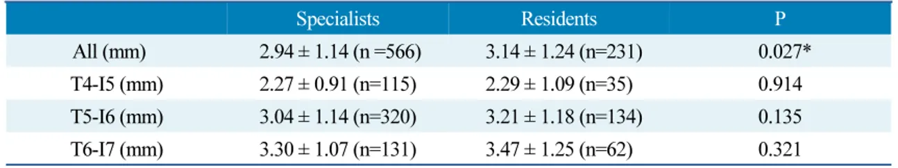
1. 자연치 후방에 식립한 임플란트의 비교
자연치 후방에 식립한 임플란트 전체를 대상으로 술자에 따라 비교했을 때, 교수진이 식립한 임플란 트와 전공의가 식립한 임플란트 간에는 통계학적으로 유의한 차이가 있었다. 하지만 식립부위별로 비 교했을 때는 유의한 차이가 없었다
(Table 1).
Table 1. Distance between implant and mesial adjacent tooth
Specialists Residents P
All (mm) 2.94 ± 1.14 (n =566) 3.14 ± 1.24 (n=231) 0.027*
T4-I5 (mm) 2.27 ± 0.91 (n=115) 2.29 ± 1.09 (n=35) 0.914
T5-I6 (mm) 3.04 ± 1.14 (n=320) 3.21 ± 1.18 (n=134) 0.135
T6-I7 (mm) 3.30 ± 1.07 (n=131) 3.47 ± 1.25 (n=62) 0.321
T: natural tooth, I: implant, *: p<0.05.
Hong-Seok Lee et al. : Research of Tendency for Implant Placement According to Experience of Operator: A Retrospective Study. Implantology 2017
2. 임플란트 후방에 식립한 임플란트의 비교
임플란트 후방에 식립한 임플란트 전체를 대상으로 비교했을 때에는 술자 간 유의한 차이가 있었다.
또한 제1대구치 부위에 식립했을 때에도 술자 간 유의한 차이가 있었다. 그 외 다른 부위에서는 유의한 차이가 발견되지 않았다
(Table 2).
Table 2. Distance between implant and mesial adjacent implant
Specialists Residents P
All (mm) 3.35 ± 1.34 (n=382) 3.62 ± 1.41 (n=127) 0.047*
I4-I5 (mm) 2.56 ± 1.30 (n=66) 2.58 ± 0.91 (n=21) 0.943
I5-I6 (mm) 3.30 ± 1.14 (n=121) 3.76 ± 1.10 (n=38) 0.028*
I6-I7 (mm) 3.64 ± 1.37 (n=195) 3.87 ± 1.55 (n=68) 0.262 I: implant, *: p<0.05.
Hong-Seok Lee et al. : Research of Tendency for Implant Placement According to Experience of Operator: A Retrospective Study. Implantology 2017
IV. 총괄 및 고찰
자연치 후방에 식립한 경우 1.5-2 mm 이상의 간격이 권장되는데 교수진의 경우 2.94 ± 1.14 mm, 전 공의의 경우 3.14 ± 1.24 mm로 유의한 차이를 보였다. 식립부위 별로 보았을 때는 유의한 차이를 보이 지는 않았지만 전체적으로 보았을 때, 교수진이 더 적절한 간격에 가깝게 식립한 것으로 볼 수 있다.
임플란트 후방에 식립한 경우 3 mm 이상의 간격이 권장되는데 교수진의 경우 3.35 ± 1.34 mm, 전공 의의 경우 3.62 ± 1.41 mm로 유의한 차이를 보였다. 또한 제1대구치 부위에 식립한 경우에도 각각 3.30 ± 1.14 mm, 3.76 ± 1.10 mm로 유의한 차이를 보였으며, 다른 부위에서는 유의한 차이를 보이지
임플란트를 식립할 때, 인접 자연치나 임플란트와의 간격에 대한 연구는 interproximal papilla의 소 실이 발생하지 않도록 일정 간격 이상이 될 것을 강조한다
10-12
. 반면 간격이 너무 멀 때 생기는 단점에 대해 강조한 연구는 상대적으로 수가 적다. 따라서 수련의가 임플란트를 식립하게 될 경우, 인접 자연 치나 임플란트와의 간격이 너무 짧아질 것을 두려워하여 교수진에 비해 유의하게 먼 거리에 식립하게 되었을 것으로 생각된다.다양한 수련기관에서 전공의나 임플란트 교육 프로그램 이수자가 식립한 임플란트에 대한 연구를 시행한 바 있다. Melo 등은 전공의가 식립한 175개의 임플란트에 대해 수련기간에 따른 생존률을 비교 하였는데 유의차가 없다고 보고하였다
16
. 또한 Starr 등은 전공의가 식립한 790개의 임플란트를 대상 으로 조사하였을 때 96.6%의 생존률을 보여 기존의 숙련된 임상가들이 발표한 논문에 비견될 만큼의 수치를 보였다고 하였다17
. Vidal 등은 전공의가 즉시 식립한 62개의 임플란트를 1년 간 관찰했을 때 100%의 성공률을 보였다고 하였으며18
, 전공의가 식립한 하악 overdenture를 위한 임플란트 100개를 대상으로 2년 간 관찰하여 97.7%의 성공률 보였다고 보고하였다19
. Bell 등은 University of Texas Health Science Center의 치과대학 학생을 대상으로 임플란트 교육 프로그램을 4년 간 운영하였는데 임플란트 식립, 보철물 제작, 유지관리까지 학생들이 직접 시행한 120개의 임플란트를 대상으로 관찰 한 결과 실패한 경우가 한 건도 없었다고 보고하였다20
. Hussaini 등은 임플란트 교육 프로그램 이수자 가 식립한 217개의 임플란트와 숙련자가 식립한 299개의 임플란트를 비교한 결과, 생존률이 각각 93.5%와 96%로 유의한 차이가 없었다고 보고하였다21
. 기존의 연구들을 봤을 때 초심자와 전문가가 식립한 임플란트의 성공률이나 생존률을 비교한 결과는 유의한 차이가 없는 경우가 대부분이었으나, 본 연구에서 임플란트 식립 간격을 비교하였을 때에는 유의한 차이가 있다는 결과가 나왔다.임플란트 직경이나 식립 개수를 고려하지 않았고 상, 하악이나 좌우 구분 없이 조사한 점이 이 연구 의 한계점이라 할 수 있다. 이러한 점까지 고려된 전향적 연구가 진행된다면 더 의미 있는 결과를 얻을 수 있을 것으로 생각된다.
V. 결론
본 연구의 결과를 바탕으로 다음의 결론을 얻을 수 있다.
1. 자연치 후방에 임플란트를 식립 하였을 때 전공의의 경우 3.14 ± 1.24 mm, 교수진의 경우 2.94 ± 1.14 mm로 전공의가 교수진보다 유의적으로 더 먼 위치에 식립하였다. 식립부위 별 유의차는 없었다.
2. 임플란트 후방에 임플란트를 식립 하였을 때 전공의의 경우 3.62 ± 1.41 mm, 교수진의 경우 3.35 ± 1.34 mm로 전공의가 교수진보다 유의적으로 더 먼 위치에 식립하였다. 제1대구치 부위에 식립한
경우에도 전공의가 3.76 ± 0.110 mm, 교수진이 3.30 ± 0.114 mm 로 전공의가 교수진보다 유의적 으로 더 먼 위치에 식립하였다. 그 외 부위에서의 유의차는 없었다.
References